Pulmonary PE
advertisement
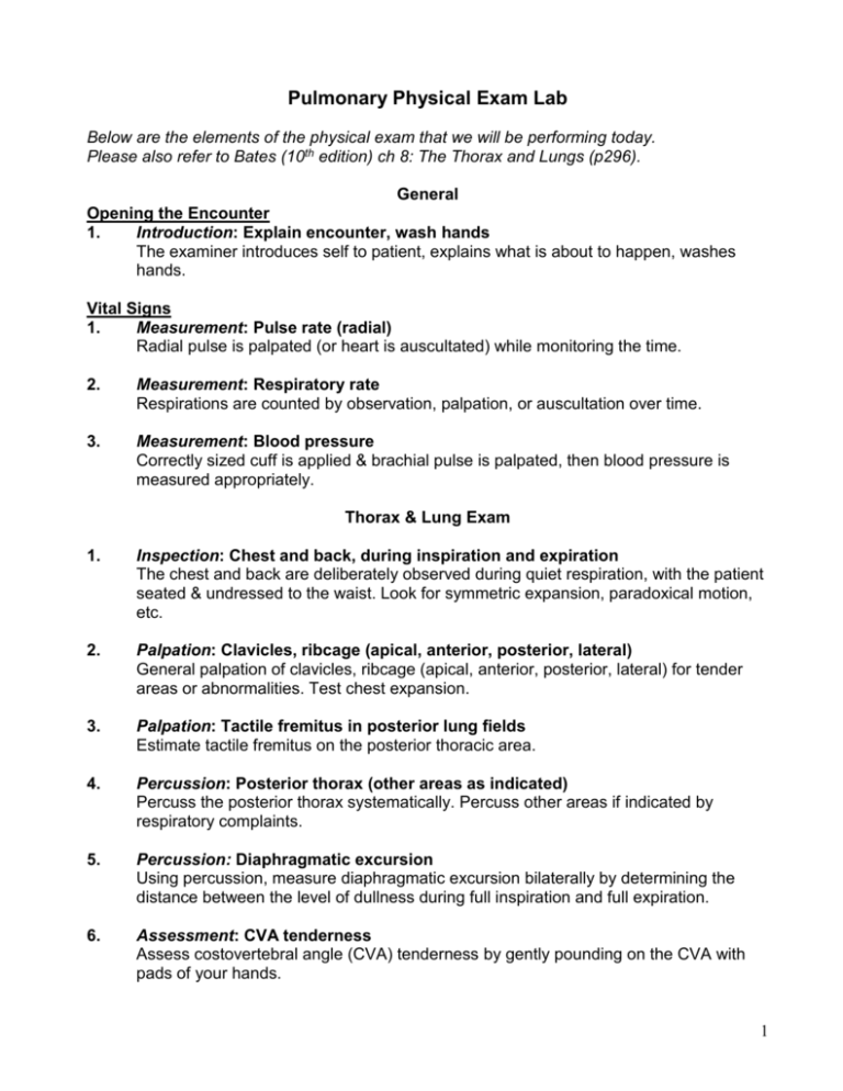
Pulmonary Physical Exam Lab Below are the elements of the physical exam that we will be performing today. Please also refer to Bates (10th edition) ch 8: The Thorax and Lungs (p296). General Opening the Encounter 1. Introduction: Explain encounter, wash hands The examiner introduces self to patient, explains what is about to happen, washes hands. Vital Signs 1. Measurement: Pulse rate (radial) Radial pulse is palpated (or heart is auscultated) while monitoring the time. 2. Measurement: Respiratory rate Respirations are counted by observation, palpation, or auscultation over time. 3. Measurement: Blood pressure Correctly sized cuff is applied & brachial pulse is palpated, then blood pressure is measured appropriately. Thorax & Lung Exam 1. Inspection: Chest and back, during inspiration and expiration The chest and back are deliberately observed during quiet respiration, with the patient seated & undressed to the waist. Look for symmetric expansion, paradoxical motion, etc. 2. Palpation: Clavicles, ribcage (apical, anterior, posterior, lateral) General palpation of clavicles, ribcage (apical, anterior, posterior, lateral) for tender areas or abnormalities. Test chest expansion. 3. Palpation: Tactile fremitus in posterior lung fields Estimate tactile fremitus on the posterior thoracic area. 4. Percussion: Posterior thorax (other areas as indicated) Percuss the posterior thorax systematically. Percuss other areas if indicated by respiratory complaints. 5. Percussion: Diaphragmatic excursion Using percussion, measure diaphragmatic excursion bilaterally by determining the distance between the level of dullness during full inspiration and full expiration. 6. Assessment: CVA tenderness Assess costovertebral angle (CVA) tenderness by gently pounding on the CVA with pads of your hands. 1 7. Auscultation: Systematically, bilaterally, including RML Auscultation systematically, bilaterally (superior and inferior, posterior and anterior), including right middle lobe (RML). If abnormal lung sounds, ask patient to cough. 8. Auscultation: Assess bronchophony (99), whispered pectoriloquy (99), egophony (E-to-A). Assess for bronchophony (voices sounds are louder and clearer), whispered pectoriloquy (patient speaks or whispers “99-99-99”. If abnormal, 99s are amplified), and egophony (patient says “Eeeeee”. If abnormal, sounds like “Aaaay”). Special Maneuvers: Forced Expiratory Time. Ask Patient to take a deep breath and then breathe out as quickly and completely as possible through their mouth. Cardiovascular Exam (as time allows) Heart 1. Inspection and Palpation: Heaves, thrills, and PMI. The total precordial area is inspected & palpated for heaves & thrills. The exact location of the PMI is palpated, in left lateral recumbent position if necessary. 2. Auscultation: 4 areas for heart sounds. Note apical pulse. Auscultate for transmitted cardiac sounds in four areas (Aortic, Pulmonic, Tricuspid, Mitral), using the diaphragm & bell, while the patient is seated & supine. Note the apical pulse (S1) & correlate with the peripheral (radial) pulse. 3. Inspection: Neck vein distention and pulsations at 45 degrees The neck veins are observed for distention or pulsations with the patient lying at a 45° angle. 4. Auscultation: Carotids, (using bell) The carotids are auscultated using the bell while the patient pauses in breathing. Extremities 5. Inspection: Extremities. Inspect skin & hair distribution on extremities (observing rashes, swelling, & color). Fingernails are examined for clubbing, & capillary refill. Hands are examined (observing size, symmetry, swelling, and color). 6. Palpation: Differences in skin temperature. Assess edema. Deliberately palpate the skin on extremities, using dorsum of the hand to note discrepancies of skin temperature. Edema should be assessed in the lower extremities or dependent regions. 7. Palpation: Peripheral Pulses Bilaterally Palpate the radial, brachial, femoral, popliteal, posterior tibial, & dorsalis pedis pulses. Compare bilaterally. 2
