TTO Protocol
advertisement
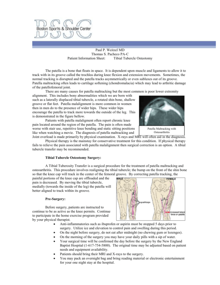
Paul P. Weitzel MD Thomas S. Pacheco PA-C Patient Information Sheet: Tibial Tubercle Osteotomy The patella is a bone that floats in space. It is dependent upon muscle and ligaments to allow it to track with in its groove called the trochlea during knee flexion and extension movements. Sometimes, the normal tracking is disrupted and the patella tracks asymmetrically or even subluxes out of its groove. Patella maltracking often leads to cartilage softening (chondromalacia) which may lead to arthritic damage of the patellofemoral joint. There are many causes for patella maltracking but the most common is poor lower extremity alignment. This includes bony abnormalities which we are born with such as a laterally displaced tibial tubercle, a rotated shin bone, shallow groove or flat feet. Patella malalignment is more common in women then in men do to the presence of wider hips. These wider hips encourage the patella to track more towards the outside of the leg. This is demonstrated in the figure bellow. Patients with patella malaligment often report chronic knee pain located around the region of the patella. The pain is often made Patella Maltracking with worse with stair use, repetitive knee bending and static sitting positions Osteoarthritis like when watching a movie. The diagnosis of patella maltracking and Joint overload is made primarily by physical examination. X-rays and MRI will often aid in the diagnosis. Physical therapy is the mainstay for conservative treatment for this condition. If physical therapy fails to relieve the pain associated with patella malalignment then surgical correction is an option. A tibial tubercle transfer may be recommended. Tibial Tubercle Osteotomy Surgery: A Tibial Tuberosity Transfer is a surgical procedure for the treatment of patella maltracking and osteoarthritis. This procedure involves realigning the tibial tubercle; the bump on the front of the shin bone so that the knee cap will track in the center of the femoral groove. By correcting patella tracking, the painful portions of the knee cap are offloaded and the pain is decreased. By moving the tibial tubercle, medially (towards the inside of the leg) the patella will better aligned to track within its groove. Pre-Surgery: Before surgery, patients are instructed to continue to be as active as the knee permits. Continue to participate in the home exercise program provided by your physical therapist. Anti-inflammatories such as ibuprofen or aspirin must be stopped 5 days prior to surgery. Utilize ice and elevation to control pain and swelling during this period. On the night before surgery, do not eat after midnight (no chewing gum or lozenges). On the morning of the surgery you may have your daily pills with a sip of water. Your surgical time will be confirmed the day before the surgery by the New England Baptist Hospital (1-617-754-5800). The original time may be adjusted based on patient needs and equipment availability. Patients should bring their MRI and X-rays to the surgery. You may pack an overnight bag and bring reading material or electronic entertainment for your one night stay at the hospital. Surgery: When you arrive at the hospital and register, a nurse will bring you into the pre-op area were you will have an IV placed and meet your anesthesiologist. General anesthesia is utilized to assure a comfortable surgery. This Example of Incision means that you will be “asleep” and completely unaware of the surgery until you wake up in the recovery area. Most patients will have a small tube placed in there windpipe, formal intubation may not be required. Local blocks are also available to supplement pain control. Your anesthesiologist will discuss this with you prior to surgery. The length of the procedure is approximately 90 minutes. The procedure will involve a knee arthroscopy to inspect the inside of the knee joint. Arthroscopic surgery involves using a video camera and small instruments through small incisions to see the anatomy of the knee joint. The video camera allows us to visually inspect the knee joint and specifically evaluate the patella cartilage and assess active patella tracking. Pictures of this will be taken and reviewed during your first post-op visit. The tibial tubercle transfer will be conducted through an incision in the front of your leg below the patella. A surgical fracture is made in the proximal tibia. This bone portion is then mobilized in a position to assure proper tracking of the patella. Screws are then utilized to secure the mobilized bone in its new place. These screws may need to be removed in the future if they become a source of irritation. Risks to tibial tubercle Femur osteotomy surgery are rare and involve: Compartment syndrome, blood clots (DVT), infection and delayed bone healing. You may have decreased sensation of the skin around the incision region. The procedure is also available to view online at www.bostonssc.com Post-Surgery: Tibial Tubercle After the surgery is completed, you will awaken in the operating room and be moved to the recovery area. Pain Control: After mobilization of bone, the tubercle is Femoral Nerve Block: Upon your consent, secured with two screws a femoral nerve block may be provided by an Anesthesiologist for pain control. This consists of an injection of marcaine (like novacaine) into the region around the femoral nerve and may decrease leg pain for up to 12 hours. PCA pump: Shortly after you are transferred to the hospital unit, you may be provided a pain button that will deliver a set amount of pain medication at your control. This pump will be discontinued the next morning. Oral pain medicine: Oral pain medicine will be provided once you are able to tolerate oral medications. A pain pill will be selected that best controls your pain. A pain medication prescription will be provided to you prior to discharge. You may take the prescribed medication as directed. You should expect to experience minimal to moderate knee discomfort for several days and even weeks following the surgery. Patients often only need prescription narcotics for a few days following surgery and then can switch to over-the-counter medications such as Tylenol or Ibuprofen. Ice bags and elevation should be utilized both in the hospital and after discharge to decrease swelling and pain. Keep ice on for 20 minutes and off for 45 minutes every 4 hours. Utilize ice as much as you can during the first 10 days after surgery. Be careful not to burn your skin with excessive cold exposure. Special cooling devices (called Cryo/Cuffs) are also available for patients to purchase thru our office. This can be used in the place of the ice bags. See the included information sheets if you would like to purchase one of these cooling devices At the completion of surgery, you will have a brace placed on your leg. The brace should be maintained in the locked position with all ambulation. The brace may only be removed when sitting with your leg elevated in an extended position and when in the CPM (Continuous Passive Motion) unit. You will receive a CPM unit at home. Utilize this device to for 3, two hour sessions daily to increase the range of motion of the operative knee. Progress the CPM up to 90°. Physical Therapy: You will receive PT prior to discharge from the hospital. PT will work on ambulation, functional mobility and leg exercises. You should be comfortable walking independently with crutches before leaving the hospital. You will be able to put as much weight as tolerated on your knee with brace locked in extension. You should participate in the home exercise program provided at the end of this packet until outpatient physical therapy is started. If the bandage is draining, reinforce it with additional dressings for the first 48 hours. After 48 hours remove the bandage leave the steri-strips in place. If drainage continues or restarts after 3 days please call Dr. Weitzel’s office. You may shower on post up day one. Keep incision covered when showering for up to three days post-op. You may shower with the wound exposed once the wound is completely dry. Skin numbness often occurs around the incision. This usually returns but may be permanent Take one 325 mg (full strength) aspirin daily for 21 days (unless otherwise instructed) to prevent blood clots. Eat a regular diet as tolerated and please drink plenty of fluids. First post-op appointment is 10-14 days after the surgery. You may drive once you establish full control of your extremity (able to perform a straight leg raise, etc.). If your right knee was operated on, this may take a week or more to achieve Call office for Temperature >102 degrees, excessive swelling, pain or redness around the incision sites. Plan at least 1-2 weeks away from work or school. Utilize this time to decrease swelling and participate in your home exercise program. You may be able to resume work once the pain and swelling resolves (this varies based on job activity). Outcomes/Expectations: It may take from nine months to a year before patients notice significant improvement from their pre-operative condition. Most patients report 80-90% pain relief at this time. Post-op Rehabilitation Protocol – Tibial Tubercle Osteotomy Phase 1 (Weeks 0-2): Goals: Minimize effusion, Progress range of motion, Utilize brace Treatment plan: 1) Utilize CPM unit, progress range of motion to full 2) Swelling Control with ice and compression wrap 3) Brace in place and locked in extension with all ambulation 4) Initiate quadriceps and hamstring muscle activation and general leg control Quad setting, SLR, heel slides, isometric hamstring/quadriceps contraction Ankle pumps 5) WBAT with crutches Phase 2 (Weeks 2-6): Goals: Full knee ROM in extension and flexion, progress quadriceps/hamstring strengthening, independent mobility Treatment plan: 1) Continue with swelling control 2) Full knee ROM (half to full revolution on exercise bike) 3) PRE (go easy with direct quadriceps strengthening until 6 weeks post-op) 4) Continue brace locked in extension with all ambulation until post-op week 6 Phase 3 (Weeks 6-12): Goals: Full lower extremity strengthening/conditioning program, Full activity in gym avoiding open chain full arc exercises Treatment plan: 1) Progress CKC strengthening 2) Progress dynamic balance training Early Post-operative Exercises Start the following exercises as soon as you are able. You can begin these in the recovery room shortly after surgery. You may feel uncomfortable at first, but these exercises will speed your recovery and actually diminish your post-operative pain. Quad Sets - Tighten your thigh muscle. Try to straighten your knee. Hold for 5 to 10 seconds. Repeat this exercise approximately 10 times during a two minute period, rest one minute and repeat. Straight Leg Raises - Tighten the thigh muscle with your knee fully straightened on the bed, as with the Quad set. Lift your leg several inches. Hold for five to 10 seconds. Slowly lower. Repeat until your thigh feels fatigued. Ankle Pumps - Move your foot up and down rhythmically by contracting the calf and shin muscles. Perform this exercise periodically for two to three minutes, two or three times an hour in the recovery room. Continue this exercise until you are fully recovered and all ankle and lower-leg swelling has subsided. Knee Straightening Exercises - Place a small rolled towel just above your heel so that it is not touching the bed. Tighten your thigh. Try to fully straighten your knee and to touch the back of your knee to the bed. Hold fully straightened for five to 10 seconds. Repeat until your thigh feels fatigued. Bed-Supported Knee Bends - Bend your knee as much as possible while sliding your foot on the bed. Hold your knee in a maximally bent position for 5 to 10 seconds and then straighten. Repeat several times until your leg feels fatigued or until you can completely bend your knee.

