hippo_guidelines
advertisement
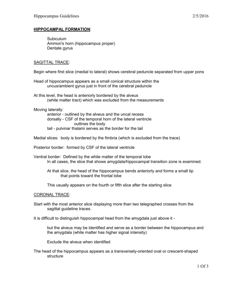
Hippocampus Guidelines 2/5/2016 HIPPOCAMPAL FORMATION: Subiculum Ammon's horn (hippocampus proper) Dentate gyrus SAGITTAL TRACE: Begin where first slice (medial to lateral) shows cerebral peduncle separated from upper pons Head of hippocampus appears as a small conical structure within the uncus/ambient gyrus just in front of the cerebral peduncle At this level, the head is anteriorly bordered by the alveus (white matter tract) which was excluded from the measurements Moving laterally: anterior - outlined by the alveus and the uncal recess dorsally - CSF of the temporal horn of the lateral ventricle outlines the body tail - pulvinar thalami serves as the border for the tail Medial slices: body is bordered by the fimbria (which is excluded from the trace) Posterior border: formed by CSF of the lateral ventricle Ventral border: Defined by the white matter of the temporal lobe In all cases, the slice that shows amygdala/hippocampal transition zone is examined. At that slice, the head of the hippocampus bends anteriorly and forms a small tip that points toward the frontal lobe This usually appears on the fourth or fifth slice after the starting slice CORONAL TRACE: Start with the most anterior slice displaying more than two telegraphed crosses from the sagittal guideline traces. It is difficult to distinguish hippocampal head from the amygdala just above it but the alveus may be identified and serve as a border between the hippocampus and the amygdala (white matter has higher signal intensity) Exclude the alveus when identified The head of the hippocampus appears as a transversely-oriented oval or crescent-shaped structure 1 Of 3 Hippocampus Guidelines 2/5/2016 Lateral: temporal horn of the lateral ventricle serves as border The edge between gm and wm is sometimes blurred on the tissue classified image; need to use T1- and T2- weighted images as reference Dorsal: Follow the line defined by the telegraphed crosses Exclude the alveus if visible Uncal sulcus usually obliterated on the most anterior slices When the uncal sulcus becomes visible, the uncal recess serves as the dorsal border Medial: telegraphed crosses provide an orientation on the most anterior slices. Tracing includes parts of the ambient gyrus, and on some slices the entorhinal sulcus serve as a border. Ventral: Laterally defined by the WM of the temporal lobe Medially the subiculum has to be cut off from the cortex of the parahippocampal gyrus. Do this by following the horizontal line that is defined by the subiculum/WM border BODY [on CORONAL]: Comes into view at about the level of the red nucleus Lateral: Marked by the temporal stem and inferior horn of the lateral ventricle Dorsal: Exclude the alveus and fimbria. Note the CSF of the lateral ventricle Medial: CSF of the ambient cistern and the crus cerebri serve as border Ventral: Defined by the WM of the temporal lobe. Exlude the parahippocampal gyrus Tail: Starts at the level where the brainstem becomes separated from the midbrain (level of the superior colliculus/pineal gland) Lateral: Demarcated by the ascending crus of the fornix and CSF of the atrium of the lateral ventricles. 2 Of 3 Hippocampus Guidelines 2/5/2016 It is difficult to differentiate the intraventricular part of the tail from ventricular CSF (use the telegraphed crosses as guideline) Dorsal: border defined by the pulvinar of the thalamus Separation of the hippocampal head may be difficult due to partial voluming effects - sag plane should be used as reference On the most caudal slice, dorsal border is marked by lower part of the splenium of the corpus callosum (This part of hippocampal tail sometimes referred to as the subsplenial gyrus) Medial: CSF of the quadrigeminal cistern serves as border Terminal slices: Include the fasciola cinerea and the gyrus fasciolaris as well as the gyrus of Andreas Retzius Ventral border: Defined by the WM of the temporal lobe (parahippocampal gyrus) 3 Of 3
