Periodontal Regeneration Lecture Handout
advertisement
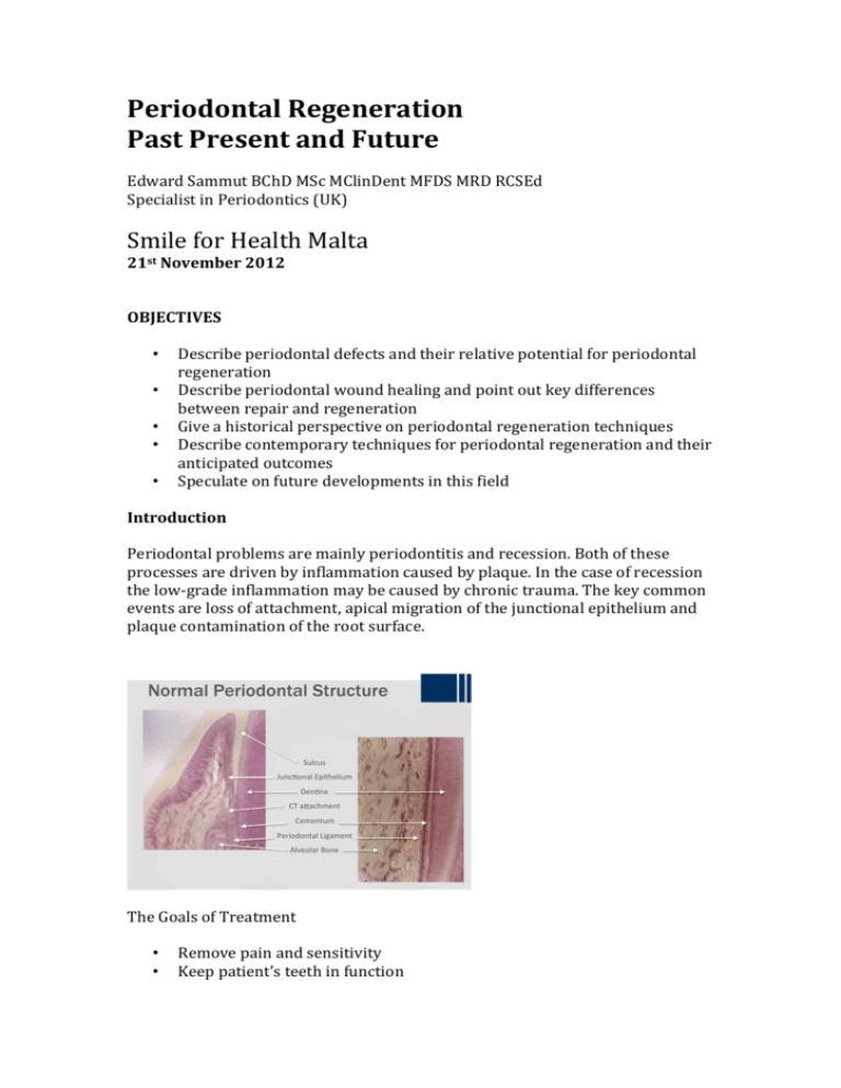
Periodontal Regeneration Past Present and Future Edward Sammut BChD MSc MClinDent MFDS MRD RCSEd Specialist in Periodontics (UK) Smile for Health Malta 21st November 2012 OBJECTIVES • • • • • Describe periodontal defects and their relative potential for periodontal regeneration Describe periodontal wound healing and point out key differences between repair and regeneration Give a historical perspective on periodontal regeneration techniques Describe contemporary techniques for periodontal regeneration and their anticipated outcomes Speculate on future developments in this field Introduction Periodontal problems are mainly periodontitis and recession. Both of these processes are driven by inflammation caused by plaque. In the case of recession the low-grade inflammation may be caused by chronic trauma. The key common events are loss of attachment, apical migration of the junctional epithelium and plaque contamination of the root surface. Normal Periodontal Structure Sulcus Junc onal Epithelium Den ne CT a achment Cementum Periodontal Ligament Alveolar Bone The Goals of Treatment • • Remove pain and sensitivity Keep patient’s teeth in function • Satisfy aesthetics Prevent further attachment loss Remove cause of inflammation and establish conditions for it not to return Regenerate the lost attachment? Cover root surface? A case was presented showing extreme attachment loss in an intrabony defect mesial to LR6. Patient had pulp symptoms from the tooth and extraction was considered due to the cost of RCT, perio surgery and crown being similar to a dental implant. The patient declined and simple root surface debridement was performed repeatedly. The patient demonstrated reduction in pocket depths, increase in the clinical attachment and radiographic bone fill of the defect over a two-year period. Bone regeneration is not a new idea. In 1957: Prichard, J. published: The infrabony technique as a predictable procedure. J. Periodontology, 28: 202-216 Surgically excised all tissue in the pocket Tin-foil placed to keep out the wound dressing Denuded bone was allowed to granulate over – much like a socket Predictable bone fill seen radiographically Later on, another key paper in this field, which is special because it includes reentry surgery: • 1976: Rosling B, Nyman S, Lindhe J, and Jern, B: The Healing potential of the periodontal tissues following different techniques of periodontal surgery in plaque free dentitions. J Clin Periodontol 1976 3:233-250 Different surgical procedures to treat intrabony defects Re-entry surgery to confirm results Access and apically repositioned flaps showed bone fill >80% Periodontal defects may be SUPRABONY or INTRABONY Suprabony defects refer to horizontal bone loss with circumferential attachment loss around a tooth. It is not currently possible to consider periodontal regeneration in the suprabony situation. Intrabony or angular defects can be classified into 1, 2 or 3 walled defects. These are graphically represented below. Periodontal regeneration can be considered in intrabony defects. There is a good supply of cells from both the PDL and the bone which can populate the defect and regenerate missing tissue. A true regeneration does not result only in the growth of new bone in the defect – it also includes growth of new cementum on the root surface and new periodontal ligament fibres joining the root surface to the new bone. Classic animal models of healing following periodontal surgery show us that there is a very small amount of true regeneration and most healing is by repair in which a long junctional epithelium lines the root surface and separates it from the newly formed bone One-wall defect Two-wall defect Three-wall defect Classic Animal Models • • • J Clin Periodontol. 1976 3:54-58. Osseous repair of an infrabony pocket without new attachment of connective tissue Caton JG, Zander HA J Periodontol. 1979 Sep;50(9):462-6. The attachment between tooth and gingival tissues after periodic root planing and soft tissue curettage. Caton JG, Zander HA. J Periodontal Res 1979 14:520-525 Periodontal repair after reduction of inflammation. Polson AM, Kantor ME, Zander HA. From: Clinical Periodontology and Implant Den stry 4th Ed. Lindhe, Karring, and Lang. Blackwell Munksgaard. The outcomes which we measure clinically – that is probing depth, clinical attachment level and radiographic bone gain may be no different in periodontal regeneration and in periodontal repair. However, regeneration is a more desirable outcome because it restores full and functional periodontal structure around a tooth. REPAIR REGENERATION Bone fill Root surface covered with “long” junctional epithelium No new ligament No new cementum Bone fill New attachment forms New ligament forms New cementum covers previously “diseased” root surface Although PDL is very rich in stem cells and PDL stem cells have the ability to form all tissues required for regeneration – this doesn’t happen. • Why doesn’t this happen? Epithelial down-growth along the root surface is quick Lack of adherence of PDL cells to root surface dentine Karring, Lindhe and co-workers did a series of studies to look at healing processes between root surface, bone, ligament and gingival connective tissue. The Karring Story 1980-1984 • Beagle dog series of experiments • Experimental periodontitis - roots with attachment loss were cleaned, extracted and buried under flaps • • • Part of root still covered in PDL reattached to bone or gingival tissue Other “denuded” part of root surface developed ankylosis and resorption where in contact with bone or gingival tissue PDL grew up the root in a third experiment where the root was not extracted but covered by the flap Following these experiments, they performed a human experiment in a landmark 1982 paper: A patient who was due to have extraction of LL2 was consented to having a GTR procedure using a Millipore membrane, and a notch was cut in the root surface to indicate the level of the attachment loss. Three months later the tooth was removed en bloc together with surrounding tissue. Histology confirmed presence of new attachment coronal to the notch on the root, demonstrating the principle of GTR. Guided Tissue Regeneration Nyman S, Lindhe J, Karring T and Rylander H. (1982) New a achment following surgical treatment of human periodontal disease. J Clin Periodontol 9: 290-296 • Various membranes used over the years Non-resorbable Gore-tex Gore-tex titanium reinforced Resorbable Synthetic – eg Vicryl or Resolute Collagen – eg Bio-Gide Filler material can be used below membrane to help maintain space Issues with the non-resorbable membranes were that they sometimes became infected and they also required a second operation to remove them. For this reason resorbable membranes became more popular. The definitive review is: Needleman IG, Worthington HV, Giedrys-Leeper E, Tucker RJ. Guided tissue regeneration for periodontal infra-bony defects. Cochrane Database Syst Rev. 2006 Apr 19;(2):CD001724. • Large variability between studies • Mean gain of clinical attachment (averaged out over the various studies) over OFD (open flap debridement) was 1.2mm • Number Needed To Treat of 8 patients – before the advantage of GTR was observed. Various bone filler materials have been used over the years • Patient’s own bone (Autogenous graft) • From a cadaver (Allograft, eg Rocky Mountain) • From an animal (Xenograft, eg Bio-oss) • Coralline Calcium Carbonate • Synthetic Hydroxyapatite Bioactive glass Beta-tricalcium phosphate Clinically these results in an average reduced PPDs and improved CALs (1-2mm) over open flap debridement. They also produce nice radiographs because the materials are typically radio-opaque. The problem is one cannot be sure if the image is of the radio-opaque material or any newly formed bone. Xenograft and synthetic particles tend to encapsulate in fibrous tissue - unless covered by a GTR membrane – in which case the filler is a simple space maintainer and the procedure should be viewed as a GTR procedure. Trombelli L, Heitz-Mayfield L, Needleman I, Moles D, Scabbia A: A systematic review of graft materials and biological agents for periodontal intraosseous defects. J Clin Periodontol 2002; 29(Suppl. 3): 117–135. A completely different approach was taken at by Swedish researchers in the late 1990s. In experiments looking at tooth development dentine on its own was insufficient stimulus for follicular cells to differentiate into cementoblasts. This challenged the understanding of the process of tooth development as it was thought that once Hertwig root sheath disintegrated the presence of the dentine stimulated follicular cells to lay down cementum. In actual fact, it was found that deposition of enamel matrix proteins onto a developing root surface is an essential step preceding formation of acellular cementum. A thin layer of ”enamel” can still be histologically seen between cementum and dentine. The formation of cementum is a critical step in the attachment of periodontal ligament and formation of alveolar bone – whether its in tooth development or in periodontal regeneration, so enamel proteins were looked at for potential to stimulate periodontal regeneration. A proof of principle study extracted animal teeth, cut cavities in the root surface, then treated the same cavities with enamel matrix proteins and re-planted the teeth. Acellular cementum formed on the root surfaces in the cavity and re-established periodontal support. Within a short time, Enamel Matrix Derivative (EMD) was marketed as Emdogain by Biora in Sweden. EMD is purified acidic extract of proteins from pig enamel matrix: • 90% Amelogenin protein – hydrophobic aggregates – highly conserved • 10% mixture of other proteins – not completely defined or understood but includes proline-rich non-amelogenins, tuftelin, tuft protein, serum proteins as well as ameloblastin and amelin Emdogain was acquired by Straumann in 2003. The effects of EMD in vitro include • Proliferation and growth of PDL fibroblasts • Inhibition of epithelial cells • Increase protein synthesis by PDL fibroblasts • Formation of mineralized nodules in PDL fbs • Growth of mesenchymal cells • Release of autocrine growth factors from PDL fibroblasts • Macrophages stimulated to produce BMPs A case was then shown, showing successful periodontal regeneration in a 16 year old girl lateral incisor. The regenerated periodontal tissues were able to accept orthodontic tooth movement and space closure with almost no loss of papillary architecture. The key reviews on EMD are • • • • Palmer RM, Cortellini P. Periodontal tissue engineering and regeneration: Consensus report of the Sixth European Workshop of Periodontology. J Clin Periodontol 2008; 35 (Suppl.8): 83 – 86 Bosshardt DD. Biological mediators and periodontal regeneration: a review of enamel matrix proteins at the molecular and cellular levels. J Clin Periodontol 2008:35 (Suppl 8):87 – 105. Trombelli L, Farina R. Clinical outcomes with bioactive agents alone or in combination with grafting or guided tissue regeneration. J Clin Periodontol 2008;35 (Suppl 8):117 – 135. Cairo F, Pagliaro U, Nieri M. Treatment of gingival recession with coronally advanced flap procedures: a systematic review. J Clin Periodontol 2008;35 (Suppl 8):136 – 162. Future developments Improvement of surgical techniques – surgery is becoming more and more minimal – this progression shows us the development of flap designs over 20 years. • Papilla Preservation (Takei, 1985) • Modified/Simplified Papilla Preservation (Cortellini, 1999) • Minimally Invasive Surgical Technique (Cortellini, 2007) • Single Flap Approach (Trombelli, 2009) Minimally invasive surgery has the following advantages • Reduction in surgical trauma • Increase of flap and wound stability • Reduction in surgical time • Increase in surgical accuracy • Reduced postoperative morbidity Interestingly if one looks at the control groups of studies looking at OFD alone vs OFD + EMD application shows that the results from the OFD procedure by itself are improving with time. We are getting better at surgery J Clin Periodontol. 2008 Feb;35(2):139-46. Epub 2007 Dec 13. Is there a temporal trend in the reported treatment efficacy of periodontal regeneration? A metaanalysis of randomized-controlled trials. Tu YK, Tugnait A, Clerehugh V. Another improvement which was help better periodontal regeneration treatment is the 3D diagnosis and measurement of defects using CBCT imaging. This comes an increased investigation cost and a radiation dose. The images also suffer from metal-restoration artefacts. One also needs special software to carry out 3D bone defect measurements. The future probably lies in combinations of active substances with biomaterials and cell cultures to create “Endogenous Regenerative Technology” systems. • Active Substances Growth Factors Platelet Rich Plasma Enamel Matrix Derivative • Biomaterials Scaffolds Bone Graft/Fillers Membranes • Autologous cell cultures Systems with combinations of active substances with biomaterials are already in use. These include GEM21 – a combination of recombinant human Platelet Derived Growth Factor in a beta-TCP matrix. This product has licencing issues in the EU at the minute. Another one is OP-1 Implant which consists of 3.3mg rhBMP-7 with 1g purified bovine Type I collagen. This product is used in spinal fusion and fracture repair and has promising lab and animal periodontal models show bone, cementum and ligament formation. In this exciting and technological treatment, its important not to lose sight of the basics – periodontal regeneration demands patients have good control of plaque, bleeding, do not smoke and are not excessively stressed. They must also be able to cope with the follow-up regime. At the tooth level one must chose defects which are amenable to treatment, on teeth with favourable anatomy.
