PREVALENCE AND SIGNIFICANCE OF
advertisement
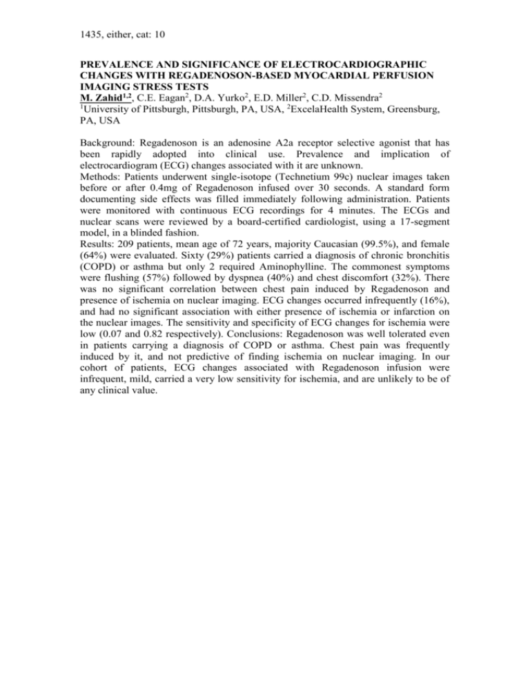
1435, either, cat: 10 PREVALENCE AND SIGNIFICANCE OF ELECTROCARDIOGRAPHIC CHANGES WITH REGADENOSON-BASED MYOCARDIAL PERFUSION IMAGING STRESS TESTS M. Zahid1,2, C.E. Eagan2, D.A. Yurko2, E.D. Miller2, C.D. Missendra2 1 University of Pittsburgh, Pittsburgh, PA, USA, 2ExcelaHealth System, Greensburg, PA, USA Background: Regadenoson is an adenosine A2a receptor selective agonist that has been rapidly adopted into clinical use. Prevalence and implication of electrocardiogram (ECG) changes associated with it are unknown. Methods: Patients underwent single-isotope (Technetium 99c) nuclear images taken before or after 0.4mg of Regadenoson infused over 30 seconds. A standard form documenting side effects was filled immediately following administration. Patients were monitored with continuous ECG recordings for 4 minutes. The ECGs and nuclear scans were reviewed by a board-certified cardiologist, using a 17-segment model, in a blinded fashion. Results: 209 patients, mean age of 72 years, majority Caucasian (99.5%), and female (64%) were evaluated. Sixty (29%) patients carried a diagnosis of chronic bronchitis (COPD) or asthma but only 2 required Aminophylline. The commonest symptoms were flushing (57%) followed by dyspnea (40%) and chest discomfort (32%). There was no significant correlation between chest pain induced by Regadenoson and presence of ischemia on nuclear imaging. ECG changes occurred infrequently (16%), and had no significant association with either presence of ischemia or infarction on the nuclear images. The sensitivity and specificity of ECG changes for ischemia were low (0.07 and 0.82 respectively). Conclusions: Regadenoson was well tolerated even in patients carrying a diagnosis of COPD or asthma. Chest pain was frequently induced by it, and not predictive of finding ischemia on nuclear imaging. In our cohort of patients, ECG changes associated with Regadenoson infusion were infrequent, mild, carried a very low sensitivity for ischemia, and are unlikely to be of any clinical value.

