surgical results of pars plana vitrectomy combined with small
advertisement
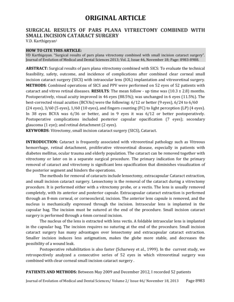
ORIGINAL ARTICLE SURGICAL RESULTS OF PARS PLANA VITRECTOMY COMBINED WITH SMALL INCISION CATARACT SURGERY V.D. Karthigeyan1 HOW TO CITE THIS ARTICLE: VD Karthigeyan. “Surgical results of pars plana vitrectomy combined with small incision cataract surgery”. Journal of Evolution of Medical and Dental Sciences 2013; Vol. 2, Issue 46, November 18; Page: 8983-8988. ABSTRACT: Surgical results of pars plana vitrectomy combined with SICS: To evaluate the technical feasibility, safety, outcome, and incidence of complications after combined clear corneal small incision cataract surgery (SICS) with intraocular lens (IOL) implantation and vitreoretinal surgery. METHODS: Combined operations of SICS and PPV were performed on 52 eyes of 52 patients with cataract and vitreo retinal diseases. RESULTS: The mean follow - up time was (10.3 ± 2.8) months. Postoperatively, visual acuity improved in 46 eyes (88.5%); was unchanged in 6 eyes (11.5%). The best-corrected visual acuities (BCVAs) were the following: 6/12 or better (9 eyes), 6/24 to 6/60 (24 eyes), 3/60 (5 eyes), 1/60 (10 eyes), and fingers counting (FC) to light perception (LP) (4 eyes). In 38 eyes BCVA was 6/36 or better, and in 9 eyes it was 6/12 or better postoperatively. Postoperative complications included posterior capsular opacification (7 eyes); secondary glaucoma (1 eye); and retinal detachment (2 eyes). KEYWORDS: Vitrectomy, small incision cataract surgery (SICS), Cataract. INTRODUCTION: Cataract is frequently associated with vitreoretinal pathology such as Vitreous hemorrhage, retinal detachment, proliferative vitreoretinal disease, especially in patients with diabetes mellitus, ocular trauma and elderly population. The cataract can be removed together with vitrectomy or later on in a separate surgical procedure. The primary indication for the primary removal of cataract and vitrectomy is significant lens opacification that diminishes visualization of the posterior segment and hinders the operations. The methods for removal of cataracts include lensectomy, extracapsular Cataract extraction, and small incision cataract surgery. Lensectomy is the removal of the cataract during a vitrectomy procedure. It is performed either with a vitrectomy probe, or a vectis. The lens is usually removed completely, with its anterior and posterior capsule. Extracapsular cataract extraction is performed through an 8-mm corneal, or corneoscleral, incision. The anterior lens capsule is removed, and the nucleus is mechanically expressed through the incision. Intraocular lens is implanted in the capsular bag. The incision must be sutured at the end of the procedure. Small incision cataract surgery is performed through a 6mm corneal incision. The nucleus of the lens is extracted with lens vectis. A foldable intraocular lens is implanted in the capsular bag. The incision requires no suturing at the end of the procedure. Small incision cataract surgery has many advantages over lensectomy and extracapsular cataract extraction. Smaller incision induces less astigmatism, makes the globe more stable, and decreases the possibility of a wound leak. Postoperative rehabilitation is also faster (Scharwey et al., 1999). In the current study, we retrospectively analyzed a consecutive series of 52 eyes in which vitreoretinal surgery was combined with clear corneal small incision cataract surgery . PATIENTS AND METHODS: Between May 2009 and December 2012, I recorded 52 patients Journal of Evolution of Medical and Dental Sciences/ Volume 2/ Issue 46/ November 18, 2013 Page 8983 ORIGINAL ARTICLE (52 eyes) who had pars plana vitrectomy (PPV) combined with clear corneal small incision cataract surgery. Visual and surgical results, as well as complication rates in these 52 consecutive cases, were retrospectively analyzed. The following preoperative information was obtained for each patient: age, sex, visual acuity, intraocular pressure (IOP), and indication for vitreoretinal surgery. Keratometry and axial length measurements were performed on the eye to be operated on whenever possible. If this was not possible, the data were taken from the fellow eye. Intraocular lens calculation was performed using the Binkhorst formula. The type of cataract extraction and all posterior segment procedures were noted, including IOL style, haptic location, and type of anesthesia. Information regarding best-corrected visual acuity, refractive error, and ophthalmic findings were recorded by slit - lamp microscopy, tonometry and ophthalmoscopy were recorded before and after surgery. Postoperative data included length of followed - up, reasons for PPV, best - corrected visual acuity (BCVA), and subsequent postoperative procedure (e.g., vitreoretinal reoperation, yttrium aluminum – garnet (YAG) laser capsulotomy). In all patients, surgery was performed using peribulbar anesthesia. In all 52 cases, cataract extraction preceded vitreoretinal surgery. A 6.0 mm wide and 1.5 ~ 2.0 mm long clear corneal tunnel was created at the temporal limbus, a 5.0 to 6.0 mm curvilinear capsulorhexis was completed, and small incision cataract surgery and cortex removal were performed. The anterior chamber and capsular bag were filled with viscomet, and the corneal tunnel was temporarily closed with a single 10 - 0 nylon suture. Subsequently, a standard 3-port pars plana vitrectomy was performed using a 20 - gauge vitreous cutter and hand - held light pipe. Sclerotomies were placed 3.5 mm posterior to the limbus in the superotemporal, superonasal and inferotemporal quadrants. The infusion cannula was sutured in the inferotemporal sclerotomy site. After the vitreoretinal surgery was completed and before intraocular tamponade was performed, the foldable silicone IOL was implanted in 10 cases. During implantation, the sclerotomies were left open. The corneal suture was removed and a foldable silicone IOL was implanted through a 3.5 mm corneal incision. The corneal incision was water - sealing and the internal tamponade was performed. Sclerotomies and conjunctive were sutured, and subconjunctival gentamicin sulfate (20 mg) and dexamethasone sodium phosphate (4 mg) were administered. RESULTS: Follow - up duration - The follow - up was between 6 and 41 months (means (10.3 ± 2.8) months). Patients demographics - Fifty - two eyes of 52 patients who had pars plana vitrectomy combined with clear corneal small incision cataract surgery were recruited and analysed. The baseline demographics of the patients are summarized in Table 1. Demographics Age (Years) Gender Male Female Duration of symptoms (d) All eyes (N = 52) 55.4 (34~77) N=28 N=24 322.1 Journal of Evolution of Medical and Dental Sciences/ Volume 2/ Issue 46/ November 18, 2013 Page 8984 ORIGINAL ARTICLE (2 ~1825) Diseases RRD with PVR N=18 Macular hole N=9 Diabetic retinopathy N=12 Trauma N=13 BCVA LP~FC Table 1: Baseline demographics of 52 patients who had combined SICS with PPV RRD: Rhegmatogenous retinal detachment; PVR: Proliferative vitreoretinopathy; BCVA: Best - corrected visual acuity; LP: Light perception; FC: Fingers counting Clinical course of the patients: Vitrectomy was combined with membrane removal in 18 eyes (34.6%), endolaser photocoagulation in 23 eyes (44.2%), scleral buckling in 34 eyes (65.4%), and removal of an intraocular foreign body embedded in the retina in 4 eyes (7.7%). Gas tamponade in 16 eyes (30.8%), and silicone oil tamponade in 9 eyes (17.3%) (Table 2). Clinical course of the patients Number of eyes N / % Vitrectomy combined with membrane removal 18 / 34.6 Endolaser 23 / 44.2 Buckling 34 / 65.4 Intraocular foreign body removal 4 / 7.7 Gas tamponade 16 / 30.8 Silicone oil tamponade 9 / 17.3 Table 2 Visual acuity: Postoperatively, visual acuity improved in 46 eyes (88.5%); was unchanged in 6 eyes (11.5%) because of 1 with long - standing (5 years) retinal detachment, 1 with macular hole, 1 with severe trauma, 3 with diabetic retinopathy VI. The best corrected visual acuity (BCVA) were the following: 6/12 or better (9 eyes), 6/24 to 6/36 (24 eyes), 6/60 (5 eyes), 3/60 (10 eyes), and fingers counting (FC) to light perception (LP) (4 eyes). In 38 eyes BCVA was 6/60 or better, and in 9 eyes it was 6/12 or better postoperatively (Table 3). BCVA Preoperative (N) Postoperative (N) LP ~ FC 52 4 3/60 0 10 6/60 0 5 0 24 6/36 – 6/18 6/12-6/6 0 9 Table 3: Preoperative and postoperative BCVA of the patients Journal of Evolution of Medical and Dental Sciences/ Volume 2/ Issue 46/ November 18, 2013 Page 8985 ORIGINAL ARTICLE COMPLICATIONS: No hyphema and fibrin transudation occurred in anterior chamber. Seven eyes developed posterior capsule opacification 3 months, 5 months, 6 months, 12 months, 13 months postoperatively. An Nd:YAG capsulotomy was performed in all. One eye with retinal detachment 3 weeks postoperatively, the retinal hole was the bed of the retinal foreign body, requiring a retinal reattachment, one eye with retinal redetachment 4 months after silicone oil removal. Secondary glaucoma occurred 5 weeks after silicone oil tamponade, requiring silicone oil removal. DISCUSSION: Vitreoretinal pathology is frequently associated with cataract, and can accelerate the development of cataract. As the same time, cataract interferes with safe performance of vitrectomy, postoperative observation and postoperative treatment: retinal photocoagulation, separate anterior and posterior segment surgeries are the traditional methods. Kokame et al. (1989) reported a method of pars plana lensectomy in which the anterior lens capsule is left in place, allowing insertion of a posterior chamber IOL in the ciliary sulcus. Several other techniques for cataract removal during vitreoretinal surgery have been advocated, including intracapsular cataract extraction (ICCE), extracapsular cataract extraction (ECCE), both of which require a large incision, increase the risk of wound dehiscence caused by globe manipulation during posterior segment procedures. Both methods may also increase postoperative ocular inflammation. Combined surgery comprising small incision cataract surgery (SICS), intraocular lens (IOL) implantation, and pars plana vitrectomy (PPV) has been regarded as a safe and effective procedure. This type of combined surgery is now considered a standard procedure for selected patients with clinically significant cataract and vitreoretinal diseases (Pinter and Sugar, 1999; Chang et al., 2005). I used the small incision cataract surgery (SICS) - vitrectomy - IOL insertion combined operation sequence. A clear corneal incision was made for cataract removal and IOL insertion and this incision was sutured before the pars plana vitrectomy was done. There were no complications related directly to IOL implantation at the time of vitreoretinal surgery (Scharwey et al., 1999; Honjo and Ogura, 1998). In my experience, clear corneal small incision cataract surgery can be safely combined with vitreoretinal surgery. This cataract extraction technique is rapid and does not increase operating time significantly. As the incision is performed in avascular tissue, there is no additional bleeding into the anterior chamber, and a postoperative inflammatory reaction is minimal. The incision is small, watertight and very resistant to increase IOP and globe manipulations during subsequent vitreoretinal surgery. In contrast to scleral tunnel incision, corneal incision does not interfere with sclerotomies, even if sclerotomy has to be enlarged. Endothelial opening of the corneal incision is remoter from the iris, reducing the risk of iris incarceration in the cataract incision. Trauma to the iris with the SICS is also minimized, decreasing the risk of intraoperative miosis. Intraocular lens implantation should be delayed until the end of posterior segment surgery to maintain the advantages of small, self - sealing corneal incision and to avoid disturbing light reflexes from the IOL rim (causing difficulties in visualization of the far retinal periphery). According to my clinical experience, the operation that combines cataract extraction, IOL implantation, and vitreous surgery is a safe and desirable option in patients with significant lens opacities and vitreoretinal pathology. And the main advantage of combined procedure is more rapid visual rehabilitation with a single operation, reducing costs and patient discomfort. Journal of Evolution of Medical and Dental Sciences/ Volume 2/ Issue 46/ November 18, 2013 Page 8986 ORIGINAL ARTICLE Development or progression of cataract is a frequent postoperative complication after Parsplana Vitrectomy (PPV) for macular holes, the epiretinal membrane and diabetic retinopathy in the elderly. Combined pars plana vitrectomy and cataract surgery is a safe and effective procedure that allows the surgeon to avoid a second operation, but adequate treatment is recommended to all patients over 60 years. Combining PPV and cataract surgery may be indicated, especially in older patients. PPV offered good visual outcomes and that ambulatory surgery is possible with the technique. There is no influence of IOL diameter on visual outcome, and no increased risk of cataract when used in conjunction with a gas or silicone oil endotamponade. The risk of cataract formation after PPV was 74%, but that rose with age. For patients 60 or older, there was a 100% occurrence of cataract after PPV, but for patients below 40 there was an extremely low risk. The combined PPV and cataract surgery required a modified technique for cataract extraction. The anterior chamber is opened via a long corneo - scleral tunnel or a long corneal tunnel. This approach tolerates a pressure increase and allows a sufficiently large opening for cataract surgery. CONCLUSION: My experience with combined surgery is encouraging and by proper patient selection, a faster visual rehabilitation can be provided and multiple surgeries can be avoided. I acknowledge that the present study is limited by its retrospective nature and heterogeneity in diagnosis. REFERENCES: 1. Freeman WR, Azen SP, Kim JW, el - haig W, Mishell DR, Bailey I. Vitrectomy for the treatment of full - thickness stage 3 or 4 macular holes. Arch Ophthalmol 1997; 115:11-21. 2. Chung TY, Chung H, Lee JH. Combined surgery and sequential surgery comprising phacoemulsification, pars plana vitrectomy and intraocular lens implantation: Comparison of clinical outcomes. J Cataract Refract Surg 2002; 28 : 2001 - 5. 3. Lahey JM, Francis RR, Fong DS, Kearney JJ, Tanaka S. Combining phacoemulsification with vitrectomy for treatment of macular holes. Br J Ophthalmol 2002; 86: 876-8. 4. Koenig SB, Han DP, Mieler WF, Abrams GW, Jaffe GJ, Burton TC. Combined phacoemulsification and pars plana vitrectomy. Arch Ophthalmol 1990; 108 : 362 - 4. 5. Demetriades AM, Gottsch JD, Thomsen R, Azab A, Stark WJ, Campochiaro PA, et al . Combined phacoemulsification, intraocular lens implantation and vitrectomy for eyes with coexisting cataract and vitreoretinal pathology. Am J Ophthalmol 2003; 135 : 291 - 6. Page 11 of 13 6. Lam DS, Young AL, Rao SK, Cheung BT, Yuen CY, Tang HM. Combined phacoemulsification, pars plana vitrectomy and foldable intraocular lens implantation. J Cataract Refract Surg 2003; 29 : 1064 - 9. 7. Lahey JM, Francis RR, Kearney JJ. Combining phacoemulsification with pars plana vitrectomy in patients with proliferative diabetic retinopathy: A series of 223 cases. Ophthalmology 2003; 110 : 1335 - 9. 8. Lahey JM, Francis RR, Kearney JJ, Cheung M. Combining phacoemulsification and vitrectomy in patients with proliferative diabetic retinopathy. Curr Opin Ophthalmol 2004; 15 : 192 - 6. Journal of Evolution of Medical and Dental Sciences/ Volume 2/ Issue 46/ November 18, 2013 Page 8987 ORIGINAL ARTICLE 9. Heiligenhaus A, Holtkamp A, Koch J, Schilling H, Bornfeld N, Losche CC, et al . Combined phacoemulsification and pars plana vitrectomy: Clear corneal versus sclera incisions: Prospective randomized multicenter study. J Cataract Refract Surg 2003; 29 : 1106 - 12. 10. Androudi S, Ahmed M, Fiore T, Brazitikos P, Foster CS. Combined pars plana vitrectomy and phacoemulsification to restore visual acuity in patients with chronic uveitis. J Cataract Refract Surg 2005; 31 : 472 - 8. 11. Brazitikos PD, Androudi S, Christen WG, Stangos NT. Primary pars plana vitrectomy versus scleral buckle surgery for the treatment of pseudophakic retinal detachment: A randomized clinical trial. Retina 2005; 25 : 957 - 64. 12. Melberg NS, Thomas MA. Nuclear sclerotic cataract after vitrectomy in patients younger t 102 : 1466 - 71. 13. Smiddy WE, Stark WJ, Michels RG, Maumenee AE, Terry AC, Glaser BM. Cataract extraction after vitrectomy. Ophthalmology 1987; 94 : 483 - 7. 14. Koenig SB, Mieler WF, Han DP, Abrams GW. Combined phacoemulsification, pars plana vitrectomy and posterior chamber intraocular lens insertion. Arch Ophthalmol 1992; 110 : 1101 - 4. 15. Hurley C, Barry P. Combined endocapsular phacoemulsification, pars plana vitrectomy and intraocular lens implantation. J Cataract Refract Surg 1996; 22 : 462 - 6. 16. Benson WE, Brown GC, Tasman W, McNamara JA. Extracapsular cataract extraction, posterior chamber lens insertion and pars plana vitrectomy in one operation. Ophthalmology 1990; 97 : 918 - 21. 17. Hainsworth DP, Chen SN, Cox TA, Jaffe GJ. Condensation on polymethylmethacrylate, acrylic polymer and silicone intraocular lenses after fluid – air exchange in rabbits. Ophthalmology 1996; 103 : 1410 - 8. 18. Meyers SM, Klein R, Chandra S, Myers FL. Unplanned extracapsular cataract extraction in post - vitrectomy eyes. Am J Ophthalmol 1978; 86 : 624 - 6. 19. Sneed S, Parrish RK 2nd, Mandelbaum S, O'Grady G. Technical problems of extracapsular cataract extractions after vitrectomy. Arch Ophthalmol 1986; 104 : 1126 - 7. 20. Cheung CM, Hero M. Stabilization of anterior chamber depth during phacoemulsification cataract surgery in vitrectomized eyes. J Cataract Refract Surg 2005; 31 : 2055-7. 21. Scharwey K, Pavlovic S, Jacobi KW. Combined clear corneal phacoemulsification, vitreoretinal surgery and intraocular lens implantation. J Cataract Refract Surg 1999; 25 : 693 - 8. AUTHORS: 1. V.D. Karthigeyan PARTICULARS OF CONTRIBUTORS: 1. Consultant, Department of Ophthalmology, Hindu Mission Hospital, Chennai. NAME ADDRESS EMAIL ID OF THE CORRESPONDING AUTHOR: Dr V.D Kartigeyan 16B, TKC Street, New Perungalathur, Chennai 63. Email – vd.karthigeyan@gmail.com Date of Submission: 30/10/2013. Date of Peer Review: 31/10/2013. Date of Acceptance: 10/11/2013. Date of Publishing: 14/11/2013 Journal of Evolution of Medical and Dental Sciences/ Volume 2/ Issue 46/ November 18, 2013 Page 8988

