Larger Lentigo Maligna Lesions and Invasive
advertisement

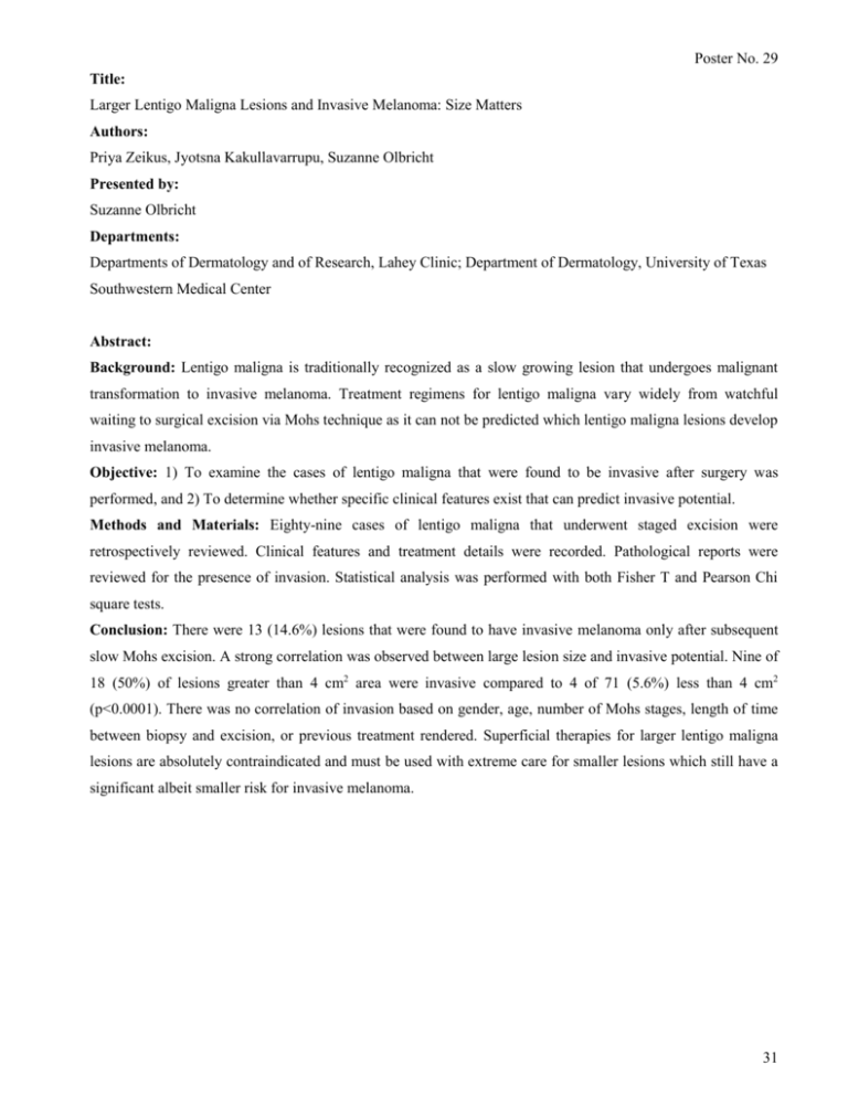
Poster No. 29 Title: Larger Lentigo Maligna Lesions and Invasive Melanoma: Size Matters Authors: Priya Zeikus, Jyotsna Kakullavarrupu, Suzanne Olbricht Presented by: Suzanne Olbricht Departments: Departments of Dermatology and of Research, Lahey Clinic; Department of Dermatology, University of Texas Southwestern Medical Center Abstract: Background: Lentigo maligna is traditionally recognized as a slow growing lesion that undergoes malignant transformation to invasive melanoma. Treatment regimens for lentigo maligna vary widely from watchful waiting to surgical excision via Mohs technique as it can not be predicted which lentigo maligna lesions develop invasive melanoma. Objective: 1) To examine the cases of lentigo maligna that were found to be invasive after surgery was performed, and 2) To determine whether specific clinical features exist that can predict invasive potential. Methods and Materials: Eighty-nine cases of lentigo maligna that underwent staged excision were retrospectively reviewed. Clinical features and treatment details were recorded. Pathological reports were reviewed for the presence of invasion. Statistical analysis was performed with both Fisher T and Pearson Chi square tests. Conclusion: There were 13 (14.6%) lesions that were found to have invasive melanoma only after subsequent slow Mohs excision. A strong correlation was observed between large lesion size and invasive potential. Nine of 18 (50%) of lesions greater than 4 cm2 area were invasive compared to 4 of 71 (5.6%) less than 4 cm2 (p<0.0001). There was no correlation of invasion based on gender, age, number of Mohs stages, length of time between biopsy and excision, or previous treatment rendered. Superficial therapies for larger lentigo maligna lesions are absolutely contraindicated and must be used with extreme care for smaller lesions which still have a significant albeit smaller risk for invasive melanoma. 31


