Nanotechnology Platforms in Targeted Photodynamic Therapy and
advertisement

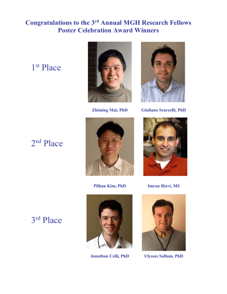
Congratulations to the 3rd Annual MGH Research Fellows Poster Celebration Award Winners 1st Place Zhiming Mai, PhD Giuliano Scarcelli, PhD Pilhan Kim, PhD Imran Rizvi, MS Jonathan Celli, PhD Ulysses Sallum, PhD 2nd Place 3rd Place Nanotechnology Platforms in Targeted Photodynamic Therapy and Imaging of Ovarian Cancer Zhiming Mai*, Benjamin A. Teply† and Tayyaba Hasan* * Wellman Center for Photomedicine, Massachusetts General Hospital, Harvard Medical School. Boston, MA, USA; and †Department of Chemical Engineering, Harvard–MIT Center of Cancer Nanotechnology Excellence, Massachusetts Institute of Technology, Cambridge, MA, USA Purpose: Poor survival rates associated with advanced ovarian cancer disseminated in the abdominal cavity as well as resistance to standard chemotherapeutic drugs and radiation necessitate the need for a drastic change of approach to the disease management. The overall goal of this study is to develop nanoparticle platforms utilizing targeted photodynamic therapy and imaging modalities to achieve high selectivity combined with the most effectiveness against disseminated chemo- and/or radiation-resistant ovarian cancer, as well as to perform early diagnosis and monitor treatment outcomes. Materials & Methods: Figure 1 cartoons the concept of a nanoparticle platform for targeted photodynamic therapy. Targeted nanoparticles synthesized here are multicomponent structures with a carrier system that forms the core and contains the therapeutic or imaging payload, surface modifiers to reduce reticuloendothelial system uptake and enhance biodistribution, and a targeting component. Polymers used in the preparation of nanoparticles were poly (lactic-co-glycolide)-poly(ethylene glycol) (PLGA-PEG), biodegradable and FDA-approved drug carriers. The photosensitizer-bearing naoparticles (CMA-NP) were made by encapsulation of the chlorin e6 monoethylene diamine disodium salt (CMA) into PLGA-PEG. Finally, the targeting CMA-NP was obtained through conjugation of CMA-NP with targeting ligands called aptamers for EGFR-family receptors overexpressed on ovarian cancer cells. Using such nanoparticle platforms, we have initiated photosensitizer uptake, photodestruction, and imaging assays to characterize their multifunctional capacities in management of ovarian cancer. Results: The synthesized nanoparticles were in monodisperse with an average size of 202.0 nm and negative surface charge at -30 mV. The payload of photosensitizers in nanoparticles was at 0.41 mM or 0.279 mg/ml of equivalent CMA. Results of photosensitizer uptake measurement suggested that lower CMA-NP accumulation in normal tissues/cells was due to their overall negative charge which mediates the repulsion between the nanoparticles and cellular membranes innately bearing negative charge. This feature comprises one of the aspects in selection, thus minimizing side effects of drug toxicity to normal tissue/cells. Confocal fluorescence microscope imaging indicated that specific ligand-bearing CMA-NP had a high selectivity for OVCAR-5 and OVCAR-3 ovarian cancer cells as compared with control cells, consisting of another aspect in selection. Also, the fluorescent property of CMA in the nanoparticle complex formed the basis of imaging analysis for both diagnosis and treatment monitoring. Photodestruction studies showed that the photosensitizers in nanoparticle core have a higher potency in cancer cell photodynamic killing than free CMA. Conclusion: To our knowledge, this novel nanoparticle platform is the first using nanobiotechnology and aptamer-based targeting PDT and imaging. The nanoparticle complex facilitates the delivery of therapeutic photosensitizers, and will provide a model for a highly selective, effective, and predicable therapy, an approach that can be adapted for other targets as reagents become available. Author Contact: Zhiming Mai; Dermatology; 617-726-6995; zmai@partners.org In vivo Biomechanical Microscopy of Tissue and Biomaterials Giuliano Scarcelli and Seok Hyun Yun Wellman Center for Photomedicine, Massachusetts General Hospital and Harvard Medical School Purpose: The mechanical properties of biological tissues and biomaterials are closely related to their functional abilities, and thus play significant roles in many areas of medicine. For example, hardened coronary arteries by calcification can cause heart problems; changes in the elasticity of crystalline lens and cornea are thought to be central in the development of ocular disorders such as cataracts, presbyopia and corneal ectasia; biomechanical compatibility is crucial in tissue engineering procedures; and, the stiffness of extra-cellular matrix influences drug delivery and cell motility. However, measuring such biomechanical properties remains a significant challenge due to a dearth of non-invasive technologies. Our goal is to develop a novel diagnostic technique, Brillouin confocal microscopy[1], to probe the biomechanical properties of tissue in vivo without contact, quantitatively, and with high spatial resolution. Materials & Methods: Brillouin light scattering arises from the interaction between light and sound waves inside material. Such interaction induces a small frequency shift in the scattered light that is directly related to the viscosity and elasticity of samples. We have developed a high-resolution optical spectrometer that is able to measure such tiny frequency shift with unprecedented detection efficiency and we integrated it with a home-built confocal microscope. Results: Using Brillouin microscopy, we have obtained the first 3D images that use elasticity as contrast mechanism (Fig.1a); we monitored fast dynamic changes in elastic modulus during polymer cross-linking (Fig.1c); and we performed the first in vivo measurement of crystalline lens in the mouse eye (Fig.1b). We have also obtained the first in vivo demonstration of age-related stiffening of lenses (not shown). Fig. 1: (a) Brillouin images of an intraocular lens (ellipse) immersed in fluid (blue). (b) In situ measurement of the elastic modulus of a mouse crystalline lens. (c) Real-time monitoring of elasticity variation in a polymer due to UV-induced cross-linking. Conclusion: Brillouin confocal microscopy can indeed characterize in vivo the biomechanics of tissue and biomaterials non-invasively and with micron-scale resolution. The first areas of biomedical applications we are exploring are in ophthalmology where Brillouin microscopy may enable measuring changes in corneal and lens elasticity by aging, by the progression of disease, or in response to treatment and drugs; and in tissue engineering for the optimization of procedures by mapping and monitoring in situ and in real time the micromechanical properties of host and implanted tissue. [1] G. Scarcelli & S. H. Yun, Brillouin confocal microscopy for 3D mechanical imaging, Nature Photonics 2, 39-43 (2008). Author Contact: Giuliano Scarcelli, Dermatology, 617-724-6798, gscarcelli@partners.org Longitudinal cellular imaging of murine internal organs by in vivo endomicroscopy Pilhan Kim1, Georges Tocco2, Euiheon Chung3, Cavit D. Kant2, Hiroshi Yamashita3, Dai Fukumura3, Rakesh K. Jain3, Gilles Benichou2, and Seok H. (Andy) Yun1 1 Wellman Center for Photomedicine, Massachusetts General Hospital and Harvard Medical School 2 Surgery / Transplantation Unit, Massachusetts General Hospital 3 Edwin L. Steele Lab., Department of Radiation Oncology, Massachusetts General Hospital and Harvard Medical School Purpose: Intravital fluorescence microscopy has emerged as a powerful tool for visualizing the cellular processes within small animals. However, due to the large size of standard microscope lenses, imaging the internal organs of small animals, such as mice, remains a challenge, requiring invasive procedures. The first specific aim of our research is to develop a miniature confocal and multiphoton endoscopic imaging system. The second specific aim is to apply this novel technique to visualize and quantify immune cell trafficking in chronic heart rejection and angiogenesis in orthotopic colorectal tumors, following the same animals over weeks. Materials & Methods: Using gradient-index (GRIN) lenses, we fabricated front- and side-viewing types of imaging probes that are 1 mm in diameter and 15 to 40 mm in length. To avoid motion-induced image blurring, we integrated the probe into a 15-gauge tube that was designed to hold the tissue locally by gentle vacuum pressure. We then coupled the endoscope into the real-time video-rate confocal and multiphoton microscope system we have developed previously. The image resolution was 1 μm in transverse and 15 μm in depth. To measure the kinetics of antigen presenting cells trafficking (from donor or recipient origin), hearts were harvested from MHC class-II:GFP+ mice and transplanted into the abdomen of wildtype and aly/aly mice, and the heart, spleen, and draining lymph nodes were imaged by laparotomy from day 0 till several weeks after transplantation. To study the angiogenesis in colorectal cancer, green fluorescent protein (GFP) expressing human colon adenocarcinoma cells were surgically implanted to the wall of colon in athymic nude mice, and the tumor and vasculature were imaged by rotational-pullback colon-microscopy at post-operative day 6, 14, and 34, Results: Initially, right after heart transplantation, a large number of MHC class-II+ cells were observed in the heart. After 3 days, in a wildtype recipient mouse, 93% of these cells in heart were no longer present in the graft, and donor-derived MHC class II+ cells were observed in various lymph nodes and spleen. In contrast, in aly/aly recipient mice (lacking lymph nodes) after 1 week, we observed that 96% of donor MHC class II+ cells are still present in the heart graft, and interestingly a similar number of MHCclass II+ cells as the wildtype recipient was observed in the spleen. At 2 weeks post operation, we found 10 times increased number of MHC class II+ cell in the spleen of same aly/aly recipient mouse, while 92% of donor MHC class II+ cells are still present in the grafted heart, suggesting the antigen presentation in the spleen of aly/aly recipient mouse. In the colon cancer xenograft model, we observed the increase in size of the implanted tumor with time. Supplying peri-tumor vessels for tumor growth and the elevated leakage at tumor sites were visualized at the boundary of tumor. Dilated and distorted vessels compared to normal vessels in healthy region were observed at tumor sites. Conclusion: We have developed a novel microendoscopy system for longitudinal in vivo imaging of various viscera organs in mice. Using home-built endoscopes, we have demonstrated the first cellular imaging in a beating heart, quantitative immune cell trafficking following organ transplantation, and wide-area tumor imaging in the colon mucosa. We expect in vivo mouse endomicroscopy to prove useful in a variety of other biomedical investigations using experimental animal models. Author Contact: Pilhan Kim, Wellman Center for Photomedicine, 617-643-2908, pkim5@partners.org Imaging Drug Penetration and Metabolic Activity in 3D Ovarian Cancer Models Imran Rizvi1,2, Conor L. Evans1, Daina Burnes1, Jonathan Celli1, Johannes de Boer1 and Tayyaba Hasan1 1 Wellman Center for Photomedicine, Massachusetts General Hospital, Boston, MA 02114 2 Thayer School of Engineering, Dartmouth College, Hanover, NH 03755 Purpose: Our lab has published promising pre-clinical results using photodynamic therapy (PDT), an emerging light-based modality, in combination with Erbitux®, an antibody targeted against the epidermal growth factor, to synergistically treat advanced ovarian carcinomatosis. As this therapeutic strategy moves into clinical trials, we are investigating the mechanisms underlying the observed synergism to optimize this combination regimen. The purpose of the current study is to characterize heterogeneities in drug penetration and metabolic activity using 3D ovarian cancer models, and optical imaging technologies, to elucidate the mechanisms of synergistic treatment response. Materials & Methods: Based on breast cancer models described by Drs. Mina Bissell and Joan Brugge, we have developed 3D cultures for ovarian carcinoma as our in vitro research platform to conduct these mechanistic studies. Ovarian cancer cells seeded on Growth Factor Reduced (GFR) Matrigel™ beds spontaneously form 3D acinar structures that more closely resemble in vivo tumor nodules than cells in monolayer. We are using confocal microscopy of benzoporphrin derivative (BPD) and Erbitux® along with multiphoton microscopy of intrinsic fluorophores to nonpurtubatively investigate the patterns of drug penetration and metabolic activity in these acinar structures. Results: As with other treatments, preliminary experiments suggest that the 3D acini are less sensitive to PDT than cells in monolayer. This differential treatment response could be attributed to heterogeneity in drug penetration as well differences in signaling and architectural cues between the two systems. In this study, we have shown that BPD and Erbitux® are unevenly distributed in 3D acinar structures (Figure 1). Furthermore, an unexpected distribution pattern of metabolic and apoptotic activity is observed in the ovarian cancer acini following multiphoton microscopy of intrinsic fluorophores. Conclusion: Development of novel research platforms to complement, or replace, traditional model systems could significantly impact our ability to conduct reliable mechanistic studies and predict the efficacy of therapeutic strategies. Our current research demonstrates heterogeneity in drug distribution and metabolic activity in 3D ovarian cancer acini, which could have implications for optimizing PDT + Erbitux®-based combination treatment of ovarian carcinomatosis. A B C D Figure 1 Confocal and multiphoton fluorescence images of 3D acinar cultures of ovarian cancer: A) BPD and B) Erbitux distributions; C) Intrinsic fluorescence excited at 720nm; D) Volumetric rendering of BPD and intrinsic fluorescence. Figures C and D are presented in false-color intensity scale. The scan volume for Figure D is 110µm x 105µm x 100µm. Author Contact: Imran Rizvi, Wellman Center for Photomedicine, 617-726-3991 IRIZVI@PARTNERS.ORG Minimally invasive fluorescence endoscope for detection and treatment monitoring of ovarian cancer Jonathan Celli, Wei Zhong, Imran Rizvi, Conor Evans, Zhiming Mai, Johannes deBoer, Seok-Hyun Yun, Tayyaba Hasan Wellman Center for Photomedicine, Massachusetts General Hospital, Harvard Medical School Purpose: The goal of this work is to develop a minimally invasive imaging system with sufficiently high resolution and selectivity at the cellular level to detect ovarian cancer (OVCA) micrometastases at an early stage and monitor their response to treatment. The dismal mortality rate for patients with ovarian cancer is often associated with these small, disseminated metastases that pose serious challenges to current diagnostic and treatment assessment techniques such as white light laparscopy. These modalities lack adequate sensitivity for detecting OVCA at the sub-clinical stage or for detecting recurrent disease. Materials & Methods: We have developed a custom micro-endoscope which achieves 9 μm spatial resolution. This instrument employs epi-fluorescence illumination to image through a flexible optical fiber coupled to a microscope objective onto a low light sensitive CCD camera with on chip multiplication gain. Using this system we conducted imaging experiments on a murine animal model of OVCA previously established in the laboratory. In these studies the photosensitizer BPD-MA, used here both as a fluorescent contrast agent and in some cases additionally for photodynamic therapy (PDT) treatment, was injected into mice 1 hour prior to imaging. Images of tumor nodules were compared to histology in order to establish the specificity and sensitivity of the instrument. In some mice PDT treatment was conducted at the time of the initial imaging. These mice were imaged again 5-7 days following treatment to monitor the treatment efficacy. A recent modification to the micro-endoscope system includes the addition of optical coherence tomography (OCT) imaging, a highly sensitive, crosssectional imaging technique (analogous to ultrasound) capable of visualizing the microscopic details of biological tissues to provide additional 3D structural information. Results: We demonstrate that the endoscope can detect in vivo tumor nodules tens of microns in size, an order of magnitude smaller than previously reported. By comparison of imaging data to histology we have determined the system’s sensitivity and specificity for detecting tumor nodules, to be 86% and 53%, respectively. In experiments in which diseased mice were imaged with the endoscope immediately before and seven days following PDT treatment, a significant decrease in tumor burden is observed relative to control mice and confirmed by surgical resection and determination of tumor burden by weight. Liver 100 μm 100 μm Small Bowel Tumor Left: Fluorescence image of the peritoneal cavity of an OVCA mouse 1 hour after injection of BPD-MA. Center: H & E stain of a 5 μm section from the same region. Arrows indicate tumor nodules visible in each image. Right: en bloc resection showing advanced metastatic disease in OVCA mouse model. Conclusion: We have developed and characterized a fluorescence endoscope for minimally invasive imaging and demonstrated its capability for detecting OVCA tumor nodules in vivo, an order of magnitude smaller than those reported previously. Author Contact: Jonathan Celli, Wellman Center for Photomedicine, 617-726-3991, jcelli1@partners.org An Enzyme Activated Photodynamic Prodrug for the Photodynamic Therapy of Drug Resistant Bacterial Infections Ulysses W. Sallum, Xiang Zheng, Sarika Verma, Conor L. Evans, Humra Athar, Tayyaba Hasan Abstract: Antimicrobial photodynamic therapy (PDT) is an emergent approach for the treatment of drug resistant infections. The use of pathogen specific enzyme activated photodynamic prodrugs could enhance the specificity of antimicrobial PDT. In this proof of principle work, a ß-lactamase specific, enzyme activated photodynamic prodrug (ß-LEAPP) was designed, synthesized, and characterized. ß-LEAPP was activated specifically by purified ß-lactamase and ß-lactamase producing whole cell bacterial suspensions, but not by non-induced reference bacteria or human keratinocytes and fibroblasts cell cultures. A specific photodynamic toxicity toward ß-lactam resistant bacteria was achieved while a dark toxic effect of ß-LEAPP on ß-lactam susceptible bacteria was observed.


