Cervical Spinal Surgery
advertisement
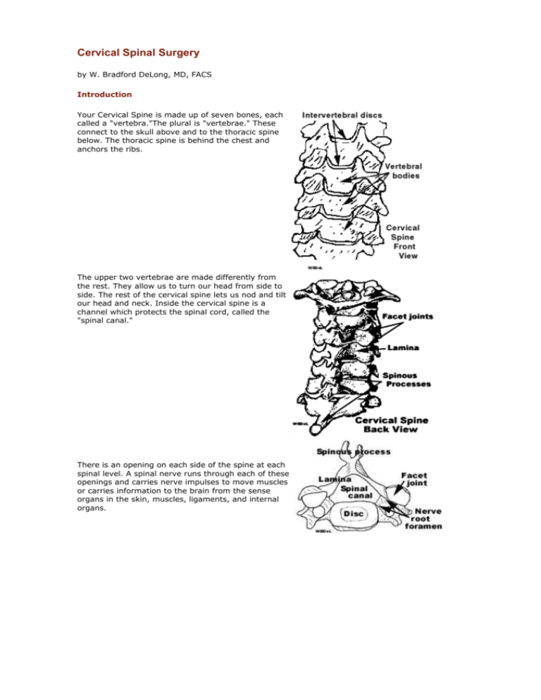
Cervical Spinal Surgery by W. Bradford DeLong, MD, FACS Introduction Your Cervical Spine is made up of seven bones, each called a "vertebra."The plural is "vertebrae." These connect to the skull above and to the thoracic spine below. The thoracic spine is behind the chest and anchors the ribs. The upper two vertebrae are made differently from the rest. They allow us to turn our head from side to side. The rest of the cervical spine lets us nod and tilt our head and neck. Inside the cervical spine is a channel which protects the spinal cord, called the "spinal canal." There is an opening on each side of the spine at each spinal level. A spinal nerve runs through each of these openings and carries nerve impulses to move muscles or carries information to the brain from the sense organs in the skin, muscles, ligaments, and internal organs. An intervertebral disc sits between every two vertebrae, and cushions the spine. The discs are pads of connective tissue and cartilage (sort of like the tough tissue in the breast bone of a chicken). They act like shock absorbers between the vertebrae. Sometimes, pieces of the disc can push backward into the spinal canal and press on the nerves or spinal cord. Inflammation from the disc can sometimes irritate the nerves or spinal cord. Other times calcium deposits can build up on the back of the vertebra and push on the nerves or spinal cord. One of these calcium deposits is called a "spur." The medical name is "osteophyte," which means a bony projection. Your Cervical Spine is made up of seven bones, each called a "vertebra."The plural is "vertebrae." These connect to the skull above and to the thoracic spine below. The thoracic spine is behind the chest and anchors the ribs. The upper two vertebrae are made differently from the rest. They allow us to turn our head from side to side. The rest of the cervical spine lets us nod and tilt our head and neck. Inside the cervical spine is a channel which protects the spinal cord, called the "spinal canal." There is an opening on each side of the spine at each spinal level. A spinal nerve runs through each of these openings and carries nerve impulses to move muscles or carries information to the brain from the sense organs in the skin, muscles, ligaments, and internal organs. An intervertebral disc sits between every two vertebrae, and cushions the spine. The discs are pads of connective tissue and cartilage (sort of like the tough tissue in the breast bone of a chicken). They act like shock absorbers between the vertebrae. Sometimes, pieces of the disc can push backward into the spinal canal and press on the nerves or spinal cord. Inflammation from the disc can sometimes irritate the nerves or spinal cord. Other times calcium deposits can build up on the back of the vertebra and push on the nerves or spinal cord. One of these calcium deposits is called a "spur." The medical name is "osteophyte," which means a bony projection. Herniated Cervical Disc If You Have a Herniated Cervical Disc causing neck and arm pain, and if you have not improved from a good program of conservative care, then you might consider having surgery. A herniated disc develops when a disc between the vertebrae breaks down. The back part of the disc becomes weak and the disc pushes backward against the nerves or even against the spinal cord. Problems can also arise from bone spurs which can develop around the cervical discs. These spurs can pinch the nerves to the arms or can press on the spinal cord itself, causing pain, weakness, or numbness. Sometimes surgery is necessary to remove them. Simple Cervical Disc Surgery The simplest type of herniated disc is one which is herniated only on one side and is causing pain on that side. If that's what you have, you are probably a candidate for a simple microsurgical disc excision without a fusion. The surgery can be done through the back of your neck (a "posterior approach") or through the front (an "anterior approach"). I usually recommend the anterior approach because it is a relatively comfortable approach for the patient. If the disc is pushing backward from the middle of the disc (a "central herniation"), or on both sides, then more of the disc needs to be removed. If that is the case, then a bone fusion may be recommended. A fusion involves taking a small piece of bone and putting it where the disc was between the vertebrae. This is called an "interbody fusion" because the bone is located between the vertebral bodies in the front part of the cervical spine. The piece of bone can either come from the bone bank or from your own iliac crest (the top of the pelvic bone). A Percutaneous Discectomy A Percutaneous Discectomy, is a "closed" operation, because the surgeon does not actually have to make an incision to get at the disc. The central part of the herniated disc is suctioned out with the instruments. The procedure is roughly comparable to arthroscopic surgery of the knee, except that the surgeon "sees" into the disc by means of fluoroscopy rather than by looking directly through an arthroscope. Techniques are being developed which will allow the surgeon to look into the disc directly with an arthroscope and remove the center of the disc directly either with small instruments or with laser surgery. However, neither Percutaneous Discectomy nor Arthroscopic Discectomy allows the surgeon to remove pieces of the disc which have become stuck in the spinal canal. If you have a piece of the disc pushing on a nerve or the spinal cord, or if you have significant bone spurs, then you need an "open" operation. Anterior Cervical Surgery As I mentioned above, I usually recommend that cervical spine surgery be done from the front. If the problem is a simple single herniated disc or bone spur, then only one level will need to be approached. On the other hand, if discs are herniated at two or more levels, or if bone spurs are creating problems at several levels, then more than one level will need surgery. Sometimes the surgery has to be quite extensive if bone spurs are narrowing the spinal canal and pressing on several nerves or on the spinal cord. In those cases, the surgeon needs to drill away the vertebral bodies in the front of the cervical spine. This is necessary to open up the spinal canal and give the spinal cord and nerves more room. In these more extensive cases, the bone which is drilled away is replaced with a piece of bone. This bone graft supports the spine over the two or more levels which have been opened up. We often fasten one or two little metal plates onto the bone graft and onto the front part of the spine. These plates support the spine and protect the graft from slipping out. Postoperative Care You will be watched closely by the nursing staff the night of the operation to make sure that postoperative swelling doesn't cause any problem with your breathing. The next day you may be able to go home, depending on how you're feeling. You will need to wear a cervical brace for several weeks. The kind of brace you need and how long you wear it will depend on how extensive the surgery has been. For simpler operations, you can wear a Philadelphia collar, a Somi brace, or some similar type of brace. You will need the brace for six to twelve weeks, depending on how fast the fusion heals. If you haven't needed a fusion, you can probably remove the brace after two weeks. For operations which involve removing three or more discs, you will probably need a heavier brace, for increased safety (a "Halo" brace). You must not drive until you're out of the brace. We encourage you to walk at least one-half hour a day, and you should start doing simple range of motion exercises for your shoulders about two weeks after the surgery. After the fusion is solid, we usually involve you in more active rehabilitative physical therapy. Your Own Bone Graft or Bank Bone? You have a choice of using bone from your own iliac crest or using bone from the bone bank. I recommend bank bone. If you use your own bone, then the surgeon must make a separate incision over your iliac crest. This incision is usually more uncomfortable than the neck incision, and the discomfort usually persists considerably longer than the discomfort from the neck. Taking bone from your own iliac crest raises the possibility of another set of complications, such as infection. When we use bank bone, we obtain the bone only from a well-established reputable tissue bank. The bone is carefully screened and sterilized against AIDS and hepatitis. Bank bone fuses very well. In our experience, the fusion rates for bank bone from a good tissue bank and for the patient's own bone are the same. Bank bone takes a little longer to fuse, but most people think it is worth the wait to avoid the discomfort of the iliac crest incision. Using bank bone is especially important if we need to use a piece of bone to support the spine after we have drilled out the front part of the vertebral bodies. The piece of bone we can obtain from the tissue bank is stronger than the bone we can obtain from the patient. However, if a patient has strong feelings about using their own bone, we will certainly honor those feelings and use their own bone. Possible Complications Neck surgery is surgery that we do very commonly, but we take it very seriously. Complications can occur. Before you make the final decision concerning cervical spinal surgery, the surgeon will discuss with you in detail the possible complications and what they mean, along with other risks, limitations, and alternatives associated with the procedure. Conservative Care Before Surgery Most people who have symptoms from a herniated cervical disc or from bone spurs get better without surgery, provided they are involved in a good program of conservative care. Effective conservative care includes physical therapy and/or chiropractic care. I advise against the use of chiropractic adjustments in the neck if a patient has a significantly herniated disc or severe bone spurs, but there are many other forms of chiropractic treatment which can be helpful. Spinal adjustments can still be carried out to maintain alignment of the thoracic and lumbosacral regions. Overhead cervical traction can help. It fits over a door, with the patient facing the door and the pulley. Conservative care in some cases can include injections of medication to treat the inflamed nerve roots or ligaments. My philosophy is that surgery should be done only if conservative measures don’t help enough. Surgery is only part of the overall program. After surgery, it is very important that the patient be involved in a good program of rehabilitation.
