wcb-MRIGOD
advertisement
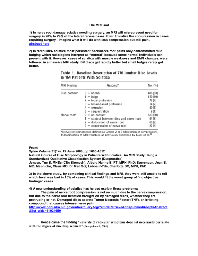
The MRI God 1) In nerve root damage sciatica needing surgery, an MRI will misrepresent need for surgery in 28% to 29% of the lateral recess cases. It will trivialize the compression in cases requiring surgery - imagine what it will do with less compression but still pain. abstract here 2) In radiculitis- sciatica most persistent back/nerve root pains only demonstrated mild bulging which radiologists interpret as “normal” because some normal individuals can present with it. However, cases of sciatica with muscle weakness and EMG changes, were followed in a massive MRI study. Bif discs get rapidly better but small bulges rarely got better. From: Spine Volume 31(14), 15 June 2006, pp 1605-1612 Natural Course of Disc Morphology in Patients With Sciatica: An MRI Study Using a Standardized Qualitative Classification System [Diagnostics] Jensen, Tue S. MHSc (Clin Biomech); Albert, Hanne B. PT, MPH, PhD; Soerensen, Joan S. MD; Manniche, Claus MD, Dr Med Sci; Leboeuf-Yde, Charlotte DC, MPH, PhD 3) In the above study, by combining clinical findings and MRI, they were still unable to tell which level was bad in 10% of cases. This would fit the worst group of "no objective findings" cases. 4) A new understanding of sciatica has helped explain these problems: The pain of nerve root compression is not so much due to the nerve compression, but due to the nerve root irritation brought on by damaged discs, whether they are protruding or not. Damaged discs secrete Tumor Necrosis Factor (TNF), an irritating compound that causes intense nerve pain. http://www.ncbi.nlm.nih.gov/entrez/query.fcgi?cmd=Retrieve&db=pubmed&dopt=Abstract &list_uids=11924650 Hence came the finding " severity of radicular symptoms does not necessarily correlate with the degree of disc displacement"( Karppinen J, 2001) CT SCAN IN : Diagnostic imaging for back pain Michael Yelland Australian Family Physician Vol. 33, No. 6, June 2004 Article here "Computerised tomography scanning has virtually no role in the assessment of uncomplicated back pain and only a very limited role in the assessment of low back pain with sciatica." Nerve Studies One review of nerve studies found considerable variation in findings, and that many were incomplete. Dr. Bogdug comments they are only useful for some entrapments like carpal tunnel. Hence for the vast majority of chronic pain patients, normal nerve studies means nothing. The worst cases are thoracic outlet syndrome, where one would think there would be findings but there are not in the vast majority of cases. Worse yet, come the conclusions of Dr. N. Bogduk, one of the father’s of cervical radiofrequency neurotomy and researcher: “In most cases, causes for chronic low back pain cannot be found using conventional investigations, such as radiography and magnetic resonance imaging (MRI), with fewer than 10% of cases diagnosed by these means.” ….” When diagnostic joint blocks are used, the source of pain can be traced to the sacroiliac joint in about 20% of patients,14 while lumbar zygapophysial joint pain is found in about 15% of injured workers,15 and as many as 40% of people with chronic back pain in older populations.16 CT discography reveals internal disc disruption in at least 40% of patients.17” These figures belie the assertion that 80% of patients with chronic low back pain cannot be diagnosed. This is true if investigations are limited to CT or MRI, but a diagnosis becomes possible if diagnostic blocks and discography are used “ (my emphasis) Given that none of above tests have been routinely used here, we would be at the 80% not diagnosed. We do have a new back doctor in town and that could be changing; this will not help people in harm's way of WCB up to now.
