Pediatric Skin Assessment
advertisement
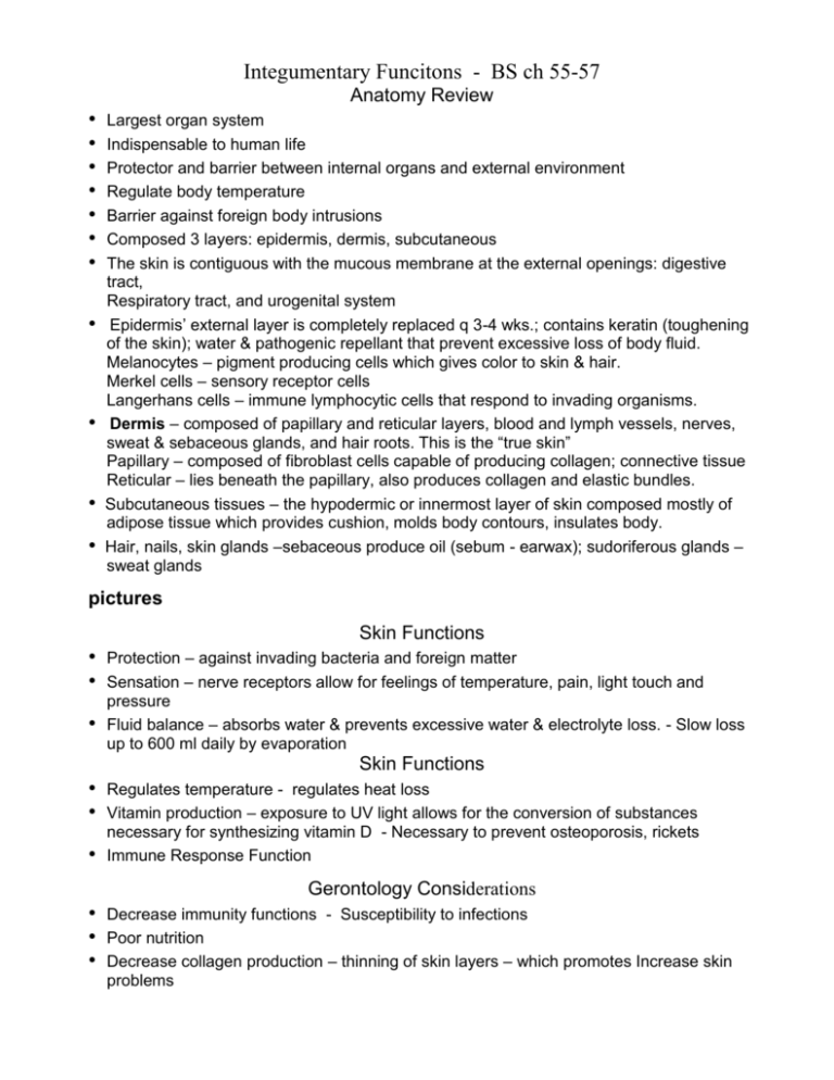
Integumentary Funcitons - BS ch 55-57 Anatomy Review • • • • • • • • • • • Largest organ system Indispensable to human life Protector and barrier between internal organs and external environment Regulate body temperature Barrier against foreign body intrusions Composed 3 layers: epidermis, dermis, subcutaneous The skin is contiguous with the mucous membrane at the external openings: digestive tract, Respiratory tract, and urogenital system Epidermis’ external layer is completely replaced q 3-4 wks.; contains keratin (toughening of the skin); water & pathogenic repellant that prevent excessive loss of body fluid. Melanocytes – pigment producing cells which gives color to skin & hair. Merkel cells – sensory receptor cells Langerhans cells – immune lymphocytic cells that respond to invading organisms. Dermis – composed of papillary and reticular layers, blood and lymph vessels, nerves, sweat & sebaceous glands, and hair roots. This is the “true skin” Papillary – composed of fibroblast cells capable of producing collagen; connective tissue Reticular – lies beneath the papillary, also produces collagen and elastic bundles. Subcutaneous tissues – the hypodermic or innermost layer of skin composed mostly of adipose tissue which provides cushion, molds body contours, insulates body. Hair, nails, skin glands –sebaceous produce oil (sebum - earwax); sudoriferous glands – sweat glands pictures Skin Functions • • • Protection – against invading bacteria and foreign matter Sensation – nerve receptors allow for feelings of temperature, pain, light touch and pressure Fluid balance – absorbs water & prevents excessive water & electrolyte loss. - Slow loss up to 600 ml daily by evaporation Skin Functions • • • Regulates temperature - regulates heat loss Vitamin production – exposure to UV light allows for the conversion of substances necessary for synthesizing vitamin D - Necessary to prevent osteoporosis, rickets Immune Response Function Gerontology Considerations • • • Decrease immunity functions - Susceptibility to infections Poor nutrition Decrease collagen production – thinning of skin layers – which promotes Increase skin problems • Taking more medications; and Excessive environmental exposure Visible changes if the Skin • Cyanosis • Color changes –Hypopigmentation –Eczema – Infection –Vitiligo (failure of melanin formation) • Assess the vascularity & hydration of skin • Nails – configuration, consistency, color • Hair – color and distribution • • • • • • • • Assessing light to dark skin Assessing Lesions Vary in size, shape and cause Primary – initial lesions characteristic of the disease itself Secondary – result from external causes: scratching, trauma, infections, changes in wound healing. Dermatosis = any erruptions, lesions or disorder of the skin Systemic Disease Affecting Skin Diabetic dermapathy Stasis dermatitis Skin infections Leg/foot ulcers Cuteneous signs of HIV disease • • • Clinical photographs Taken to document the nature & extent Dertermine progress Track the status of a mole if the characteristics are changing Pediatric Skin Assessment • • • • Involves inspection & palpation Color, texture, temp., moisture, turgor Light color skin - milky white and rose to a deep hue of pink Dark color skin – various brown, yellow, olive green & bluish tones Color changes of racial groups Description Cyanosis – bluish tones seen through skin; reflects deoxygenation Pallor – paleness; may indicate anemia, chronic disease, edema or shock Erythema – may indicate increased blood flow from climatic conditions, local inflammation, infection, skin irritation, allergy or other dermatoses.; or may be caused by increased numbers of RBC as compensatory response to chronic hypoxia Ecchymosis – large, diffuse, black or blue area, caused by hemorrhage of blood into the skin resulting from injuries Petechia – same as ecchymosis except for size. Small distinct, pinpoint hemorrhages 2 mm or less in size, can denote some type of blood disorder such as leukemia. Jaundice – yellow staining of the skin usually caused by bile pigments Light skin appearance Bluish tinge, especially in palpebral conjuctiva, nail beds, earlobes, lips oral membranes, soles, palms Loss of rosy glow in skin, especially in the face Redness easily visible anywhere on the body Dark skin appearance Ashen gray lips and tongue Ashen gray in black skin color; More yellowish brown color in brown skin Much more difficult to assess; Rely on palpation for warmth or edema Purplish to yellow-green areas; may be seen anywhere on the skin Very difficult to see unless in mouth or conjuctiva Purplish pinpoint markings most easily seen anywhere on the skin. Usually invisible except in oral mucosa, conjunctiva of eyelids, and conjuctiva covering eyeball Yellow staining seen in sclera of eyes, skin, soles, palms, fingernails, oral mucosa. Mostly reliably assessed in sclera, hard palate, palms and soles. Skin Characteristics • • • Normally young children’s skin texture is smooth, slightly dry, not oily or clammy. Evaluate skin temp. by symmetrically feeling and comparing each body parts and upper areas with lower areas. Determine skin turgor – best indicator for dehydration • • • • • • Integumentary Functions Anatomy Review Largest organ system; It’s indispensable to human life Protector and barrier between internal organs and external environment Regulate body temperature Barrier against foreign body intrusions Transmits sensation 3 layers: epidermis, dermis, subcutaneous • • • • • • Skin Functions Protection – against invading bacteria and foreign matter Sensation – nerve receptors allow for feelings of temperature, pain, light touch and pressure Fluid balance – absorbs water & prevents excessive water & electrolyte loss. –Slow loss up to 600 ml daily by evaporation Regulates temperature - regulates heat loss Vitamin production – exposure to UV light allows for the conversion of substances necessary for synthesizing vitamin D –Necessary to prevent osteoporosis, rickets Immune Response Function - Gerontology Considerations Be aware of the significant changes of aging: • Decrease immunity functions; Susceptibility to infections • Poor nutrition; Thinning of epidermal skin layers • Decrease collagen production – loss of subcutaneous • Increase skin problems; Excessive environmental exposure • Taking more medications • Dryness, wrinkling, Uneven pigmentation • Various proliferative lesions • Visible changes if the Skin: Cyanosis & Color changes; Hypopigmentation, Eczema, Infection; Vitiligo (failure of melanin formation) • Assess the vascularity & hydration of skin • Nails – configuration, consistency, color • Hair – color and distribution • • • • • • • • • • • • Assessing light to dark skin Assessing Lesions Vary in size, shape and cause Primary – initial lesions characteristic of the disease itself Secondary – result from external causes: scratching, trauma, infections, changes in wound healing. Dermatosis = any erruptions, lesions or disorder of the skin Pediatric Skin Assessment Involves inspection & palpation Color, texture, temp., moisture, turgor or elasticity Skin turgor is one of the best estimates of adequate hydration and nutrition Skin Color in Children Light color skin – milky white and rose to a deep hue of ping Dark color skin – various brown, yellow, olive green & bluish tones Skin Characteristics Normally young children’s skin texture is smooth, slightly dry, not oily or clammy. Evaluate skin temp. by symmetrically feeling and comparing each body parts and upper areas with lower areas. Determine skin turgor – best indicator for dehydration Dermatologic Problems in Newborns Erythema Toxicum Neonatorum • Flea bite dermatitis (benign newborn rash) • Skin erruption ususally appear 1-2 days of life;duration 5-7 days • 1-3 mm lesions • Firm, pale yellow, white papules • May have pustules which resemble flea bites. • Appears on face, extremities, trunk and buttocks. • Cause unknown • Treatment not necessary Candidiasis Albicans • • • • • Yeast (fungal) infection Acquired from maternal vaginal infection Also person-to-person transmission Oral infection is known as Thrush Infants may refuse to suck bottle because of pain in os. P.315 • • • • HERPES A serious viral infection in newborns Up to 60% mortality Calssified according to types (locations) Antiviral therapy – Acyclovir Bullous Impetigo • Superficial skin condition caused by Staphylococcus aureus • Buttocks, perineum, trunk, face, extremities • Vary in size • Dx. by gram stain & blister cultures • Tx, = Mupirocin (Bactroban); isolation; prevent from scratching; Hyperbilirubinemia • Excessive levels of accumulated bilirubin in the blood • Jaudince or icterus • Usually benign; Caused by unconjugated/conjugated bilirubin • Immature heaptic function, increase bilirubin loard from RBC hemolysis Therapy • Increase frequency of feedings; Avoid supplements • Monitor bilirubin levels; Phototherapy lights • Perform risk assessment Disorders Affecting the Skin Skin Lesions p.755 • Caused by etiologic factors: –Infectious agents –Toxic chemicals: skin irritation –Physical trauma: burns, lacerations –Hereditary factors –External factors: allergens, environmental, contact dermitis –Systemic diseases: measles, lupus, nutritional deficiency Skin Lesions: Nursing Care: –Assessment: descriptions; pt. history, causative factors –Evaluation of skin –Nursing Diagnosis p.764 –Interventions for skin care to promote healing and prevent further injury –Pain management & comfort –Infection control –Nursing evaluation & reassessment Skin Cancers Melanoma • Melanomas have the (ABCD rule) characteristics –A: Asymmetry; two sides of pigmented area do not match –B: Irregular border, exhibits indentations –C: Color (pigmented area) is black, brown, tan, and sometimes red or blue –D: Diameter is > 6 mm (size of pencil eraser) Rule of Nines
