Issues with Analysis & Interpretation
advertisement
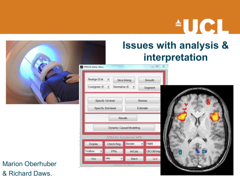
Issues with analysis & interpretation Marion Oberhuber & Richard Daws. 30000 25000 20000 fMRI 15000 EEG 10000 5000 0 1985 1990 1995 2000 2005 2010 2015 2020 Recap - Hypothesis testing H0: con1 = con2 HA: con1 ≠ con2 The Test Statistic T Computed at each voxel Summarises evidence about H0 Null Distribution of T We need to know the distribution of T under the null hypothesis Significance level α u Set a priori (e.g. 0.05) choose threshold uα to obtain acceptable false positive rate α P-value A p-value summarises evidence against H0 This is the chance of observing value more extreme than t under the null hypothesis. Null Distribution of T t p(T t | H 0 ) P-val The conclusion about the hypothesis We reject H0 in favour of H1 hypothesis if p(H0) < uα Null Distribution of T Type I/type II error Each voxel can be classified as one of four types Truly active Declared active Declared inactive ✔ Type II error Truly inactive False positives uβ Type I error ✔ False negatives u specificity: 1- u = proportion of actual negatives which are correctly identified sensitivity (power): 1- uβ = proportion of actual positives which are correctly identified Effect of shifting α uu Multiple comparisons u u u t t t t t “Using the same threshold for datasets with 10.000 voxels and datasets with 60.000 voxels would mean to accept the same probability/proportion of false positives - cannot be appropriate” Bennett et al. 2009 “Naive thresholding of 100000 voxels at 5% threshold is inappropriate, since 5000 false positives would be expected in null data” Nichols et al. 2003 Multiple comparisons Studies published in 2008 who reported multiple comparisons correction: • • • • • NeuroImage 74% of the studies (193/260) Cerebral Cortex 67.5% (54/80) Social Cognitive and Affective Neuroscience 60% (15/25) Human Brain Mapping 75.4% (43/57) Journal of Cognitive Neuroscience 61.8% (42/68) Poster sessions less consistent Bennett 2010 Limiting family-wise-error-rate (FWER) • FWER of 0.05 – 5% chance of 1 or more false positives across the whole set of statistical tests Bonferroni: α=PFWE/n • Divides desired p-threshold by the number of tests • Assumes spatial independence between voxels BUT # independent values < # independent voxels • Loss of statistical power Random Field Theory (RFT): α = PFWE ≒ E[EC] • Applied to smoothed data (Gaussian kernel, FWHM) • Default option when using “corrected p-threshold” in SPM Limiting false discovery rate (FDR) • FDR of 0.05 – no more than 5% of the detected results are false positives (=controlling fraction of false positives) • FDR control adapts to level of signal that is present in the data Benjamini & Hochberg, 1995 • Blue: areas significant under uncorrected threshold of p < 0.001 with 10 voxel extent criteria. • Orange: corrected threshold of FDR = 0.05. Bennett 2009 a. Raw data b. Bonferroni correction (2 voxel FWHM gaussian kernel) c. FDR correction Logan et al., 2008 a. b. c. Multiple comparisons correction Large volume of imaging data Multiple comparison problem Mass univariate analysis Uncorrected p value Too many false positives Never use this. FWER CORRECTION FDR CORRECTION Bonferroni Corrected p value FDR Less conservative than FWE Better balance between multiple comparisons correction and statistical power RFT Corrected p value • • Simultaneous correction Control probablility of EVER reporting false positives • • Selective correction Control proportion of false positives The “costs” of focussing on controlling type I error • Increased Type II errors • Bias towards studying large effects over small • Bias towards sensory/motor processes rather than complex cognitive/affective processes • Deficient meta-analyses Liebermann 2009 It’s all about balance… • Larger # of subjects/scans • Taking replication and meta-analyses into account • Careful designing of tasks Liebermann 2009 Ways of assessing statistic images Cluster-Extent Based Thresholding Woo et al., 2013 Woo et al., 2013 Some suggestions • Think about choice of thresholding method (cluster extent based thresholding good if moderate effect/sample size. For studies with good power voxel-wise corrections such as FWER and FDR better) • Primary threshold • Reporting strategies • Lower threshold as default in analysis packages Woo et al., 2013 3mm fMRI Voxel What is inside an fMRI Voxel? Neurones: ~630,000 ~4 x Glial cells: 3 mm Blood Vessels 3 mm 3 mm http://miny.ir/EAaZv What are we seeing? Non-independent selective analysis 1. Testing H1 2. Find an active region 3. Draw a ROI around activation 4. Perform Secondary Statistical Analysis 5. Correlate with task Associated beh. measure Vul et al. (2009); Kriegeskorte et al. (2010) Double dipping / Non-independent selective analysis. •• Non-Independent Double dipping gives the analysis: illusion ofActivations providing an presented extra result.on a blob map are voxels that already correlate with your model! • Resulting scatter plot is biased, inflated and • Computing secondary cannot inform of the true statistics on active neuronal relationship, if voxels is problematic due one exists. to intrinsic noise favouring the correlation. Vul et al. (2009) Ochsner et al. (2006) How have so many double dipping papers been published? Eisenberger, N.I., Lieberman, M.D., & Williams, K.D. (2003). Does rejection hurt? An FMRI study of social exclusion. Science, 302, 290-292. Hooker, C.I., Verosky, S.C., Miyakawa, A., Knight, R.T., & D'Esposito, M. (2008). The influence of personality on neural mechanisms of observational fear and reward learning. Neuropsychologia, 466(11), 2709-2724. Takahashi, H., Matsuura, M., Yahata, N., Koeda, M., Suhara, T., & Okubo, Y. (2006). Men and women show distinct brain activations during imagery of sexual and emotional in.delity. Neuroimage, 32, 1299-1307. Canli, T., Amin, Z., Haas, B., Omura, K., & Constable, R.T. (2004). A double dissociation between mood states and personality traits in the anterior cingulate. Behavioral Neuroscience, 118, 897-904. Canli, T., Zhao, Z., Desmond, J.E., Kang, E., Gross, J., & Gabrieli, J.D.E. (2001). An fMRI study of personality influences on brain reactivity to emotional stimuli. Behavioral Neuroscience, 115, 33-42. Eisenberger, N.I., Lieberman, M.D., & Satpute, A.B. (2005). Personality from a controlled processing perspective: an fMRI study of neuroticism, extraversion, and self-consciousness. Cognitive, Affective & Behavioral Neuroscience, 5, 169-181. Takahashi, H., Kato, M., Matsuura, M., Koeda, M., Yahata, N., Suhara, T., & Okubo Y.(2008). Neural correlates of human virtue judgment. Cerebral Cortex, 18(9), 1886-1891. Sander, D., Grandjean, D., Pourtois, G., Schwartz, S., Seghier, M.L., Scherer, K.R., & Vuilleumier, P. (2005). Emotion and attention interactions in social cognition: Brain regions involved in processing anger prosody. Neuroimage, 28, 848–858. Najib, A., Lorberbaum, J.P., Kose, S., Bohning, D.E., & George, M.S. (2004). Regional brain activity in women grieving a romantic relationship breakup. American Journal of Psychiatry,161, 2245–2256. Amin, Z., Constable, R.T., & Canli, T. (2004). Attentional bias for valenced stimuli as afunction of personality in the dot-probe task. Journal of Research in Personality, 38(1), 15-23. Ochsner, K.N., Ludlow, D.H., Knierim, K., Hanelin, J., Ramachandran, T., Glover, G.C., & Mackey, S.C. (2006). Neural correlates of individual differences in pain-related fear and anxiety. Pain, 120, 69-77. Goldstein, R.Z., Tomasi, D., Alia-Klein, N., Cottone, L.A., Zhang, L., Telang, F., & Volkow, N.D. (2007a). Subjective sensitivity to monetary gradients is associated with frontolimbic activation to reward in cocaine abusers. Drug and Alcohol Dependence, 87(2–3), 233-240. ... Vul et al. (2009): Why is this overwhelming trend present in fMRI? • This sort of analysis would not be tolerated in behavioural science papers. • This overwhelming trend in fMRI is/was a new technique. • Reviewers unfamiliarity with the techniques & complexity of the analyses. Resting state fMRI • It’s free-thinking, not rest. • Consistent Instructions. • Task hangover effects. • Method reviews Murphy et al. (2013) Duncan et al. (2012) Biswal et al. (1995) General things to bear in mind •What was the H1? •Is the task appropriate for the H1? •How many people involved? •Acquisition. •Do the findings allow an appropriate discussion? All models are wrong, but some are useful. George Box Emily Martin • Asks, ‘Why has the blood gone missing?’ • She criticises neuroscientists using fMRI for not providing enough emphasis on blood flow. • She argues the importance of neurovasculature being considered a part the brain . Martin (2013) Emily Martin interviewing anon Neuroscientist EM: [Why is it that 999 out of 1,000 pictures of the brain don’t show anything about the blood?] Neuroscientists couldn’t care less about the blood. EM: [Why not?] If you were to show pictures of a city and all of the things taking place – the mayor’s office, the policemen’s office, the schools, all the activities everybody is doing that make up the sort of neural network of the city – would you show the water supply and the sewer supply? Media Just like every fMRI experiment, every media article on “neuro – X” should come with a caveat. Especially if printed by the mail... Thank you for your attention… And thanks to Tom FitzGerald! References Bennett, C. M., Wolford, G. L. and Miller, M. B. (2009). "The principled control of false positives in neuroimaging." Soc Cogn Affect Neurosci 4(4): 417-422. Lieberman, M. D. and Cunningham, W. A. (2009). "Type I and Type II error concerns in fMRI research: re-balancing the scale." Soc Cogn Affect Neurosci 4(4): 423-428. Logan, B. R., Geliazkova, M. P. and Rowe, D. B. (2008). "An evaluation of spatial thresholding techniques in fMRI analysis." Hum Brain Mapp 29(12): 1379-1389. Nichols & Hayasaka (2003), "Controlling the familywise error rate in functional neuroimaging: a comparative review," Statistical Methods in Medical Research 12, 419-446 Woo, C. W., Krishnan, A. and Wager, T. D. (2014). "Cluster-extent based thresholding in fMRI analyses: Pitfalls and recommendations." Neuroimage. Previous MfD slides http://imaging.mrc-cbu.cam.ac.uk/imaging/PrinciplesMultipleComparisons Calculating contents of fMRI voxel http://miny.ir/EAaZv Biswal, B., Zerrin Yetkin, F., Haughton, V. M., & Hyde, J. S. (1995). Functional connectivity in the motor cortex of resting human brain using echo‐planar mri.Magnetic resonance in medicine, 34(4), 537-541.Martin (2013) Blood and the Brain. J Royal Anthropological Institute PracticalfMRI.blogspot.co.uk Mouraux A, Diukova A, Lee MC, Wise RG, Iannetti GD. A multisensory investigation of the functional significance of the "pain matrix". Neuroimage. 2011 Feb 1;54(3):2237-49. Murphy, K., Birn, R. M., & Bandettini, P. A. (2013). Resting-state FMRI confounds and cleanup. NeuroImage. Ochsner, K. N., Ludlow, D. H., Knierim, K., Hanelin, J., Ramachandran, T., Glover, G. C., & Mackey, S. C. (2006). Neural correlates of individual differences in pain-related fear and anxiety. Pain, 120(1), 69-77. Vul, E., Harris, C. R., Winkielman, P., Pashler, H. (2009) Puzzingly high correlations in fMRI studies of emotion, personality, and social cognition. Perspectives on Psychological Science, 4(3), 274-290.
