Here is the Original File
advertisement
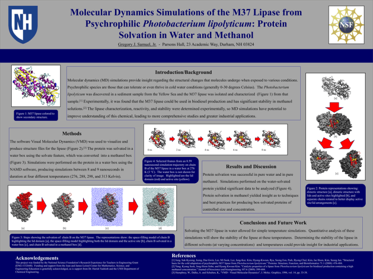
Molecular Dynamics Simulations of the M37 Lipase from Psychrophilic Photobacterium lipolyticum: Protein Solvation in Water and Methanol Gregory J. Samuel, Jr. - Parsons Hall, 23 Academic Way, Durham, NH 03824 Introduction/Background Molecular dynamics (MD) simulations provide insight regarding the structural changes that molecules undergo when exposed to various conditions. Psychrophilic species are those that can tolerate or even thrive in cold water conditions (generally 0-30 degrees Celsius). The Photobacterium lipolyticum was discovered in a sediment sample from the Yellow Sea and the M37 lipase was isolated and characterized (Figure 1) from that [a] sample.[1] Experimentally, it was found that the M37 lipase could be used in biodiesel production and has significant stability in methanol solutions.[2] The lipase characterization, reactivity, and stability were determined experimentally, so MD simulations have potential to Figure 1: M37 lipase colored to show secondary structure. improve understanding of this chemical, leading to more comprehensive studies and greater industrial applications. Methods [b] The software Visual Molecular Dynamics (VMD) was used to visualize and produce structure files for the lipase (Figure 2).[3] The protein was solvated in a 0 ns 2 ns 4 ns 6 ns 8 ns water box using the solvate feature, which was converted into a methanol box (Figure 3). Simulations were performed on the protein in a water box using the NAMD software, producing simulations between 8 and 9 nanoseconds in duration at four different temperatures (276, 288, 298, and 313 Kelvin). Figure 4: Selected frames from an 8.59 nanosecond simulation trajectory on chain B of the M37 lipase in a water box at 276 K (3 °C). The water box is not shown for clarity of image. Highlighted are the lid domain (red) and active site (yellow). Results and Discussion Protein solvation was successful in pure water and in pure [c] methanol. Simulations performed on the water-solvated protein yielded significant data to be analyzed (Figure 4). Protein solvation in methanol yielded insight as to techniques and best practices for producing box-solvated proteins of Figure 2: Protein representations showing: dimeric structure [a], dimeric structure with lids and active sites highlighted [b], and separate chains rotated to better display active site/lid arrangements [c]. controlled size and concentration. Conclusions and Future Work [a] [b] [c] [d] Figure 3: Steps showing the solvation of chain B on the M37 lipase. The representations show: the space-filling model of chain B highlighting the lid domain [a], the space-filling model highlighting both the lid domain and the active site [b], chain B solvated in a water box [c], and chain B solvated in a methanol box [d]. Acknowledgements This project was funded by the National Science Foundation’s Research Experience for Teachers in Engineering Grant (ENG-1132648). Funding and support from the Joan and James Leitzel Center for Mathematics, Science, and Engineering Education is gratefully acknowledged, as is support from Dr. Harish Vashisth and the UNH Department of Chemical Engineering. Solvating the M37 lipase in water allowed for simple temperature simulations. Quantitative analysis of these simulations will show the stability of the lipase at these temperatures. Determining the stability of the lipase in different solvents (at varying concentrations) and temperatures could provide insight for industrial applications. References [1] Jung, Suk‐Kyeong, Jeong, Dae Gwin, Lee, Mi Sook, Lee, Jung-Kee, Kim, Hyung-Kwoun, Ryu, Seong Eon, Park, Byong Chul, Kim, Jae Hoon, Kim, Seung Jun. "Structural basis for the cold adaptation of psychrophilic M37 lipase from Photobacterium lipolyticum." Proteins: Structure, Function, and Bioinformatics 71.1 (2008): 476-484. [2] Yang, Kyung Seok, Jung-Hoon Sohn, and Hyung Kwoun Kim. "Catalytic properties of a lipase from Photobacterium lipolyticum for biodiesel production containing a high methanol concentration." Journal of bioscience and bioengineering 107.6 (2009): 599-604. [3] Humphrey, W., Dalke, A. and Schulten, K., ''VMD - Visual Molecular Dynamics", J. Molec. Graphics, 1996, vol. 14, pp. 33-38.
