Chapter 8
Laura Coronado
Bio 10
Chapter 8
Biology and Society:
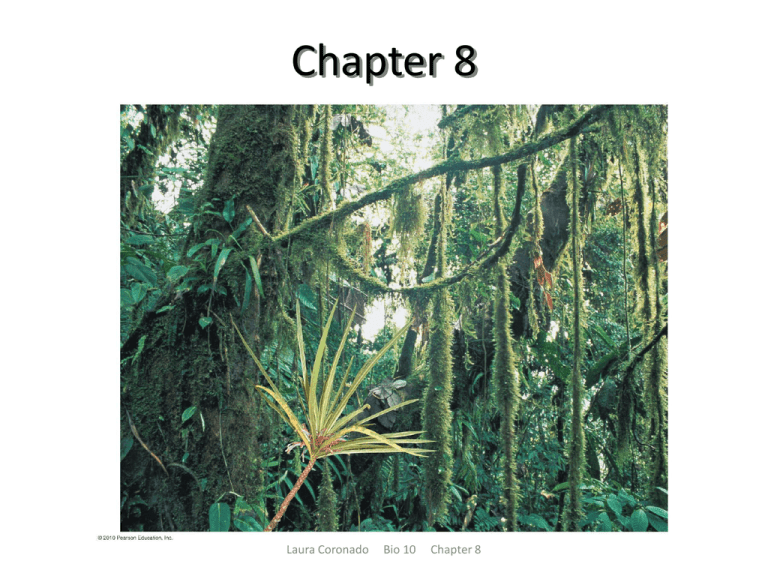
Rain Forest Rescue
– Endangered species of plants that normally
reproduce sexually can be propagated by asexual
reproduction.
– Cell division is at the heart of organisms
reproduction, whether by sexual or asexual means.
Laura Coronado
Bio 10
Chapter 8
WHAT CELL REPRODUCTION ACCOMPLISHES
– Reproduction:
• May result in the birth of new organisms
• More commonly involves the production
of new cells
• When a cell undergoes reproduction, or
cell division, two “daughter” cells are
produced that are genetically identical to
each other and to the “parent” cell.
Laura Coronado
Bio 10
Chapter 8
Cell Division
– Cell division plays important roles in the lives
of organisms.
• Replaces damaged or lost cells
• Permits growth
• Allows for reproduction
Laura Coronado
Bio 10
Chapter 8
LM
Colorized TEM
FUNCTIONS OF CELL DIVISION
Cell Replacement
Growth via Cell Division
Human kidney cell
Early human embryo
Laura Coronado
Bio 10
Chapter 8
Figure 8.1a
Asexual Reproduction
• Single-celled organisms reproduce by simple cell
division
• There is no fertilization of an egg by a sperm
• The parent and its offspring have identical genes.
• Mitosis is the type of cell division responsible for:
– Asexual reproduction
– Growth and maintenance of multicellular organisms
– Some multicellular organisms, such as sea stars, can grow new
individuals from fragmented pieces.
– Growing a new plant from a clipping
Laura Coronado
Bio 10
Chapter 8
LM
FUNCTIONS OF CELL DIVISION
Asexual Reproduction
Amoeba
Sea stars
Laura Coronado
Bio 10
Chapter 8
African Violet
Figure 8.1b
Sexual Reproduction
– Sexual reproduction requires fertilization of
an egg by a sperm using a special type of cell
division called meiosis.
– Thus, sexually reproducing organisms use:
• Meiosis for reproduction
• Mitosis for growth and maintenance
Laura Coronado
Bio 10
Chapter 8
LM
Chromosomes
Laura Coronado
Bio 10
Chapter 8
Figure 8.3
Chromosomes
– Chromosomes:
• Are made of chromatin, a combination of DNA
and protein molecules
• Are not visible in a cell until cell division occurs
– Before a parent cell splits into two, it duplicates its
chromosomes, the structures that contain most of
the organism’s DNA.
– During cell division, each daughter cell receives
one set of chromosomes.
Laura Coronado
Bio 10
Chapter 8
Number of chromosomes
in body cells
Species
Indian muntjac deer
6
Koala
16
Opossum
22
Giraffe
30
40
Mouse
46
Human
Duck-billed platypus
54
60
Buffalo
Dog
78
102
Red viscacha rat
Laura Coronado
Bio 10
Chapter 8
Figure 8.2
Eukaryotic Cell Genetic Information
– Most genes are located on chromosomes in the cell
nucleus
– A few genes are found in DNA in mitochondria and
chloroplasts
– Each chromosome contains one very long DNA
molecule, typically with thousands of genes.
– The number of chromosomes depends on the
species.
– The DNA in a cell is packed into an elaborate,
multilevel system of coiling and folding.
– Histones are proteins used to package DNA.
– Nucleosomes consist of DNA wound around histone
molecules.
Laura Coronado
Bio 10
Chapter 8
DNA double helix
Histones
TEM
“Beads on
a string”
Nucleosome
Tight helical fiber
Looped domains
Laura Coronado
TEM
Duplicated chromosomes
(sister chromatids)
Bio 10
Centromere
Chapter 8
Figure 8.4
Chromosomes
– Before a cell divides, it duplicates all of its
chromosomes, resulting in two copies called sister
chromatids.
– Sister chromatids are joined together at a narrow
“waist” called the centromere.
– When the cell divides, the sister chromatids separate
from each other.
– Once separated, each chromatid is:
• Considered a full-fledged chromosome
• Identical to the original chromosome
Laura Coronado
Bio 10
Chapter 8
Chromosome (one
long piece of DNA)
Centromere
Sister
chromatids
Duplicated chromosome
Laura Coronado
Bio 10
Chapter 8
Figure 8.UN2
Chromosome
duplication
Sister
chromatids
Chromosome
distribution to
daughter cells
Laura Coronado
Bio 10
Chapter 8
Figure 8.5
The Cell Cycle
– A cell cycle is the orderly sequence of events that
extend from the time a cell is first formed from a
dividing parent cell to its own division into two cells.
– The cell cycle consists of two distinct phases:
• Interphase
• The mitotic phase
Laura Coronado
Bio 10
Chapter 8
Interphase
– Most of a cell cycle is spent in interphase.
– During interphase, a cell:
• Performs its normal functions
• Doubles everything in its cytoplasm
• Grows in size
Laura Coronado
Bio 10
Chapter 8
S phase (DNA synthesis;
chromosome duplication)
Interphase: metabolism and
growth (90% of time)
G1
Mitotic (M) phase:
cell division
(10% of time)
Cytokinesis
(division of
cytoplasm)
G2
Mitosis
(division of nucleus)
Laura Coronado
Bio 10
Chapter 8
Figure 8.6
Mitosis
– The mitotic (M) phase includes two overlapping
processes:
• Mitosis, in which the nucleus and its contents
divide evenly into two daughter nuclei
• Cytokinesis, in which the cytoplasm is divided in
two
Laura Coronado
Bio 10
Chapter 8
Mitosis and Cytokinesis
– Mitosis consists of four distinct phases:
• (A) Prophase
• (B) Metaphase
• (C) Anaphase
• (D) Telophase
– Cytokinesis typically:
• Occurs during telophase
• Divides the cytoplasm
• Is different in plant and animal cells
Laura Coronado
Bio 10
Chapter 8
INTERPHASE
Centrosomes
(with centriole pairs)
PROPHASE
Early mitotic
spindle
Chromatin
Plasma
membrane
Centromere
Chromosome, consisting
of two sister chromatids
Spindle
microtubules
LM
Nuclear
envelope
Centrosome
Fragments of
nuclear envelope
Laura Coronado
Bio 10
Chapter 8
Figure 8.7.a
METAPHASE
ANAPHASE
TELOPHASE AND CYTOKINESIS
Nuclear
envelope
forming
Spindle
Cleavage
furrow
Daughter
chromosomes
Laura Coronado
Bio 10
Chapter 8
Figure 8.7b
Mitosis and Cytokinesis
– During mitosis the mitotic spindle, a footballshaped structure of microtubules, guides the
separation of two sets of daughter chromosomes.
– Spindle microtubules grow from two centrosomes,
clouds of cytoplasmic material that in animal cells
contain centrioles.
Laura Coronado
Bio 10
Chapter 8
SEM
Cleavage
furrow
Cleavage furrow
Contracting ring of
microfilaments
Laura Coronado
Bio 10
Daughter cells
Chapter 8
Figure 8.8a
Cell plate
forming
Daughter
nucleus
LM
Wall of
parent cell
Cell wall
Vesicles containing
cell wall material
Laura Coronado
Cell plate
Bio 10
Chapter 8
New cell wall
Daughter cells
Figure 8.8b
Laura Coronado
Bio 10
Chapter 8
Figure 8.10
Homologous Chromosomes
– Different individuals of a single species have the
same number and types of chromosomes.
– A human somatic cell:
• Is a typical body cell
• Has 46 chromosomes
– A karyotype is an image that reveals an orderly
arrangement of chromosomes.
– Homologous chromosomes are matching pairs of
chromosomes that can possess different versions of
the same genes.
Laura Coronado
Bio 10
Chapter 8
LM
Pair of homologous
chromosomes
Centromere
Sister
chromatids
One duplicated
chromosome
Laura Coronado
Bio 10
Chapter 8
Figure 8.11
Human Chromosomes
– Humans have:
• Two different sex chromosomes, X and Y
• Twenty-two pairs of matching chromosomes,
called autosomes
– Humans are diploid organisms in which:
• Their somatic cells contain two sets of
chromosomes
• Their gametes are haploid, having only one set of
chromosomes
Laura Coronado
Bio 10
Chapter 8
Gametes and the Life Cycle of a Sexual Organism
– The life cycle of a multicellular organism is the
sequence of stages leading from the adults of one
generation to the adults of the next.
Laura Coronado
Bio 10
Chapter 8
Haploid gametes (n 23)
Egg cell
n
n
Sperm cell
FERTILIZATION
MEIOSIS
Multicellular
diploid adults
(2n 46)
2n
MITOSIS
and development
Laura Coronado
Bio 10
Chapter 8
Diploid
zygote
(2n 46)
Key
Haploid (n)
Diploid (2n)
Figure 8.12
Meiosis
– Humans are diploid organisms in which:
• Their somatic cells contain two sets of chromosomes
• Their gametes are haploid, having only one set of
chromosomes
– In humans, a haploid sperm fuses with a haploid egg
during fertilization to form a diploid zygote.
– Sexual life cycles involve an alternation of diploid and
haploid stages.
– Meiosis produces haploid gametes, which keeps the
chromosome number from doubling every
generation.
Laura Coronado
Bio 10
Chapter 8
Homologous
chromosomes
separate.
Chromosomes
duplicate.
Sister
chromatids
separate.
Pair of homologous
chromosomes in
diploid parent cell
Duplicated pair of
homologous
chromosomes
Sister
chromatids
MEIOSIS I
INTERPHASE BEFORE MEIOSIS
Laura Coronado
Bio 10
Chapter 8
MEIOSIS II
Figure 8.13-3
The Process of Meiosis
– In meiosis:
•
•
•
Haploid daughter cells are produced in diploid
organisms
Interphase is followed by two consecutive divisions,
meiosis I and meiosis II
Crossing over occurs
Laura Coronado
Bio 10
Chapter 8
MEIOSIS I: HOMOLOGOUS CHROMOSOMES SEPARATE
INTERPHASE
Centrosomes
(with centriole
pairs)
Nuclear
envelope
Chromatin
Chromosomes
duplicate.
PROPHASE I
Sites of
crossing over
Spindle
Sister
chromatids
Pair of
homologous
chromosomes
Homologous
chromosomes
pair up and
exchange
segments.
Laura Coronado
METAPHASE I
ANAPHASE I
Microtubules
attached
to chromosome
Sister chromatids
remain attached
Centromere
Pairs of
homologous
chromosomes
line up.
Bio 10
Chapter 8
Pairs of
homologous
chromosomes
split up.
Figure 8.14a
MEIOSIS II: SISTER CHROMATIDS SEPARATE
TELOPHASE I
AND
CYTOKINESIS
PROPHASE II
METAPHASE II
ANAPHASE II
TELOPHASE II
AND
CYTOKINESIS
Sister
chromatids
separate
Haploid
daughter
cells forming
Cleavage
furrow
Two haploid
cells form;
chromosomes
are still
doubled.
During another round of cell division, the sister
chromatids finally separate; four haploid
daughter cells result, containing single
chromosomes.
Laura Coronado
Bio 10
Chapter 8
Figure 8.14b
LM
Laura Coronado
Bio 10
Chapter 8
Figure 8.14bc
Review: Comparing Mitosis and Meiosis
– In mitosis and meiosis, the chromosomes duplicate
only once, during the preceding interphase.
– The number of cell divisions varies:
• Mitosis uses one division and produces two diploid
cells
• Meiosis uses two divisions and produces four
haploid cells
– All the events unique to meiosis occur during meiosis
I, while meiosis II is the same as mitosis since it
separates sister chromatids.
Laura Coronado
Bio 10
Chapter 8
MEIOSIS
MITOSIS
Prophase I
Prophase
Chromosome
duplication
Chromosome
duplication
Duplicated chromosome
(two sister chromatids)
MEIOSIS I
Parent cell
(before chromosome duplication)
2n 4
Homologous
chromosomes come
together in pairs.
Site of crossing over
between homologous
(nonsister) chromatids
Metaphase I
Metaphase
Homologous pairs
align at the middle
of the cell.
Chromosomes
align at the
middle of the
cell.
Anaphase I
Telophase I
Anaphase
Telophase
2n
Sister
chromatids
separate
during
anaphase.
Daughter cells
of mitosis
2n
Homologous
chromosomes
separate during
anaphase I;
sister chromatids
remain together.
Chromosome with two
sister chromatids
Daughter
cells of meiosis I
Haploid
n2
MEIOSIS II
Sister chromatids
separate during
anaphase II.
n
n
n
Daughter cells of meiosis II
Laura Coronado
Bio 10
Chapter 8
n
Figure 8.15
Independent Assortment of Chromosomes
– When aligned during metaphase I of meiosis, the side-byside orientation of each homologous pair of chromosomes
is a matter of chance.
– Every chromosome pair orients independently of the
others during meiosis.
– For any species the total number of chromosome
combinations that can appear in the gametes due to
independent assortment is:
• 2n where n is the haploid number.
– For a human:
• n = 23
• 223 = 8,388,608 different chromosome combinations possible
in a gamete
Laura Coronado Bio 10 Chapter 8
POSSIBILITY 2
POSSIBILITY 1
Metaphase of
meiosis I
Metaphase
of meiosis II
Gametes
Combination a
Combination b
Laura Coronado
Combination c
Bio 10
Chapter 8
Combination d
Figure 8.16-3
Random Fertilization
– A human egg cell is fertilized randomly by one
sperm, leading to genetic variety in the zygote.
– If each gamete represents one of 8,388,608
different chromosome combinations, at
fertilization, humans would have 8,388,608 ×
8,388,608, or more than 70 trillion, different
possible chromosome combinations.
Laura Coronado
Bio 10
Chapter 8
Laura Coronado
Bio 10
Chapter 8
Figure 8.17
Crossing Over
– In crossing over:
• Homologous chromosomes exchange genetic
information
• Genetic recombination, the production of gene
combinations different from those carried by
parental chromosomes, occurs
Laura Coronado
Bio 10
Chapter 8
Prophase I
of meiosis
Homologous chromatids
exchange corresponding
segments.
Duplicated pair of
homologous
chromosomes
Chiasma, site of
crossing over
Metaphase I
Sister chromatids
remain joined at their
centromeres.
Spindle
microtubule
Metaphase II
Gametes
Recombinant
chromosomes
combine genetic
information from
Recombinant chromosomes
different parents.
Laura Coronado Bio 10 Chapter 8
Figure 8.18-5
The Process of Science:
Do All Animals Have Sex?
– Observation: No scientists have ever found male
bdelloid rotifers, a microscopic freshwater
invertebrate.
– Hypothesis: Bdelloid rotifers have thrived for
millions of years using only asexual reproduction.
– Prediction: Bdelloid rotifers would display much
more variation in their homologous pairs of genes
than most organisms.
Laura Coronado
Bio 10
Chapter 8
The Process of Science:
Do All Animals Have Sex?
– Experiment: Researchers compared sequences
of a particular gene in bdelloid and nonbdelloid rotifers.
– Results:
• Non-bdelloid sexually reproducing rotifers had nearly
identical homologous genes
• Bdelloid asexually reproducing rotifers had homologous
genes that differed by 3.5–54%.
– Conclusion: Bdelloid rotifers have evolved for
millions of years without any sexual
reproduction.
Laura Coronado
Bio 10
Chapter 8
LM
Laura Coronado
Bio 10
Chapter 8
Figure 8.19
How Accidents during Meiosis Can
Alter Chromosome Number
– In nondisjunction, the members of a chromosome
pair fail to separate during anaphase, producing
gametes with an incorrect number of chromosomes.
– Nondisjunction can occur during meiosis I or II.
– If nondisjunction occurs, and a normal sperm
fertilizes an egg with an extra chromosome, the
result is a zygote with a total of 2n + 1
chromosomes.
– If the organism survives, it will have an abnormal
number of genes.
Laura Coronado
Bio 10
Chapter 8
NONDISJUNCTION IN MEIOSIS II
NONDISJUNCTION IN MEIOSIS I
Meiosis I
Nondisjunction:
Pair of homologous
chromosomes fails
to separate.
Meiosis II
Nondisjunction:
Pair of sister
chromatids
fails to separate.
Gametes
n1
n1
n–1
n–1
Number of
chromosomes
Abnormal gametes
n1
n–1
Abnormal gametes
Laura Coronado
Bio 10
Chapter 8
n
n
Normal gametes
Figure 8.20-3
Abnormal egg
cell with extra
chromosome
n1
Normal
sperm cell
Abnormal zygote
with extra
chromosome 2n 1
n (normal)
Laura Coronado
Bio 10
Chapter 8
Figure 8.21
Down Syndrome
– Down Syndrome:
• Is also called trisomy 21
• Is a condition in which an individual has an extra
chromosome 21
• Affects about one out of every 700 children
• The incidence of Down Syndrome increases with
the age of the mother.
Laura Coronado
Bio 10
Chapter 8
LM
Chromosome 21
Laura Coronado
Bio 10
Chapter 8
Figure 8.22
Infants with Down syndrome
(per 1,000 births)
90
80
70
60
50
40
30
20
10
0
20
25
30
35
40
45
50
Age of mother
Laura Coronado
Bio 10
Chapter 8
Figure 8.23
Abnormal Numbers of Sex
Chromosomes
– Nondisjunction can also affect the sex
chromosomes.
Laura Coronado
Bio 10
Chapter 8
Laura Coronado
Bio 10
Chapter 8
Table 8.1
Evolution Connection:
The Advantages of Sex
– Asexual reproduction conveys an
evolutionary advantage when plants are:
• Sparsely distributed
• Superbly suited to a stable environment
– Sexual reproduction may convey an
evolutionary advantage by:
• Speeding adaptation to a changing environment
• Allowing a population to more easily rid itself of
harmful genes
Laura Coronado
Bio 10
Chapter 8
Laura Coronado
Bio 10
Chapter 8
Figure 8.24
Cancer Cells: Growing Out of Control
– Normal plant and animal cells have a cell
cycle control system that consists of
specialized proteins, which send “stop” and
“go-ahead” signals at certain key points
during the cell cycle.
Laura Coronado
Bio 10
Chapter 8
What Is Cancer?
– Cancer is a disease of the cell cycle.
– Cancer cells do not respond normally to the cell cycle
control system.
– Cancer cells can form tumors, abnormally growing
masses of body cells.
– The spread of cancer cells beyond their original site of
origin is metastasis.
– Malignant tumors can:
• Spread to other parts of the body
• Interrupt normal body functions
– A person with a malignant tumor is said to have
cancer.
Laura Coronado Bio 10 Chapter 8
Lymph
vessels
Tumor
Blood
vessel
Glandular
tissue
A tumor grows
from a single
cancer cell.
Metastasis: Cancer
cells spread through
lymph and blood
vessels to other parts
of the body.
Cancer cells invade
neighboring tissue.
Laura Coronado
Bio 10
Chapter 8
Figure 8.9
Cancer Treatment
– Cancer treatment can involve:
• Radiation therapy, which damages DNA and
disrupts cell division
• Chemotherapy, which uses drugs that disrupt
cell division
Laura Coronado
Bio 10
Chapter 8
Cancer Prevention and Survival
– Certain behaviors can decrease the risk of cancer:
• Not smoking
• Exercising adequately
• Avoiding exposure to the sun
• Eating a high-fiber, low-fat diet
• Performing self-exams
• Regularly visiting a doctor to identify tumors
early
Laura Coronado
Bio 10
Chapter 8
Distribution via
mitosis
Duplication
of all
chromosomes
Genetically
identical
daughter
cells
Laura Coronado
Bio 10
Chapter 8
Figure 8.UN1
S phase
DNA synthesis; chromosome duplication
Interphase
Cell growth and
chromosome duplication
G2
G1
Mitotic
(M) phase
Genetically
identical
“daughter”
cells
Cytokinesis
(division of
cytoplasm)
Laura Coronado
Bio 10
Chapter 8
Mitosis
(division of
nucleus)
Figure 8.UN3
Human Life Cycle
Haploid gametes (n 23)
Key
Haploid (n)
n
Egg cell
Diploid (2n)
n
Sperm cell
MEIOSIS
FERTILIZATION
Male and female
diploid adults
(2n 46)
Diploid
zygote
(2n 46)
MITOSIS
and development
Laura Coronado
Bio 10 Chapter 8
2n
Figure 8.UN4
MEIOSIS
MITOSIS
Parent
cell (2n)
Chromosome
duplication
Parent
cell (2n)
Chromosome
duplication
MEIOSIS I
Pairing of
homologous
chromosome
Crossing over
2n
Daughter
cells
2n
MEIOSIS II
n
Laura Coronado
Bio 10
n
n
n
Daughter cells
Chapter 8
Figure 8.UN5
LM
(b)
(a)
(c)
(d)
Laura Coronado
Bio 10
Chapter 8
Figure 8.UN6

