Protein
advertisement
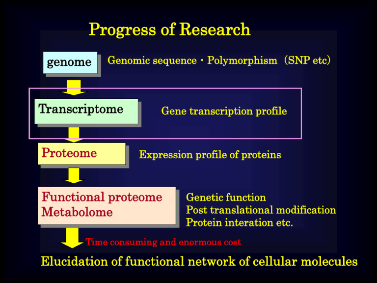
Progress of Research genome Genomic sequence・Polymorphism(SNP etc) Transcriptome Proteome Gene transcription profile Expression profile of proteins Functional proteome Metabolome Genetic function Post translational modification Protein interation etc. Time consuming and enormous cost Elucidation of functional network of cellular molecules DNA Chip Disease rat Normal rat mRNA extraction Labeled with Cy5 and Cy3 Hybridization Genes in which transcription levels are affected with the disease can be determined. Importance of RNA Species and genomic size・number of genes Genomic size(Mbp) Human C-elegance 2351 The number of genes 22287 103 19893 Drosophila 180 13676 Arabidopsis thaliana 125 25498 Cellular slime 34 Yeast 13 12500 5538 In E. Coli., 80% of genomic DNA encodes proteins. On the other hand, human genomic DNA contains only 3% for genes. However, 70-80% of human genomic DNA is transcripted! → non-coding RNA Human Accelerating Regions (HAR) The genomic region that has the most different transcription activity between human and chimpanzee. The transcript from this region was small RNA. Lung cancer: Causing by a transcriptional suppression of a microRNA which suppresses ras gene Limphoma: Causing by a overtranscription of a microRNA which suppress E2F (apoptosis inducible protein) MicroRNA controls gene expression level. (miRNA can find out the specific mRNA by using the sequence of its own.) MicroRNA also sometimes regulate protein activity with binding to it. Ex:MEI2 protein that progress meosis of fission yeastlocalize in nucleus with meiRNA. The protein localizes in cytosol without the meiRNA. Ex:7SK small nuclear RNA suppresses PolII transcription Mechanism of RNA interference by siRNA p-3’ Sense p-5’ siRNA 3’-p 5’-p 2nt Antisense 19 nt duplex dsRNA TRBP 6. Regeneration of RISC* Dicer cleavage 1. Dicer binds to dsRNA mRNA Nucleus TRBP AGO2 Dicer 3’-p 5. Activated RISC bind to mRNA p-3’ Dicer 2. Formation of RLC (RISC loading complex) 4. Activated RISC (RISC*) 3. Formation of RISC cleavage genome m-RNA Protein Non-coding RNA What is post-genomic research? Genomic research Whole sequence of human genome was determined! Individual difference(SNP) of genes are elucidated. We can access how effective of drugs or how strong of the adverse effect to individuals.(Tailor-made medicine) What is the cause of particular disease? How we can treat the medicine? How can we discover the effective drug? Gene(DNA) Expression Transcription Copy (massenger RNA) Translation Protein Progress of Research genome Genomic sequence・Polymorphism(SNP etc) Transcriptome Proteome Gene transcription profile Expression profile of proteins Functional proteome Metabolome Genetic function Post translational modification Protein interation etc. Time consuming and enormous cost Elucidation of functional network of cellular molecules Proteome The situation of whole proteins that are expressed in particular condition In living cell. To know the function of gene or life phenomena, we have to know how many proteins are expressed and what is the way of relationship of such proteins on given conditions. Proteomics techinologies Difficulties with comprehensive analysis of proteins Diversity of proteins characteristics Difficulty with development of universal techniques ⇒ case by case handling Occurrence of post-translational modification They often form complex with other proteins and molecules Expression profile is various depending on tissues or temporally Dynamic range of their expressions are very wide (1,000,000 folds difference) Identification 2D electrphresis + MS SDS PAGE Isoelectric focusing electrophoresis Restricted hydrolysis MALDI-TOF MS Dual-Channel Imaging (Detection & quantification of protein synthesis dependent on particular stimulations) + Sypro-Ruby staining Protein quantification Autoradiography imaging (35S-methionine pulse-labeling) Quantification of newly synthesized proteins Proteins with already stopping synthesis Newly synthesized proteins with the stimulation Continuously synthesizing proteins Various techniques for proteome analysis For protein indentification and differential display Peptide sequence using charged tag (SMA or SPA reagent) Isotope label ICAT assay ICAT assay with 15N-enrich medium 2-dimensional PAGE Capillary LC Identification of phosphorylation site Protein array For investigating protein-protein (ligand) interaction Base on two-hybrid system Yeast two hybrid system (Y2H) Large scale Y2H Y2H in mammalian cell Three hybrid system One hybrid system Based on protein complementation assay Using Dihydroforate reductase(DHFR) Split Ubiquitin Using protein splicing Using b-galactosidase Using rasGEF+V-src myristoylation signal Using adenylyl cyclase Other Using isotope-labeled crosslinker Protein array Assay of protein-protein interaction Yeast Two Hybrid (Y2H) system Pray bait GAL4 DNA Binding domain GAL4 transcription activating domain Reporter gene expression DNA Highly sensitive, but easy to get false-positive Not available to proteins difficult to express in yeast Only available for 1:1 interaction Costly & time consuming Apply to HSP format Conceptual scheme of Y2H Transcription activating domain : GAL4 yeast transcription factor Proteins of interest DNA binding domain Bait Prey Reporter gene expression GAL4 promoter Reporter gene a) b) Bait Pray AD c) Bait Bait DBD Binding site d) GAL4DBD Reporter gene Membrane localization factor Cellular mambrane OriP Pray Pray Pray AD AD GAL4BE TATA e) T25 T18 ATP Adenylate cyclase CAP cAMP Ras or hSos cAMP/CAP dependent promoter Schematic outlines of Two hybrid systems a) Yeast Two Hybrid System, b) effect of homodimer-forming in bait or prey in Y2H, c) two hybrid system in mammalian cell, d) protein recruitment system using Ras(RRS) or hSos(SRS), e) two hybrid system in bacterial cell Split Luciferase Assay + Protein A Protein B b) rapamycin a) FKBP Y X FRB Reconstitution of ubiquitin Cleavage with UBP Fragment of a proteinReconstitution of the protein (DHFR or β-Gal) c) X Y Luciferase or EGFP splicing Reconstitution of intein Schematic outline of protein-fragment complementation assay a:Original protein-fragment complementation assay b:Split ubiquitin assay c:Split enzyme reconstitution based on protein splicing Application of Intein-Extein system for reconstitution assay Gene A Gene B Gene A Intein extein Protein B Protein A Protein A Gene X Gene Y Luciferase’ Luciferase” Protein X Protein Y Native Luciferase a) b) AD Receptor for ligand A Receptor for ligand B A DBD Binding site DBD Binding site Reporter gene c) B Reporter gene RNA A B AD DBD Binding site AD Reporter gene Schematic outlines of one- and three-hybrid assay a:One hybrid assay for detecting DNA-protein interaction b:Three hybrid assay for detecting protein-ligand interaction c:Three hybrid assay for detecting protein-RNA interaction DNA・RNA-linkage Phage display STABLE assay DNA Target protein Biotin DNA Phage Streptavidin Coat protein Ribosome display In vitro virus RNA RNA ribosome Puromycin Panning in Phage display Washing out of unbound phages Phage library Target-coating plate Proliferation Collect of bound phages STABLE assay expression gene biotin Reverse micelle Encapsulated gene encodes fusion protein between target and streptavidin binding DNA biotin Ribosome display In vitro virus RNA RNA ribosome Expression vector without terminal codon is used for each target protein expression. Ribosome can’t detach from mRNA so that RNA-Protein fusion is obtained. Puromycin Puromycin binds to ribosome pocket when the transcription completes. Then pyromycin connects between mRNA and translated protein in covalent bonding.

