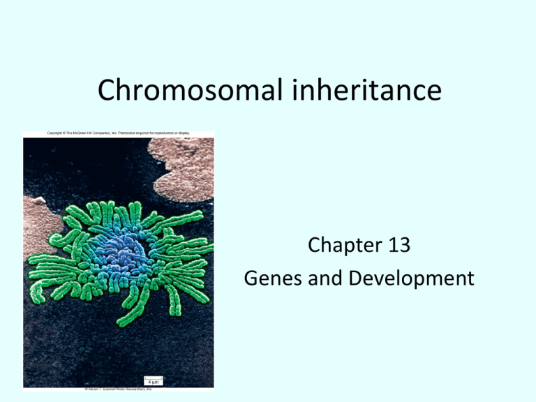
Chromosomal inheritance
Chapter 13
Genes and Development
Fig. 13.1
Fig. 13.2a
Copyright © The McGraw-Hill Companies, Inc. Permission required for reproduction or display.
Parental generation
male
Parental
generation
female
F1 progeny all had red eyes
Fig. 13.2b
Copyright © The McGraw-Hill Companies, Inc. Permission required for reproduction or display.
F1 generation
male
F1 generation
female
F2 female progeny had red eyes, only males had white eyes
Fig. 13.2c
Copyright © The McGraw-Hill Companies, Inc. Permission required for reproduction or display.
Testcross
Parental generation
male
F1 generation
female
The testcross revealed that white-eyed females
are viable. Therefore eye color is linked to the
X chromosome and absent from the Y chromosome
Fig. 13.2
Copyright © The McGraw-Hill Companies, Inc. Permission required for reproduction or display.
Parental generation
male
Parental
generation
female
F1 progeny all had red eyes
F1 generation
male
F1 generation
female
F2 female progeny had red eyes, only males had white eyes
Testcross
Parental generation
male
F1 generation
female
The testcross revealed that white-eyed females
are viable. Therefore eye color is linked to the
X chromosome and absent from the Y chromosome
Page 241
Copyright © The McGraw-Hill Companies, Inc. Permission required for reproduction or display.
X chromosome
Y chromosome
© BioPhoto Associates/Photo Researchers, Inc.
2.8 µm
Sex determination
Organism
Humans
(mammals)
System
Females Males Why males?
XX/XY
XX
XY
Drosophila
XX/XY
XX
XY
SRY on Y
One X, Y needed for
fertility
Birds, most
reptiles
ZW/ZZ
ZW
ZZ
Two Z
Some insects
XX/XO
XX
X
One X
Honey bees
Haploid/Diploid
Diploid Haploid One set of chromosomes
Fig. 13.3
Copyright © The McGraw-Hill Companies, Inc. Permission required for reproduction or display.
The Royal Hemophilia Pedigree
George III
Generation
Edward
Duke of Kent
Prince Albert
I
Frederick Victoria
III
No hemophilia
III
Queen victoria
King
Edward VII
II
Louis II
Grand Duke of Hesse
Alice
Duke of
Hesse
Alfred
Helena
Arthur
Leopold
No hemophilia
German
Royal
House
King
George V
Irene
Czar
Nicholas II
Czarina
Alexandra
Earl of
Athlone
Princess
Alice
King
George VI
Earl of
Mountbatten
Waldemar
Prussian
Royal
House
V
Queen
Elizabeth II
Prince
Philip
Prince Henry
Sigismond
Margaret
Anastasia
Russian
Royal
House
VI
?
Prince
Charles
Anne
Andrew
Edward
William
Queen
Eugenie
Henry
© Bettmann/Corbis
Alfonso
King of
Spain
?
?
Alfonso
Jamie Juan
Gonzalo
?
No evidence
of hemophilia
No evidence
of hemophilia
Spanish Royal House
British Royal House
VII
Viscount
Tremation
Leopold
King Juan
Carlos
?
Princess
Diana
Alexis
Maurice
?
?
IV
Duke of
Windsor
Prince
Henry
Beatrice
Page 243
Barr body attached to the nuclear membrane
Fig. 13.4
Copyright © The McGraw-Hill Companies, Inc. Permission required for reproduction or display.
Second gene causes patchy distribution of pigment:
white fur = no pigment, orange or black fur = pigment
Allele for black
fur is inactivated
Allele for orange
fur is inactivated
X chromosome
allele for
orange fur
X chromosome
allele for
black fur
Inactivated X
chromosome
becomes Barr body
Inactivated X
chromosome
becomes Barr body
Nucleus
© Kenneth Mason
Nucleus
Evidence for
Homologous
recombination
Harriet Creighton and
Barbara McClintock
Maize genetics
Fig. 13.5
Copyright © The McGraw-Hill Companies, Inc. Permission required for reproduction or display.
Parent
generation
F1 generation
Meiosis with
Crossing over
Meiosis without
Crossing over
Crossing
over during
prophase I
No crossing
over during
prophase I
Meiosis II
Meiosis II
Recombinant
All parental
Parental
No recombinant
Fig. 13.7
Copyright © The McGraw-Hill Companies, Inc. Permission required for reproduction or display.
recessive allele (black body)
dominant allele (gray body)
recessive allele (vestigial wings)
dominant allele (normal wings)
Using two-point
crosses to map
genes
Parental
generation
Cross-fertilization
F1 generation
Testcross
Testcross male
gamete
415 parental
wild type
(gray body,
long wing)
F1 generation
female
possible
gametes
92 recombinant
(gray body,
vestigial wing)
88 recombinant
(black body,
long wing)
405 parental
mutant type
(black body ,
vestigial wing)
180 1000 = 0.18 total recombinant offspring
18% recombinant frequency
18 cM between the two loci
Genetic Mapping
The distance between genes is proportional to the
frequency of recombination events.
recombination
=
frequency
recombinant progeny
total progeny
1% recombination = 1 map unit (m.u.)
1 map unit = 1 centimorgan (cM)
Genetic Mapping
Multiple crossovers between 2 genes can reduce the perceived
genetic distance
Progeny resulting from an even number of crossovers look like
parental offspring
Fig. 13.9
Copyright © The McGraw-Hill Companies, Inc. Permission required for reproduction or display.
A
B
C
a
b
c
A
b
C
a
B
c
Parental
Three-point testcrosses
Recombinant
Parental
Fig. 13.10
Copyright © The McGraw-Hill Companies, Inc. Permission required for reproduction or display.
Ichthyosis, X-linked
Placental steroid sulfatase deficiency
Kallmann syndrome
Chondrodysplasia punctata, X-linked recessive
Duchenne muscular dystrophy
Becker muscular dystrophy
Chronic granulomatous disease
Retinitis pigmentosa-3
Norrie disease
Retinitis pigmentosa-2
Hypophosphatemia
Aicardi syndrome
Hypomagnesemia, X-linked
Ocular albinism
Retinoschisis
Adrenal hypoplasia
Glycerol kinase deficiency
Ornithine transcarbamylase deficiency
Incontinentia pigmenti
Wiskott–Aldrich syndrome
Menkes syndrome
Androgen insensitivity
Sideroblastic anemia
Aarskog–Scott syndrome
PGK deficiency hemolytic anemia
Charcot–Marie–Tooth neuropathy
Choroideremia
Cleft palate, X-linked
Spastic paraplegia, X-linked, uncomplicated
Deafness with stapes fixation
PRPS-related gout
Anhidrotic ectodermal dysplasia
Agammaglobulinemia
Kennedy disease
Pelizaeus–Merzbacher disease
Alport syndrome
Fabry disease
Immunodeficiency , X-linked, with hyper IgM
Lymphoproliferative syndrome
Albinism–deafness syndrome
Fragile-X syndrome
Human X chromosome gene map
Lowe syndrome
Lesch–Nyhan syndrome
HPRT-related gout
Hunter syndrome
Hemophilia B
Hemophilia A
G6PD deficiency: favism
Drug-sensitive anemia
Chronic hemolytic anemia
Manic–depressive illness, X-linked
Colorblindness, (several forms)
Dyskeratosis congenita
TKCR syndrome
Adrenoleukodystrophy
Adrenomyeloneuropathy
Emery–Dreifuss muscular dystrophy
Diabetes insipidus, renal
Myotubular myopathy , X-linked
Fig. 13.11
Sickle cell anemia
Table 13.2
http://www.biotechnologyonline.gov.au/popups/img_karyotype.html
http://www.cytogenetics.org.uk/careers/PIC7.JPG
Normal human karyotypes
Abnormal human karyotype
http://www.slh.wisc.edu/cytogenetics/cases/gifs/com_karyotypes/CoMNov97karyo.gif
Fig. 13.12
Copyright © The McGraw-Hill Companies, Inc. Permission required for reproduction or display.
1
6
2
7
3
8
13
14
19
20
Abnormal human karyotype
4
9
15
21
10
16
22
5
11
17
X
12
18
Y
© Colorado Genetics Laboratory, University of Colorado Denver
What happened here?
Fig. 13.13
Incidence of Down Syndrome
per 1000 Live Births
Copyright © The McGraw-Hill Companies, Inc. Permission required for reproduction or display.
25
20
15
10
5
0
20
25
30
35
Age of Mother
40
45
Down syndrome increases with the age of the mother
Fig. 13.14
Copyright © The McGraw-Hill Companies, Inc. Permission required for reproduction or display.
Normal male
X
Triple X syndrome
Female
gametes
undergo
nondisjunction
Y
Klinefelter
syndrome
XX
XXX
XXY
XO
OY
Turner
syndrome
Nonviable
O
Fig. 13.15
Copyright © The McGraw-Hill Companies, Inc. Permission required for reproduction or display.
Uterus
Amniotic fluid
Hypodermic syringe
Amniocentesis
Fig. 13.16
Copyright © The McGraw-Hill Companies, Inc. Permission required for reproduction or display.
Cells from the chorion
Ultrasound device
Uterus
Suction tube
Chorionic villi
Chorionic villi sampling


