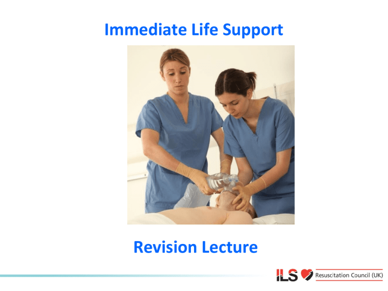
Immediate Life Support
Revision Lecture
Causes and Prevention of
Cardiac Arrest
Chain of survival
Early recognition prevents:
• Cardiac arrests and deaths
• Admissions to ICU
• Inappropriate resuscitation attempts
Early recognition of
the deteriorating patient
• Most arrests are
predictable
• Hypoxia and
hypotension are
common
antecedents
• Delays in referral to
higher levels of care
Recognition of the deteriorating patient
Example escalation protocol based on Scottish early warning score (SEWS)
Recognition of the deteriorating patient
• Several alternative systems to cardiac arrest team
• e.g. Medical emergency team (MET)
• Track changes in physiology
• e.g. Early warning scores
• Trigger a response if abnormal values:
• Call senior nurse
• Call doctor
• Call resuscitation team
The ABCDE approach to
the deteriorating patient
Airway
Breathing
Circulation
Disability
Exposure
ABCDE approach
Underlying principles:
• Complete initial assessment
• Treat life-threatening problems
• Reassessment
• Assess effects of treatment/interventions
• Call for help early
ABCDE approach
• Personal safety
• Patient responsiveness
• First impression
• Vital signs
• Respiratory rate, SpO2, pulse, BP, GCS, temperature
ABCDE approach
Airway
Causes of airway obstruction:
•
•
•
•
•
CNS depression
Blood
Vomit
Foreign body
Trauma
•
•
•
•
Infection
Inflammation
Laryngospasm
Bronchospasm
ABCDE approach
Airway
Recognition of airway obstruction:
• Talking
• Difficulty breathing, distressed, choking
• Shortness of breath
• Noisy breathing
• Stridor, wheeze, gurgling
• See-saw respiratory pattern, accessory muscles
ABCDE approach
Airway
Treatment of airway obstruction:
• Airway opening
• Head tilt, chin lift, jaw thrust
• Simple adjuncts
• Advanced techniques
• e.g. LMA, tracheal tube
• Oxygen
ABCDE approach
Breathing
Recognition of breathing
problems:
• Look
• Respiratory distress, accessory
muscles, cyanosis, respiratory
rate, chest deformity,
conscious level
• Listen
• Noisy breathing, breath
sounds
• Feel
• Expansion, percussion,
tracheal position
ABCDE approach
Breathing
Treatment of breathing problems:
• Airway
+
• Oxygen
• Treat underlying cause
• e.g. antibiotics for pneumonia
• Support breathing if inadequate
• e.g. ventilate with bag-mask
ABCDE approach
Circulation
Causes of circulation problems:
• Primary
•
•
•
•
•
•
•
Acute coronary syndromes
Arrhythmias
Hypertensive heart disease
Valve disease
Drugs
Inherited cardiac diseases
Electrolyte/acid base
abnormalities
• Secondary
•
•
•
•
•
Asphyxia
Hypoxaemia
Blood loss
Hypothermia
Septic shock
ABCDE approach
Circulation
Recognition of circulation problems:
•
•
•
•
•
Look at the patient
Pulse - tachycardia, bradycardia
Peripheral perfusion - capillary refill time
Blood pressure
Organ perfusion
• Chest pain, mental state, urine output
• Bleeding, fluid losses
ABCDE approach
Circulation
Treatment of circulation problems:
•
•
•
•
•
Airway, Breathing
Oxygen
IV/IO access, take bloods
Treat cause
Fluid challenge
+
ABCDE approach
Circulation
Acute Coronary Syndromes
• Unstable angina or myocardial infarction
• Treatment
•
•
•
•
Aspirin 300 mg orally (crushed/chewed)
Nitroglycerine (GTN spray or tablet)
Oxygen guided by pulse oximetry
Morphine (or diamorphine)
• Consider reperfusion therapy (PCI, thrombolysis)
ABCDE approach
Disability
Recognition
• AVPU or GCS
• Pupils
Treatment
• ABC
• Treat underlying cause
• Blood glucose
• If < 4 mmol l-1 give glucose
• Consider lateral position
• Check drug chart
ABCDE approach
Exposure
• Remove clothes to enable examination
• e.g. injuries, bleeding, rashes
• Avoid heat loss
• Maintain dignity
Any questions?
Summary
• Early recognition of the deteriorating patient may
prevent cardiac arrest
• Most patients have warning symptoms and signs
before cardiac arrest
• Airway, breathing or circulation problems can
cause cardiac arrest
• ABCDE approach to recognise and treat patients
at risk of cardiac arrest
Advanced Life Support Algorithm
ALS algorithm
• ILS providers should use those skills in which they
are proficient
• If using an AED – switch on and follow the
prompts
• Ensure high quality chest compressions
• Ensure expert help is coming
Adult ALS Algorithm
Unresponsive?
Not breathing or
To confirm cardiac arrest…
only occasional gasps
• Patient response
• Open airway
• Check for normal breathing
• Caution agonal breathing
• Check circulation
• Check for signs of life
Unresponsive?
Not breathing or
Cardiac arrest confirmed
only occasional gasps
Call
resuscitation team
2222
Unresponsive?
Not breathing or
Cardiac arrest confirmed
only occasional gasps
Call
resuscitation team
CPR 30:2
Attach defibrillator / monitor
Minimise interruptions
Chest compression
• 30:2
• Compressions
• Centre of chest
• 5-6 cm depth
• 2 per second (100-120 min-1)
• Maintain high quality
•
•
compressions with minimal
interruption
Continuous compressions once
airway secured
Switch compression provider
every 2 min to avoid fatigue
Shockable and Non-Shockable
START
PAUSE
Shockable
(VF / Pulseless VT)
CPR
Assess
rhythm
Non-Shockable
(PEA / Asystole)
MINIMISE INTERRUPTIONS IN CHEST COMPRESSIONS
Shockable
Shockable
(VF)
(VF)
• Bizarre irregular waveform
• No recognisable QRS
•
complexes
Random frequency and
amplitude
• Uncoordinated electrical
activity
• Coarse/fine
• Exclude artefact
• Movement
• Electrical interference
Shockable
Shockable
(VT)
(VT)
• Monomorphic VT
• Broad complex rhythm
• Rapid rate
• Constant QRS morphology
• Polymorphic VT
• Torsade de pointes
Automated External Defibrillation
• If not confident in rhythm recognition use an AED
• Start CPR whilst awaiting AED to arrive
• Switch on and follow AED prompts
AED
algorithm
Manual defibrillation
•
•
•
•
Plan all pauses in chest compressions
Brief pause in compressions to check rhythm
Do chest compressions when charging
Ensure no-one touches patient during shock
delivery
• Very brief pause in chest compressions for shock
delivery
• Resume compressions immediately after the
shock
Shockable
Shockable
(VF / VT)
(VF / VT)
RESTART
CPR
Assess
rhythm
Shockable
Shockable
(VT)
(VF / VT)
CHARGE
DEFIBRILLATOR
Assess
rhythm
Shockable
Shockable
(VF / VT)
(VF / VT)
DELIVER
SHOCK
Assess
rhythm
Shockable
Shockable
(VF / VT)
(VF / VT)
IMMEDIATELY
RESTART CPR
Assess
rhythm
Shockable
(VF / VT)
IMMEDIATELY
RESTART CPR
Shockable (VF / VT)
Assess
rhythm
MINIMISE
MINIMISE INTERRUPTIONS
INTERRUPTIONS IN
IN CHEST
CHEST COMPRESSIONS
COMPRESSIONS
Manual defibrillation energies
• Vary with manufacturer
• Check local equipment
• If unsure, deliver highest available energy
• DO NOT DELAY SHOCK
• Energy levels for manual defibrillators on this
course
If VF / VT persists
Deliver 2nd shock
CPR for 2 min
Deliver 3rd shock
CPR for 2 min
During CPR
Adrenaline 1 mg IV
Amiodarone 300 mg IV
Non-Shockable
Shockable
(VF / Pulseless VT)
Assess
rhythm
Non-Shockable
(PEA / Asystole)
MINIMISE INTERRUPTIONS IN CHEST COMPRESSIONS
Non-Shockable
Non-shockable
(Asystole)
(Asystole)
• Absent ventricular (QRS) activity
• Atrial activity (P waves) may persist
• Rarely a straight line trace
• Adrenaline 1 mg IV then every 3-5 min
Non-Shockable
Non-shockable
(Asystole)
(PEA)
• Clinical features of cardiac arrest
• ECG normally associated with an output
• Adrenaline 1 mg IV then every 3-5 min
During CPR
During CPR
Ensure high-quality CPR: rate, depth, recoil
Plan actions before interrupting CPR
Give oxygen
Consider advanced airway and capnography
Continuous chest compressions when
advanced airway in place
Vascular access (intravenous, intraosseous)
Give adrenaline every 3-5 min
Correct reversible causes
Reversible causes
Airway and ventilation
• Secure airway:
• Supraglottic airway device e.g. LMA, i-gel
• Tracheal tube
• Do not attempt intubation unless trained and
competent to do so
• Once airway secured, if possible, do not interrupt
chest compressions for ventilation
• Avoid hyperventilation
• Capnography
Immediate post-cardiac arrest treatment
Resuscitation team
• Roles planned in advance
• Identify team leader
• Importance of non-technical skills
•
•
•
•
Task management
Team working
Situational awareness
Decision making
• Structured
communication
• SBAR or RSVP
Any questions?
Summary
•
•
•
•
•
•
•
Importance of high quality chest compressions
Minimise interruptions in chest compressions
Shockable rhythms are VF/pulseless VT
Non-shockable rhythms are PEA/Asystole
Use an AED if not sure about rhythms
Correct reversible causes of cardiac arrest
Role of resuscitation team
Immediate Life Support Course
Slide set
All rights reserved
© Resuscitation Council (UK) 2010

