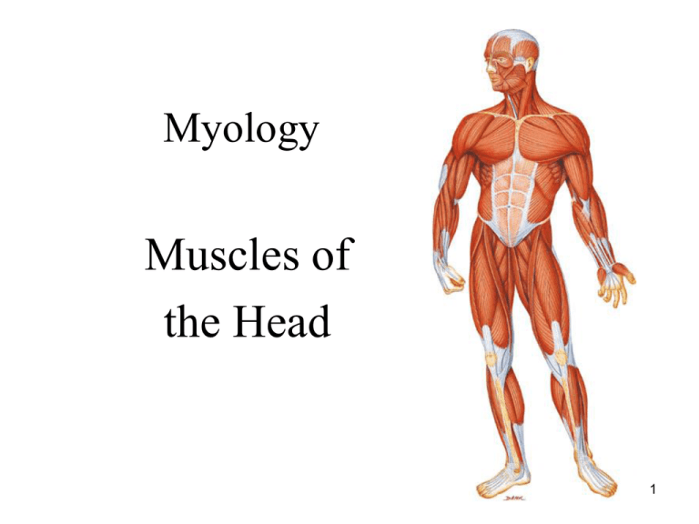
Myology
Muscles of
the Head
1
NAMING OF MUSCLES
1. Direction of muscle fibers:
a. Rectus: fibers run parallel to the midline (rectus
abdominis)
b. Transverse: fibers run perpendicular to the midline
(transverse abdominis)
c. Oblique: fibers run diagonally to the midline (external
oblique)
2. Location:
a. named for a nearby bone: (frontalis) frontal bone
b. position relative to a bone: (tibialis anterior) tibia
c. section of a muscle having more than one part (anterior
deltoid)
2
NAMING OF MUSCLES (cont)
3. Size:
a. large muscle (gluteus maximus)
b. small muscle (gluteus minimus)
c. long muscle (extensor digitorum longus)
d. short muscle (peroneus brevis)
4. Number of origins:
a. two origins: biceps femoris
b. three origins: triceps brachii
c. four origins: pronator quadratus
3
NAMING OF MUSCLES (cont)
5. Shape:
a. triangular: deltoid
b. trapezoid: trapezius
c. saw-toothed: serratus
d. rhomboid or diamond shaped: rhomboids
6. Origin and Insertion:
a. sternum, clavicle, and mastoid process: SCM
4
NAMING OF MUSCLES (cont)
7. Action:
a. decreases an angle of a joint: flexor digitorum
b. increases an angle of a joint: extensor carpi ulnaris
c. moves bone away from midline: abductor pollicis brevis
d. moves bone closer to midline: adductor longus
e. upward movement: levator scapulae
f. downward movement: depressor labii
g. turns palm up or anteriorly: supinator
h. turns palm down or posteriorly: pronator
i. decreases size of an opening: external anal sphincter
j. makes a body part more rigid: tensor fascia latae
k. moves a bone around its longitudinal axis: rotator
5
Muscles of Head Overview
Divided into 3 groups
Muscles of the Scalp
Muscles of the Face (Eyes, Nose, and Mouth)
Muscles of Mastication
6
Muscles of the Head Overview
• Muscle of head are located superficially in the fascia and
therefore move the fascia of the scalp and/or face.
– Muscles of scalp move scalp, ears, eyebrows
– Muscles of facial expression subdivided into muscles that
move:
• Skin around eyes
• Skin around nose
• Skin around mouth
– Muscles of mastication attach to mandible which is
necessary for chewing i.e. mastication.
7
Muscles of Head Overview
• Innervation:
– Muscles of scalp innervated by facial nerve
(CNVII).
– Muscles of facial expression innervated by facial
nerve (CN VII)
– Muscles of mastication innervated by trigeminal
nerve (CN V).
8
Muscles of the Scalp
• Occipitofrontalis:
– Can be considered 2 separate muscles the occipitalis and
frontalis
– Both attach into the galea aponeurotica
• Galea aponeurotica is broad flat tendon also known as the
epicranial aponeurosis
• Temporoparietalis:
– Also attaches into galea aponeurotica.
– Degree of development varies; some people it is thin other it is
non- existent.
–Occipitofrontalis and temporoparietalis are known as the
epicranius
• Auriculares:
–One, two, or all three are nonfunctional in many people
–Smallest is anterior auricularis, largest is posterior auricularis 9
Occipitalis
O: Occipital & temporal
bones
I: Galea aponeurotica
A: draws scalp posteriorly
I: CN VII (Facial nerve)
Palpation: page 15
10
Frontalis
O: Galea aponeurotica
I: Fascia and skin superior to
the eye and nose
A: draws scalp anteriorly and
elevates the eyebrows
I: CN VII (Facial nerve)
Palpation: page 15
11
Temporoparietalis
O: Fascia superior to ear
I: Lateral border of galea
aponeurotica
A: Elevates ear and Tightens
the scalp
N: CN VII (Facial nerve)
Palpation: page 18
12
Auricularis
O: Anterior: anterior fascia of the ear
Superior: lateral margin of the
galea aponeurotica
Posterior: temporal bone
I: Anterior: Spine of helix
Superior: Superior aspect of
cranial surface of the ear
Posterior: Posterior ear
A: Anterior: draws ear anteriorly
Superior: elevates ear
Posterior: draws ear posteriorly
N: CN VII (Facial nerve)
Palpation: page 20
13


