Pollak- peripheral a..
advertisement
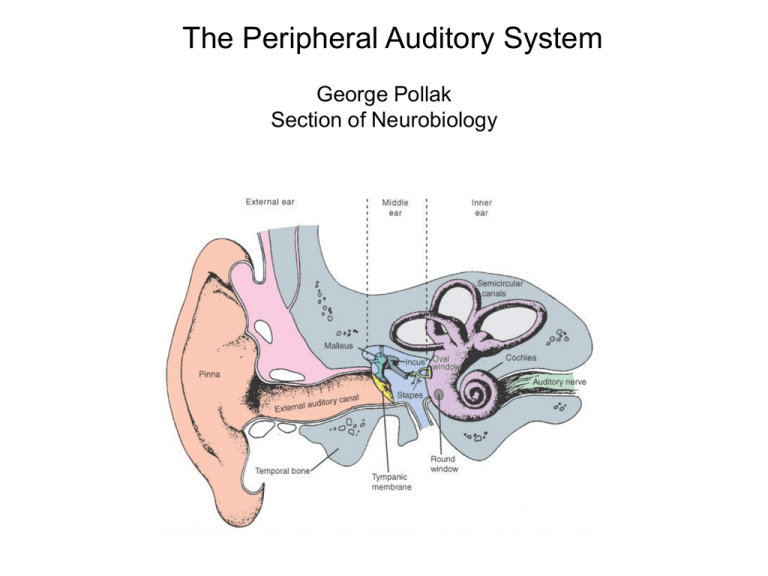
The Peripheral Auditory System George Pollak Section of Neurobiology 1 • Hair cells, the transducers of the auditory system, and how they work. Qu ickT i m e™ an d a T I FF (Un co m pr es se d) de co mp re ss or ar e n ee d ed to se e thi s p ic tu re. Organ of Corti Qu ickT i m e™ an d a T I FF (Un co m pr es se d) de co mp re ss or ar e n ee d ed to se e thi s p ic tu re. stereocillia of inner hair cells stereocillia of outer hair cells Basilar membrane stereocilia on one hair cell 9 A B C ti p link s tere ocil ia tra p door of ioni c cha nnel E F G A m odel for m ec hoele ct rica l t rans ducti on by ha ir ce ll s. In the abs ence of any s tim ul ation, at a ny i nsta nt each t ra nsduct ion channel a t a st ereoci lium 's ti p m a y be ei the r clos ed (A a nd F ) or open (E ). The grea ter proba bil ity i s that t he c hannel is c lose d. W hen the hair bundle is de flecte d w ith a posi tive sti m ulus, the spring or t ip l ink, exe rts a force on the tra p d oor and opens the c hanne l (B , C and D ). The i nflux of K + ion s into the h a ir c ell c auses it to depola rize . Pushing the hai r bundle in the oppos ite direction com pres ses th e spri ng ( tip li nk), ens uri ng t hat th e c hann el rem ai ns cl os ed (G) . Th is pr ev ent s th e in fl ux of K + i on s a nd ca us e s t he ce ll to hy pe rpo la ri ze . D endolymph Potential difference between Endolymph and cell interior K+ Hi Lo Na+ E n d ol ym p h perilymph apic al sur fac e o f hair ce ll Ek= 58 log Kout Kin = 0mV stere ocilia kin ocilium Hi K+ Lo Na+ basa l surf ace of h air ce ll tigh t ju nctions sy naptic ribb on s affe re nt ner v es F ig. 1 Pe ri ly mp h Hi Na+ Lo K+ Potential difference between Perilymph and cell interior Ek= 58 log Kout Kin = ~-70mV Rest Inhibition Excitation K+ K+ -45 mV Ca ++ K+ C a+ + K+ K+ R e st : Sm al l am o unt of K + lea ks i nto ce ll from ra tt li ng c hann els of s t ere oci li a. Th e l ea king K + de pola rize s cel l there by ope ning vol ta ge sens i tive C a + + cha nnel s res ult in g in s po nt aneous re lea s e of tra nsm it te r a nd exc it at io n of affe rent ne rve fi bers (not s how n). T h e dep ol ari za ti on ca use s K + to l eave cel l in ba sa l reg i on . E xc ita tion : S te reoci lia are bent t here by opening channe ls wh ich re sul ts in l arg er e ntry of K + int o cel l. The influx of K + ca use s a dd iti onal de polari zat ion th e reby op eni ng m ore vol tage gate d C a + + cha nnels . The ad dit iona l C a + + ch a nnels th at a re now open c au s e a l arger i nflux of C a + + i n th e b as e . T he i nfl u x of C a + + c aus e s m o re t ra ns m i t te r t o be re le as e d an d t hu s a gre at er exc ita tion of t he affere nt fibe rs (not show n ). I nh ib it ion : S tere oci li a a re bent in op posit e d irec ti on t here by cl os ing cha nnels . T hi s res ul ts i n l es s i nflu x of K + a t ch a nnel s in t h e tips o f s t ere ocil ia . K + e ffl ux oc curs at base a nd i s not rep leni sh ed b y infl ux at s te reoc ili a. C ons equ e ntl y, t he h air ce ll hy perpol ari zes t here by c losing volt age sens it iv e C a + + ch a nnels at th e bas e res ult ing in a sm all er rel ease, o r even n o re lea se o f tr ans m it te r. Di s cha rge o f a ffe rent fibe r is th ere fore re duc ed t o a rat e e ven be low th e res t ing le vel. Hi K+ low Na+ Hi Na+ low K+ Hi K+ low Na+ Hi K+ low Na+ Hi Na+ low K+ small leakage of K+ into cell K+ into cell -45 mV No K+ into cell -70 mV Next, we are going to build a cochlea Stapes Basilar membrane Sound is changed from a pressure wave in the air into mechanical movements on the basilar membrane QuickTime™ and a GIF decompressor are needed to see this picture. round window Traveling waves on basilar membrane oval window QuickTime™ and a GIF decompressor are needed to see this picture. round window The structure of the basilar membrane causes it to perform a frequency to place transformation Basilar Membrane has continuously changing dimensions along its length Apex responds maximally to low frequencies flexible wide and thin Stiff Narrow and thick Base responds maximally to high frequencies Basilar membrane converts frequency to a place of maximal response Frequency-to-Place Transformation in the Cochlea QuickTime™ and a Sor enson Video decompressor are needed to see this picture. The motion on the basilar membrane causes shearing of the cilia on hair cells and thereby causes the hair cells to depolarize and hyperpolarize in response to sound QuickTime™ and a GIF decompressor are needed to see this picture. Qu ickT i m e™ an d a T I FF (Un co m pr es se d) de co mp re ss or ar e n ee d ed to se e thi s p ic tu re. Organ of Corti Qu ickT i m e™ an d a T I FF (Un co m pr es se d) de co mp re ss or ar e n ee d ed to se e thi s p ic tu re. Basilar membrane kTime™ a basilar and membrane decompressor o see this picture. shearing of stereocillia QuickTime™ and a Animation decompressor are needed to see this picture. Organ of Corti basilar membrane QuickTime™ and a Animation decompressor are needed to see this picture. Why are there two types of hair cells? Qu ickT i m e™ an d a T I FF (Un co m pr es se d) de co mp re ss or ar e n ee d ed to se e thi s p ic tu re. Qu ickT i m e™ an d a T I FF (Un co m pr es se d) de co mp re ss or ar e n ee d ed to se e thi s p ic tu re. 98% of the fibers that project into the central auditory system are innervated by inner hair cells!! 98% What are the outer hairs doing? Answer: they act as amplifiers of the mechanical motion of the basilar membrane generated by sound Hi K++ ----depolarization hyperpolarization release of transmitter Evoked mechanical responses of isolated cochlear outer hair cells. Electromotility: OHC can change length in response to voltage change Direct evidence of an active mechanical process in the organ of Corti depolarized hyperpolarized + + + _ _ _ Qu ickTi me™ a nd a GIF deco mpressor are nee ded to see thi s picture. Dancing hair cell QuickTime™ and a YUV420 codec decompressor are needed to see this picture. 42 Outer hair cells are the only cells in the body that express prestin. Even inner hair cells do NOT have prestin. + + + + + + + + Qu ickTi me™ a nd a GIF deco mpressor are nee ded to see thi s picture. Show movie of how Prestin works QuickTime™ and a TIFF ( Uncompr essed) decompressor are needed to see this pictur e. Positive feedback loop Basilar membrane motion Sound stimuli Hair bundle deflection Change in length of hair cells OHC Membrane potential change IHC Sensory signal transmission Normal response with cochlear amplifier Apex base Basilar membrane response without cochlear amplifier Apex base How motion of basilar membrane generates tuning curves in auditory nerve fibers and thereby imparts frequency selectivity to auditory nerve fibers 50 dB SPL base apex 6 kHz 60 8 kHz Intensity (dB SPL) 7 kHz 50 40 30 20 9 kHz 10 5 10 kHz 6 7 8 9 Frequency ( kHz) 10 11 30 dB SPL base Tuning Curve The most basic feature of an auditory neuron apex 6 kHz 60 8 kHz Intensity (dB SPL) 7 kHz 50 40 30 20 9 kHz best frequency 10 5 10 kHz 6 7 8 9 Frequency ( kHz) 10 11 tuning curves tuning in animals curves with in normal no outer animals hair cells or in animals without prestin gene Sound intensity high low low frequency high How is the tonotopic organization that was first established on the basilar membrane preserved in in the central auditory system? Flow of Information Along the Central Auditory Pathway Auditory cortex Medial geniculate Medial geniculate Inferior Inferior colliculus colliculus Cochlear nucleus Auditory nerve Cochlear nucleus Superior olive Superior olive Auditory nerve The Frequency Representation on the Cochlea is Preserved in Every Nucleus of the Central Auditory System, and thus the Auditory System is Tonotopically Organized auditory cortex Auditory cortex medial geniculate Inferior colliculus cochlear nucleus Auditory nerve Cochlear nucleus Medial geniculate Medial geniculate medial geniculate Inferior Inferior colliculus colliculus superior olive Superior olive Superior olive Inferior colliculus Cochlear nucleus cochlear nucleus Auditory nerve The Frequency Representation on the Cochlea is Preserved in Every Nucleus of the Central Auditory System, and thus the Auditory System is Tonotopically Organized Auditory cortex Medial geniculate Medial geniculate Inferior Inferior colliculus colliculus Cochlear nucleus Auditory nerve Cochlear nucleus Superior olive Superior olive Auditory nerve The Frequency Representation on the Cochlea is Preserved in Every Nucleus of the Central Auditory System, and thus the Auditory System is Tonotopically Organized
