Identifying serum exosome microRNA signatures in melanoma patients
1
M.S. ,
1
M.D. ,
2
Ph.D. ,
3
M.S.
Samantha N. Barry,
Deyi Xiao,
Douglas Taylor,
Sabine Waigel,
Wolfgang
3
1
1
Zacharias, Ph.D. , Hongying Hao, M.D., Ph.D. , Kelly M. McMasters, M. D., Ph. D.
1
Department of Surgery , University of Louisville School of Medicine
2,
Department of Obstetrics, Gynecology and Women’s Health University of Louisville School of Medicine
3
Microarray Facility , University of Louisville School of Medicine
Abstract
Results
Melanoma is the 6th most common cancer in the U.S. with a median
survival rate of less than one year. Early diagnosis of melanoma is
challenging because the traditional histopathologic methods cannot
sufficiently distinguish benign from malignant lesions. Minimally
invasive and dependable determinants
that can
refine the
individualized diagnosis are needed. Exosomes are small membrane
vesicles (~30-120nm) that are secreted from cells and contain protein
and RNAs. They are implicated in cell-cell communication. Previous
studies have shown that miRNAs in tumor exosomes contribute to
progression of disease via mRNA silencing. Characterizing the
differences in expression of exosomal miRNA between melanoma
and non-melanoma patients could lead to a method for earlier and
more quantitative diagnosis. In the current study, blood exosomes
were isolated from non-melanoma subjects and Stage I melanoma
patients. miRNA array was applied to compare the miRNA
expression profiles between non-melanoma controls and Stage I
melanomas. Real time RT-PCR was performed to confirm some of
the differentially expressed miRNAs in these two groups. We showed
that a panel of exosomal miRNAs (such as hsa-miR-1228 and hsamiR-1825) has significant changes in Stage I melanoma patients vs
non-melanoma controls. These results provide a starting point for
further characterization of exosomal miRNA signatures in melanoma
patients. This research could lead to a minimally invasive method for
specific diagnosis of malignant melanoma.
•
•
noncoding
RNA
that
ANOVA analysis Stage I vs NonMelanoma
Figure 2. RT-PCR workflow.
Figure 1. Initial microarray screen.
Fold change
P-Value
miR-23a
1153.14
7.902E-10
miR-26a
1069.13
3.20E-09
miRNA in tumor exosomes have been shown to contribute to
malignant progression
let7a
711.23
1.65E-07
Analyzing exosomal miRNA signatures in melanoma patients
could identify a potential biomarkers for diagnosis & prognosis
miR-221
601.23
1.43E-08
Table 1. Selected upregulated miRNAs (stage I vs.
non-melanoma) (P<0.05, -2>FC>2)
miRNA
Fold change
P-Value
Patient Groups:
miR-940
-122.15
5.03E-07
miR-1825
-104.56
7.81E-06
miR-1228
-94.67
6.87E-06
miR-4258
-84.13
6.91E-06
miR-1281
-62.27
4.61E-05
1. Exosomes were isolated from serum using the ExoQuick protocol.
2. RNA was isolated from exosomes using a modified mirVARNA RNA
isolation protocol.
3. miRNA array was conducted using Affymetrix miRNA array 2.0.
4. Differentially expressed miRNAs between the two sample groups were
analyzed by ANOVA.
5. RT-PCR was performed to validate differentially expressed miRNAs
identified in microarray screen.
UofL Design and Print
10
4/6
28-81
52
Stage I
15
9/6
23-81
53
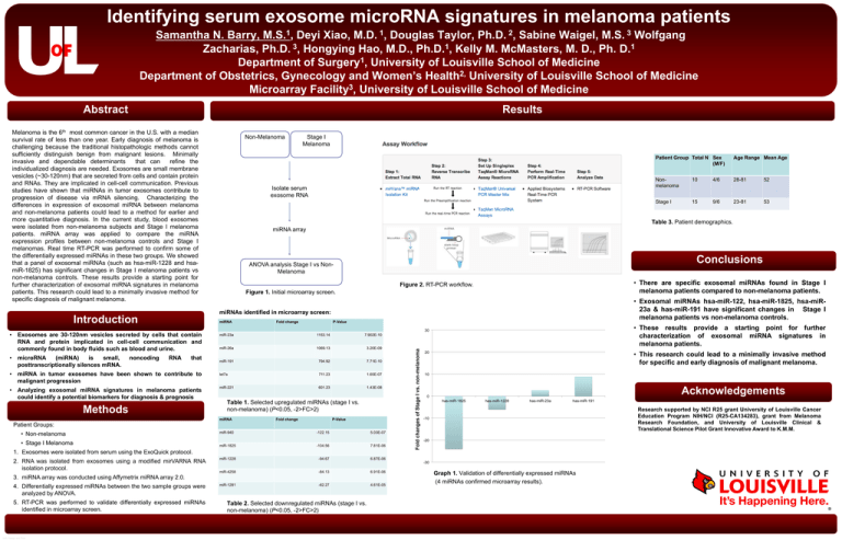
• There are specific exosomal miRNAs found in Stage I
melanoma patients compared to non-melanoma patients.
• Exosomal miRNAs hsa-miR-122, hsa-miR-1825, hsa-miR23a & has-miR-191 have significant changes in Stage I
melanoma patients vs non-melanoma controls.
7.71E-10
• Stage I Melanoma
Nonmelanoma
Conclusions
794.92
• Non-melanoma
Age Range Mean Age
miRNA array
miR-191
Methods
Patient Group Total N Sex
(M/F)
Table 3. Patient demographics.
Table 2. Selected downregulated miRNAs (stage I vs.
non-melanoma) (P<0.05, -2>FC>2)
• These results provide a starting point for further
characterization of exosomal miRNA signatures in
melanoma patients.
Fold changes of Stage I vs. non-melanoma
microRNA
(miRNA)
is
small,
posttranscriptionally silences mRNA.
Isolate serum
exosome RNA
miRNA
Exosomes are 30-120nm vesicles secreted by cells that contain
RNA and protein implicated in cell-cell communication and
commonly found in body fluids such as blood and urine.
•
Stage I
Melanoma
miRNAs identified in microarray screen:
Introduction
•
Non-Melanoma
Future Directions
• This research could lead to a minimally invasive method
for specific and early diagnosis of malignant melanoma.
Acknowledgements
Research supported by NCI R25 grant University of Louisville Cancer
Education Program NIH/NCI (R25-CA134283), grant from Melanoma
Research Foundation, and University of Louisville Clinical &
Translational Science Pilot Grant Innovative Award to K.M.M.
Graph 1. Validation of differentially expressed miRNAs
(4 miRNAs confirmed microarray results).

