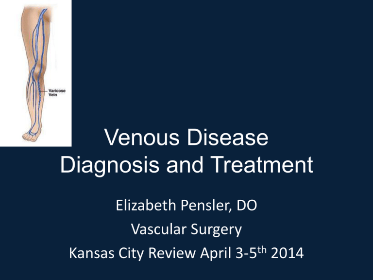
Venous Disease
Diagnosis and Treatment
Elizabeth Pensler, DO
Vascular Surgery
Kansas City Review April 3-5th 2014
Chronic venous disease
•
•
•
•
Most common vascular disorder
3 Billion US dollars spent/yr treatment
3 % of the total Heath care Budget
2 million USA work days lost per year
Abnormal Veins
Telangiectasias
<1mm
Varicose vein
Reticular veins
>3mm
1-3mm
Surgical Anatomy of the
Lower Limb Veins
I. Superficial Veins
Long & Short
saphenous veins &
their tributaries
Communicating Veins
II.Deep Veins
Tibial venae
comitantes,
popliteal & femoral
veins
Surgical Anatomy Lower Limb Veins
Superficial Veins
1. Long & short saphenous
veins and their tributaries.
2. Lie in the subcutaneous
tissue superficial to the
muscle fascia.
3. They have their own, welldeveloped muscle coat.
The Long Saphenous Vein
The longest vein in the body
Surface Anatomy
•1 cm anterior to the medial malleolus
•1 hand breadth post to med aspect of patella
•Ends on anteromedial side of femoral vein
3.5 cm below & lateral to pubic tubercle
Receives following tributaries near its termination:
•Superficial & deep external pudendal vv.
•Superficial circumflex iliac v.
•Superficial inferior epigastric v.
The Short Saphenous Vein
Anatomy
•Behind the lateral malleolus
•Pierces the deep fascia before enters popliteal vein
•Invariably terminates above popliteal fossa into the
superficial femoral vein
•Communicates with the long saphenous vein by
several channels
Surgical Anatomy Lower Limb Veins
Deep Veins
1.Accompany axial arteries.
2.Run within the muscles
deep to the muscle
fascia.
Surgical Anatomy of the Lower Limb Veins
Comunicating Veins “Perforators”
Perforate the
fascia
connecting the
superficial &
deep veins at
certain points.
Perforators
Main sites of superficial to deep venous
communication
Sapheno-femoral junction
Perforator
Mid thigh perforator
(Hunter’s canal)
Medial calf
perforators
Thigh
perforators
connect to
the long
saphenous
main trunk
Just below the knee
The lower Perforators
perforators
are joined Perforators
to form the
Perforators
Posterior
arch vein
10 cm above
Just above
Just below
Medial
malleolus
Surgical Anatomy of
the Lower Limb Veins
All lower limb veins
have valves to
direct venous return
in one direction
only
From below upwards,
and from superficial to
deep.
Venous return
The heart pump
maintaining a pressure gradient
across the veins
Venomotor tone
With dependency
Gravity
Pooling in dependent limbs may
reduce cardiac output by 2 L/min
& may cause fainting
Under control of sympathetic system
[Upright position -- dependant pooling –
dec. cardiac output -- inc. sympathetic
discharge -- inc. venous tone -- inc.
venous return.]
This is counter acted by:
Blood is pushed upwards
With calf muscle contraction
Calf muscle contraction
and prevented from retrograde
flow by competent venous valves
Competent Veno-muscular Pump
is composed of:
1. Superficial & deep veins
with competent valves.
2. Competent perforating
veins communicating the
deep & superficial systems
3. Powerful lower limb
muscles.
Definition
The ankle venous pressure
during walking is called the
“ambulatory venous
pressure”
Tibbs, DT, et al Varicose Veins, Venous Disorders,,and Lymphatic Problems in the Lower limbs, 1997, p11
A competent
veno-muscular
pump will push
the blood
towards the
heart,
thus lowering
the ambulatory
venous
pressure.
Definition
CVI collectively describes the manifestations
of impaired venous return due to abnormal
venous system function.
In the majority of cases, it is caused by
valve incompetence,
and less commonly by venous obstruction.
Patho-physiology of CVI
The main defect (problem) in the lower
limb venous system may be in the
superficial, deep or perforating veins.
This problem is usually in the valves
(reflux), but sometimes it is in the form of
obstruction. (or a combination of both
reflux & obstruction)
Patho-physiology of CVI
The defect may be:
Primary defect: related to structural
weakness of valves or venous wall, as
in primary varicose veins
Secondary defect: for example due
to previous deep venous thrombosis,
as in post-phlebitic syndrome
Patho-physiology of CVI
Whatever the cause of CVI,
eventually cause venous hypertension
of microcirculation, giving the same
symptoms & signs.
The severity of symptoms & signs depend
on the degree & duration of venous
hypertension.
Symptoms & Signs of “CVI”
Early
Persistent & Severe
1. Posture related
discomfort
4. Ankle brown
pigmentation
2. Lower limb
oedema
5. Venous eczema
3. Muscle cramps
7. Venous ulcer
6. Lipodermatosclerosis
Clinical Examination
Look for:
The patient should be standing
The extent and distribution of VV
Long saphenous
VV
Antro-lat. tributary
of LSV
Short saphenous VV
Communicating
vein varicosity
Clinical Examination
Look for:
Scars of previous op.
Lipodermatosclerosis
Some ulcers
may potentially
bleed
Eczema
Pigmentation
Ulcer
Investigations of Venous Disease
Investigations have two aims:
1. Identify the existence, site & degree
of venous reflux.
2. Confirm deep venous patency.
Etiology
• Reflux 80%
• Venous obstruction 18-28%
– Resultant edema and skin changes =
Postthrombotic syndrome
• Muscle Pump Dysfunction
Risk factors
• Age: Aging causes wear and tear.
• Sex: Women > Men. Hormonal changes during
pregnancy or menopause. Progesterone
relaxes venous walls. HRT / OCP may increase
the risk of varicose veins.
• Genetics
• Obesity: Increases venous HTN.
• Standing for long periods of time. Prolonged
immobile standing impairs venous return.
Fowkes, FG, Lee, AJ, Evans, CJ, et al. Lifestyle risk factors for lower limb venous reflux in the general population: Edinburgh Vein Study.
Int J Epidemiol 2001; 30:846.
Sadick, NS. Predisposing factors of varicose and telangiectatic leg veins. J Dermatol Surg Oncol 1992; 18:883.
Iannuzzi, A, Panico, S, Ciardullo, AV, et al. Varicose veins of the lower limbs and venous capacitance in postmenopausal women:
relationship with obesity. J Vasc Surg 2002; 36:965.
Evans, CJ, Fowkes, FG, Hajivassiliou, CA, et al. Epidemiology of varicose veins. A review. Int Angiol 1994; 13:263.
Strong familial component
• Not well studied
• Twin studies 75% identical, 52% non
identical
• If both parents VVS - 90% of children VVs
• If one parent was affected 25 percent for
men and 62 percent for women
•
Cornu-Thenard, A, Boivin, P, Baud, JM, et al. Importance of the familial factor in varicose disease.
Clinical study of 134 families. J Dermatol Surg Oncol 1994; 20:318.
Symptoms
• Achy or heavy feeling
• Burning, throbbing, muscle
cramping and swelling.
• Prolonged sitting or standing
tends to intensify symptoms.
• Pruritis
• Painful skin ulcers
Complications
• Painful ulcers form near
• Brownish pigmentation
• Bleeding
• Superficial thrombophlebitis
CEAP classification
1994 AVF Meeting
Investigation
• All get a Duplex scan
• Examines
– Deep veins
– Superficial veins
– Incompetence and
patency
Duplex scan
• Vast majority have superficial incompetence only.
• Sensitivity 95 % competence of saphenofemoral and
saphenopopliteal junctions.
• Less sensitive for identifying incompetent perforators
(40 to 60 percent)
Venous Ulcer Treatment Algorithm
Ulcer Treatment
Debride
Manage exudate
Control infection
Treat systemic problems
Compression
Multi layer elastic
Intermittent compression pumps
Rigid dressings
Healed Ulcer Care
Compression garments
Skin care
Consider surgical intervention
Treatment
• Conservative
Leg elevation
Exercise
Compression stockings
Treatment of other underlying conditions
Nothing
• 30-40mmhg Compression
Vein ablation therapies
Classified by method of vein destruction:
1. Chemical (sclerotherapy)
2. Thermal (laser or endovenous ablation)
3. Mechanical (surgical excision or
stripping)
Who gets sclerotherapy
• Small non-saphenous varicose veins (less
than 5 mm),
• Perforator veins
• Residual or recurrent varicosities following
surgery
• Telangiectasia
• Reticular veins
Sclerosing Agents
•
•
•
•
•
Sodium tetradecyl sulfate
Hypertonic Saline
Polidocanol
Monoethanolamine oleate
Glucose combinations
• Damage endothelium leading to thrombosis of
the vein.
• Pressure to try and reduce the amount of
thrombus.
Tessari, L, Cavezzi, A, Frullini, A. Preliminary experience with a new sclerosing foam in the treatment of varicose veins. Dermatol Surg
2001; 27:58.
Microsclerotherapy
• 30 g butterfly needle
• 0.2% STS
• Several courses required
benefit compression
Telangiectasias
Foam Sclerotherapy
• 1:4 Sclerosant (1% or
3%): Air
• Why foam?
– Induces spasm
– Disperses further
– Enhanced sclerosis
Breu, FX, Guggenbichler, S. European Consensus Meeting on Foam Sclerotherapy, April, 4-6, 2003,
Tegernsee, Germany. Dermatol Surg 2004; 30:709.
Spider veins
Foam Sclerotherapy:
Complications
•
•
•
•
•
•
•
•
•
Phlebitis
Skin staining
Failure
Residual lumps
Matting
Embolus (CVA)
DVT
Ulceration (rare)
Anaphylaxis (very rare)
Catheter-based Treatments
• Endovenous laser EVLA
• Radiofrequency ablation RFA
• Primarily to treat saphenous insufficiency
(great or small)
• EVLA and RFA, are equally efficacious &
have similar recanalization rates.
Boros, MJ, O'Brien, SP, McLaren, JT, Collins, JT. High ligation of the saphenofemoral junction in endovenous obliteration of varicose
veins. Vasc Endovascular Surg 2008; 42:235.
Radiofrequency ablation
Radiofrequency ablation devices (ClosureFast™, RFiTT®, ClosureRFS™) generate
a high frequency alternating current in the radio range of frequency.
Weiss, RA, Weiss, MA. Controlled radiofrequency endovenous occlusion using a unique radiofrequency catheter under duplex guidance to
eliminate saphenous varicose vein reflux: a 2-year follow-up. Dermatol Surg 2002; 28:38.
Rautio, T, Ohinmaa, A, Perala, J, et al. Endovenous obliteration versus conventional stripping operation in the treatment of primary varicose
veins: a randomized controlled trial with comparison of the costs. J Vasc Surg 2002; 35:958.
Radiofrequency ablation
Heats the tissue surrounding the catheter
electrode to a specified temperature.
Radiofrequency works well on tissue
composed primarily of collagen
Special probes have been designed for the
radiofrequency device to manage nonsaphenous and perforator veins.
Endovenous Laser
Endovenous Laser
• Devices (EVLT®, ClosurePlus™)
• Bare tipped optical fiber - applies laser light
energy to the vein.
• Therapy based on photothermolysis (light induced
thermal damage).
• Laser light heats target tissue inducing thermal
injury
Bush, RG, Shamma, HN, Hammond, K. Histological changes occurring after endoluminal ablation with two diode
lasers (940 and 1319 nm) from acute changes to 4 months. Lasers Surg Med 2008; 40:676.
Surface laser therapy
• Telangiectasias,
reticular veins and
small varicose veins
<5mm
• Not used for larger
varicose veins
Post op care
• Graduated compression stockings
• F/U duplex ultrasound is performed within
one week to evaluate for thrombus in the
common femoral vein.
• Pt recovery averages two and four days
• Significantly shorter interval than is seen
with surgical ligation and stripping
Mozes, G, Kalra, M, Carmo, M, et al. Extension of saphenous thrombus into the femoral vein: a potential complication of new endovenous ablation
techniques. J Vasc Surg 2005; 41:130.
Darwood, RJ, Theivacumar, N, Dellagrammaticas, D, et al. Randomized clinical trial comparing endovenous laser ablation with surgery for the treatment
of primary great saphenous varicose veins. Br J Surg 2008; 95:294.
Endovenous complications
• Pain, bruising, hematoma
• Skin changes: burns, induration,
pigmentation, matting, dysesthesia, &
superficial thrombophlebitis.
• Nerve injury
• DVT
• Wound infection
Mozes, G, Kalra, M, Carmo, M, et al. Extension of saphenous thrombus into the femoral vein: a potential complication of new endovenous ablation
techniques. J Vasc Surg 2005; 41:130.
VAN DEN Bos, RR, Neumann, M, DE Roos, SP, Nijsten, T. Endovenous laser ablation-induced complications: Review of the literature and new
cases. Dermatol Surg 2009;
Surgery
Saphenous vein stripping
• Cryostripping
• High saphenous ligation
• Ambulatory phlebectomy
• Stab/avulsion phlebectomy
• Transilluminated phlebectomy TIPP
• Perforator ligation
• Subfascial endoscopic perforator vein ligation
(SEPS)
Dwerryhouse, S, Davies, B, Harradine, K, Earnshaw, JJ. Stripping the long saphenous vein reduces the rate of reoperation for recurrent varicose
veins: five-year results of a randomized trial. J Vasc Surg 1999; 29:589.
Menyhei, G, Gyevnar, Z, Arato, E, et al. Conventional stripping versus cryostripping: a prospective randomised trial to compare improvement in
quality of life and complications. Eur J Vasc Endovasc Surg 2008; 35:218.


