3._Squint
advertisement
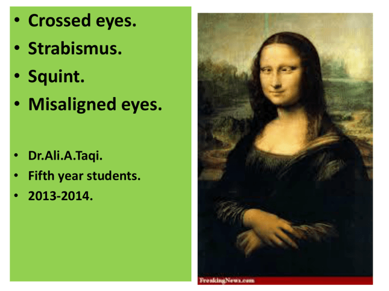
• • • • Crossed eyes. Strabismus. Squint. Misaligned eyes. • Dr.Ali.A.Taqi. • Fifth year students. • 2013-2014. What is Strabismus? • Normally visual axis of the two eyes are parallel to each other in the ‘primary position of gaze’ and this alignment is maintained in all positions of gaze. • A misalignment of the visual axes of the two eyes is called squint or strabismus. Is Intentional squinting can lead to real squint?! Anatomy of the eye muscles The extraocular muscles rotate the eyes about three axes to produce vertical (elevation and depression), horizontal (adduction and abduction), and rotational (intorsion and extorsion) movement. The horizontal recti produce purely horizontal movements; the vertical recti and the obliques have vertical, rotational, and horizontal actions. Their principal effect depends upon the horizontal position of the eye in the orbit, and therefore varies with gaze position. Anatomy and physiology of the ocular motility system extraocular muscles • A set of six extraocular muscles • (4 recti and 2 obliques) • control the movements of each eye (Fig. 13.1). • Rectus muscles are superior (SR), inferior (IR), medial (MR) and lateral (LR). • The oblique muscles include superior (SO) and inferior (IO). Nerve supply • The extraocular muscles are supplied by third, fourth and sixth cranial nerves. The third cranial nerve • (Oculomotor) supplies the superior, medial and inferior recti and inferior oblique muscles. • The fourth cranial nerve (Trochlear) supplies the superior oblique and • The sixth nerve (Abducent) supplies the lateral rectus muscle. Extra ocular muscles insertion (right eye). Extraocular muscles origin. • Structures at the orbital apex, showing muscle insertions into the annulus of Zinn and the location of various vessels and nerves. All four recti and superior oblique have their origin at the apex of the orbit, while inferior oblique has its origin at the nasal end of the anterior orbital floor. The recti insert anterior to the equator, at 7.5 mm (superior), 7.0 mm (lateral), 6.5 mm (inferior), and 5.5 mm (medial) behind the limbus. The obliques insert behind the equator; the insertion of the superior oblique tendon lies along the lateral border of superior rectus, having been reflected through the pulley of the trochlea at the anterior nasal orbital roof, and the insertion of inferior oblique lies external to the macula. The superior oblique tendon passes beneath the superior rectus, and the inferior oblique passes beneath the inferior rectus. Can Animals have squint? PRIMARY AND SECONDARY ACTIONS OF MUSCLES • The primary action of the horizontal rectus muscles is 99% horizontal. They have trivial secondary or tertiary actions. • The primary action of the vertical rectus muscles is 75% vertical, and they have secondary torsional and horizontal actions. • The primary action of the oblique muscles is 60% cyclorotation (torsion) and they have secondary vertical and horizontal actions. Eye movements • Horizontal ductions The horizontal recti are mainly responsible for adduction and abduction. • vertical ductions The vertical recti act as pure elevators and depressors in abduction. • Torsion The superior rectus and superior oblique act as intortors, and the inferior rectus and inferior oblique act as extortors. Classification of strabismus • Broadly, strabismus can be classified as below: I. Apparent squint or pseudostrabismus. II. Latent squint (Heterophoria) III. Manifest squint (Heterotropia) 1. Concomitant squint 2. Incomitant squint. Pseudostrabismus • In pseudostrabismus (apparent squint), the visual axes are in fact parallel, but the eyes seem to have a squint: 1. Pseudoesotropia or apparent convergent squint may be associated with a prominent epicanthal fold (which covers the normally visible nasal aspect of the globe and gives a false impression of esotropia) and negative angle kappa. 2. Pseudoesotropia or apparent divergent squint may be associated with hypertelorism, a condition of wide separation of the two eyes, and positive angle kappa. Disorders of ocular motility The direction of the visual axis of each eye towards a fixation point is co-ordinated by the action of the extraocular muscles. Strabismus (squint): is a failure of the coordination of binocular alignment. It leads inevitably to loss of binocular single vision. Fusion of the two images is replaced either by diplopia or suppression of one image. Strabismus may be caused by orbit, muscle, motor nerve, or brainstem pathology. Primary position Gaze straight ahead, with the visual axes parallel. Heterotropia Manifest deviation i.e. failure of the visual axes to meet at the fixation point. Manifest convergent squint is described as esotropia, and manifest divergent squint as Exotropia. Vertical squint is hypertropia and hypotropia. Heterophoria Latent deviation, i.e. failure of the visual axes to meet at the fixation point when they are dissociated e.g. by monocular occlusion. Latent convergent and divergent squint are, respectively, esophoria and exophoria. Concomitant Constant angle of deviation irrespective of the direction of gaze (nonparalytic). Incomitant Variable angle of squint, according to gaze direction, paralytic squint is Incomitant. Other Causes of acquired ocular motility disorder Neurogenic (ocular motor nerve lesion): Vascular (diabetes or hypertension). Demyelination (multiple sclerosis). Inflammatory Compressive (aneurysm or tumor) Trauma or surgery. Myogenic Myasthenia gravis Ocular myopathy Restriction Dysthyroid ophthalmopathy Trauma Inflammation Orbital Orbital mass restricting eye movement Binocular single vision • When a normal individual fixes his visual attention on an object of regard, the image is formed on the fovea of both the eyes separately; but the individual perceives a single image. This state is binocular single vision. Visual development • Binocular single vision is a conditioned reflex which is not present since birth but is acquired during first 6 months and is completed during first few years. • The process of its development is complex and partially understood. Important mile stones in the visual development are: • At birth there is no central fixation and the eyes move randomly. • By the first month of life fixation reflex start developing and becomes established by 6 months. • By 6 months the macular stereopsis and accommodation reflex is fully developed. • By 6 year of age full visual acuity (6/6) is attained and binocular single vision is well developed. Amblyopia(lazy eye) Refers to a partial loss of vision in one or both eyes, in the absence of any organic disease of ocular media, retina and visual pathway. • Pathogenesis. • Amblyopia is produced by certain Amblyogenic factors operating during the critical period of visual development (birth to 6 years of age). • The most sensitive period for development of amblyopia is first six months of life and it usually does not develop after the age of 6 years. • Amblyogenic factors include : 1---- Visual (form sense) deprivation as occurs in Anisometropia, 2---- Light deprivation e.g., due to congenital cataract, 3----Abnormal binocular interaction e.g., in strabismus. Squint types Non paralytic=concommitant squint. A-eso-deveation = esotropia= inward deviation. 1-Congenital. 2-Accommodative (refractive,nonrefractive,mixed). B-exo-deveation = exotropia =outward deviation 1-childhood-onset 2-sensory-deprivation. 3-convergence-insufficiency Paralytic strabismus 3RD( Oculomotor)cranial nerve palsy(all extraocular muscles involved except the lateral rectus & the superior oblique muscle) 6th cranial nerve (Abducent)=paralysis of lateral rectus muscle . 4th cranial nerve (Trochlear)=paralysis of superior oblique muscle Concomitant strabismus(nonparalytic) • It is a type of manifest squint in which the amount of deviation in the squinting eye remains constant (unaltered) in all the directions of gaze; and there is no associated limitation of ocular movements. Etiology • The causative factors differ in individual cases. As we know, the binocular vision and coordination of ocular movements are not present since birth but are acquired in the early childhood. The process starts by the age of 3-6 months and is completed up to 5-6 years. Therefore, any obstacle to the development of these processes may result in concomitant squint. These obstacles can be arranged into three groups, namely: sensory, motor and central. Sensory obstacles. These are the factors which hinder the formation of a clear image in one eye. These include: • Refractive errors, • Prolonged use of incorrect spectacles, • Anisometropia, • Corneal opacities, • Lenticular opacities, • Diseases of macula (e.g., central chorioretinitis), • Optic atrophy, and • Obstruction in the pupillary area due to congenital ptosis. 1-Congenital-Infantile Esotropia • • • Infants with congenital esotropia develop a large angle of esotropia at several months of age . IF it is not present at birth, some ophthalmologists prefer to name this condition infantile esotropia. The cause is unknown. It occurs in otherwise normal infants, but it is more common in infants with developmental delay and in infants with hydrocephalus. The treatment for this condition is surgery. Most physicians recess each medial rectus muscle a graded amount, depending on the angle of deviation. Some also resects one or both lateral rectus muscles for large deviations. Is squint more in mentally retarded babies? Accommodative esotropia Treatment of accommodative esotropia (inward deviated eye). Full Cycloplegics refraction measurement of the plus glass Examination Under anesthesia (EUA) 1-Cycloplegic refraction by using atropine eye drops for 3 days three times a day before the examination. 2-fundoscopy(exam. of the retina, optic nerve. Refraction and glasses Accommodative esotropia correction of accommodative squint by glasses & treatment of amblyopia by patching, so both eye now have good visual acuity and normal aligned Treatment of amblyopic eye. (lazy eye) Patching of normal eye Famous characters squint Exotropia (outward deviation) • 1-Childhood-onset Exotropia is the most common type of Exotropia. Many children develop an Exotropia that typically begins intermittently . • The average age of onset is about 2.5 years (range, 6 months-6 years), about the same time of onset as for accommodative esotropia. • The cause is unknown. It may be weakly hereditary, because few children with this condition have parents or siblings with the same condition. It is useful to think of this entity as passing through several phases or stages. 1-Childhood-onset Exotropia • The first phase of this condition is exophoria only-latent deviation not seen until one eye covered-preventing the fusion of the two images into one image. It is rarely seen because it is rarely symptomatic. • If a child happened to be examined at this time in the evolution of this condition, the examiner would find only an exophoria. Testing would reveal a latent deviation, detected only by the cover test. 1-Childhood-Onset Exotropia • Several months later, the child may progress into the • second phase: intermittent Exotropia. With fatigue, illness, or inattention and when looking at a distance of several meters or more, one of the eyes turns out for several seconds. • The child then becomes aware of diplopia and makes some unconscious effort to restore the alignment of the eyes. Alternating Exotropia? when fixate with the right eye the left deviated & vise versa, both eyes not cooperate together to form single image (no binocular single vision-BSV) 2-Sensory Exotropia • The primary etiologic factor is not a motor abnormality but some defect in the afferent or sensory system. • If two eyes do not have good binocular function, it is likely that the poorer or nondominant eye will gradually turn out • sensory deprivation = poor seeing eye(vision is a reflex !) 3-Convergence Insufficiency. • Students usually suffer from this problem. • Is an apparent weakness of convergence, called convergence insufficiency. • The entity frequently affects young adults and is a major cause of asthenopia, or tired eyes, while doing near work in this age group. • In this condition, the eyes are straight at distant fixation and without symptoms. However, at near viewing, there is an exodeveation, sometimes an exophoria or sometimes an intermittent Exotropia with transient diplopia. Paralytic (non commitant)squint Abducent nerve palsy or restrictive thyroid myopathy? Trochlear nerve palsy. Vertical deviation and vertical diplopia Squinted eye may have serious pathology?! References

