(LISST-100): Applications and limitations Lee Karp
advertisement
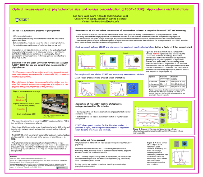
Optical measurements of phytoplankton size and volume concentration (LISST-100X): Applications and limitations Lee Karp-Boss, Laura Azevedo and Emmanuel Boss University of Maine, School of Marine Sciences Contact:lee.karp-boss@maine.edu Cell size is a fundamental property of phytoplankton • Affects metabolic rates • Determines predator-prey interactions and hence the structure of aquatic food webs • Determines sinking rates and hence the fate of primary production. • Phytoplankton span a wide range of cell sizes (few mm-few mm) Information on cell size distribution is central to the understanding of many ecological processes, yet in-situ measurements of particle size distribution (PSD) remains a challenge in oceanography. Recent developments of laser scattering techniques open new opportunities to study PSD in-situ. Measurements of size and volume concentration of phytoplankton cultures: a comparison between LISST and microscopy • LISST inversion to size was first tested with beads of known sizes (data not shown); filtered seawater (0.2mm) was used as a blank. • Scattering measurements of phytoplankton cultures were inverted to obtain size and volume concentration, using the factory algorithm. • Cells were counted and sized, using a microscope. Microscopy data were then used to calculate volumes and cross-sectional areas of cells. • Diameters of spheres of equal volume and spheres of equal cross-sectional areas were then calculated for a comparison with the LISST inversion. These diameter were than used in the microscopy based PSD. Good agreement between LISST and microscopy for species of nearly spherical shape (within a factor of 2 for concentration) Figure 1a Figure 1a-e: Size distributions of phytoplankton cultures. Red line represents sizes obtained by the LISST-100X inversion. Microscopy derived size distribution is plotted for two cases: (1) equal volume spheres (blue line) and (2) spheres of equal crosssectional area (black line). Since scattering in the geometric-optics limit is proportional to the particle’s cross-sectional area one would expect the latter to show a better fit to the LISST inversion. The choice of size parameter becomes more important as particles deviate from a spherical shape (Figure 1d). Figure 1c Figure 1b Evaluation of in-situ Laser Diffraction Particle Size Analyzer (LISST-100X) for size and concentration measurements of phytoplankton LISST measures near-forward light scattering by particles and uses a Mie-theory based inversion to obtain the PSD. It does not measure size directly. The relationship between the measured scattered light and the final PSD depends on inversion assumptions with respect to the physical and optical properties of the particles For complex cells and chains LISST and microscopy measurements deviate Species LISST “sees” cross-sectional areas of all cell orientations Figure 1d Figure 1e G. simplex # cells/ml # cells/ml (microscope) (LISST) 2330 ± 782 1584 (8<d<16 mm) A. tamarense 96 ± 11.2 110 (25<d<35 mm) 241 ± 15 124 (35<d<70 mm) L. polyedra Table 1: a summary of cell concentrations obtained by microscopy and the LISST inversion. The range of particle diameters (d) for which cell concentration was obtained is shown in brackets. Physical measurement (scattering) Inversion (Mie theory) Property descriptive of size (crosssectional area, volume) Applications of the LISST-100X to phytoplankton ecology: phytoplankton life histories A. B. translation Single length scale (SED) • During vegetative cell division mean cell size of populations of diatoms decreases over time. LISST-100x, Sequoia Scientific, Inc. photo courtesy of Jim Loftin. • The underlying assumption in converting LISST measurements into PSD is that particles are homogeneous spheres. • Near-forward light scattering by particles is dominated by diffraction and therefore is relatively insensitive to particle composition (e.g., index of refraction) • The LISST-100, which was originally designed for sediment studies, has been used successfully to detect purple sulfur bacteria in lakes (Serra et al. 2001). B a • Phytoplankton display a wide range of cell shapes. Patterns of light s scattered by non-spherical particles differ from those of spheres (Clavano i et al. c submitted). Therefore, PSD obtained from inversions computed for spheres may give poor estimate of the PSD and volume concentration of phytoplankton. References: Serra et al. 2001. Journal of Environmental Engineering 127:1023. Calvano et. al., Optical properties of non-spherical particles: from theory to observations. Submitted to Oceanography and Marine Biology, an annual review. Acknowledgements: we thank Jim Loftin, LeAnn Pritchard and Maura Thomas for assistance in the lab and Dave Townsand for the use of his microscope. The Gulf of Maine foundation provided summer support for L. Azavedo. This study was funded by NASA. • Diatoms restore cell size via sexual reproduction or vegetative cell enlargement LISST shows great promise for life histories studies; it provides a rapid, non-disruptive measurement - important when delicate life stages are involved. Same magnification (20X) was used for all photos Figure 2: Changes in the mean cell diameter in a culture of Coscinodiscus radiatus as obtained from LISST inversion (A) and microscopy (B). Field studies and future prospect • Phytoplankton of different cell sizes can be distinguished by the LISST (Figure 3) • Based on laboratory studies, the LISST shows great potential in monitoring HABs, especially those of Gymnodinium breve and L. polyedra for which blooms are often mono-specific. • The LISST may be particularly useful in lake studies, for which coulter counters are not applicable, and where Dinoflagellates (e.g., Peridinium) often form mono-species blooms. Further studies are required to evaluate its utility for monitoring phytoplankton in the field. Figure 3: A mixed culture of G. simplex and A. tamarense. LISST inversion is shown by the black line. Blue (G. simplex) and red (A. tamarense) lines show size distribution based on microscopy. Note: volume concentration of microscopy is based on stock cultures.
