Cutaneous membrane
advertisement
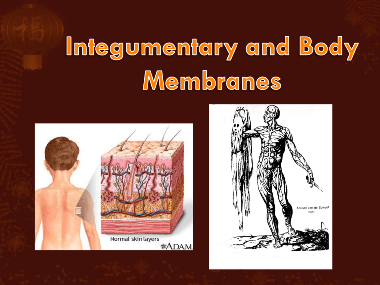
Membranes found all throughout body Functions of body membranes: Line or cover body surfaces – separate in/out Protect body surfaces Lubricate body surfaces Classified into two types: Epithelial membranes Cutaneous membrane o Mucous membrane o Serous membrane o Connective tissue membranes o Synovial membrane o Cutaneous membrane = skin Function: protect deeper body tissues dry membrane Outermost protective boundary consisting of: Superficial epidermis Contains keratin in areas of high friction Underlying dermis Mostly dense connective tissue - protection Figure 4.1a o Mucous membranes Function: protect from drying out, lubrication Lines all body cavities that open to the exterior body surface Often adapted for absorption (i.e. large intestine) or secretion (i.e. nasal cavity) o Serous membranes Function: lubrication for cushion, friction Surface simple squamous epithelium Underlying areolar connective tissue Lines open body cavities that are closed to the exterior of the body Serous layers separated by serous fluid Specific serous membranes Peritoneum • Abdominal cavity Pleura • Around the lungs Inside = visceral Outside = parietal Pericardium • Around the heart Inside = visceral Outside = parietal o Synovial membrane Function: lubrication for friction Connective tissue only Lines fibrous capsules surrounding joints Figure 4.2 Four major functions 1. Protects deeper tissues from: o Mechanical damage o Chemical damage o Bacterial damage o Thermal damage o Ultraviolet radiation o Desiccation (drying out) 2. Aids in heat regulation o 3. Aids in excretion of urea and uric acid o Maintain 98.6°F body temperature Both nitrogen-based toxins 4. Synthesizes vitamin D o Needed to help body absorb calcium for bones Comprised of two things: Skin (cutaneous membrane) Skin appendages/derivatives (objects coming from skin) o Sweat glands (sudoriferous glands) o Oil glands (sebaceous glands) o Hairs o Nails o o Epidermis – outer layer Stratified squamous epithelium (many flat layers) Often keratinized (hardened by keratin) Dermis Dense connective tissue Figure 4.3 o Under dermis is the hypodermis Not part of the skin Anchors skin to underlying organs Composed mostly of adipose (connective) tissue Where blood vessels are located Stratum corneum Shingle-like dead cells; 2530 layers Stratum lucidum Occurs only in thickened skin (calluses, corns) Stratum granulosum Stratum spinosum Stratum basale Cells undergoing mitosis Lies next to dermis Pigment melanin produced by melanocytes is present in epidermis Color varies from yellows to browns to blacks Melanocytes are mostly in the stratum basale Amount of melanin produced depends upon genetics and exposure to sunlight Papillary layer Finger-like projections just under epidermis called dermal papillae (form fingerprints) Pain receptors (nociceptors) at ends of nerves Capillary beds/loops (blood vessels) where veins & arteries meet Reticular layer Blood vessels Glands Sensory receptors (Pacinian corpuscle [pressure] and Meissner’s corpuscle [light touch]) Figure 4.4 Three things actually determine skin color: Melanin o Yellow, brown or black pigments Carotene o Orange-yellow pigment found in some vegetables that deposits itself in our skin Eating too many carrots WILL turn skin orange During the filming of the show, teenager Susan developed anorexia. She only ate carrots for weeks at a time. Eventually, directors had to stop filming because her skin was orange. Hemoglobin o Red coloring from blood cells in dermis capillaries o Oxygen content determines the extent of red coloring Light red = oxygenated (arteries) Dark red = deoxygenated (veins) Emotional stimuli and/or disease may cause alterations in skin color: Erythema: redness due to blood vessel dilation o Blushing, hypertension, inflammation, allergy Pallor: blanching (loss of color) of skin o Emotional stress, low blood pressure, low hemoglobin Jaundice: yellowing o Excess bile due to liver disorder Hematomas: bruises Many appendages/derivatives of skin. Four major ones: 1. Sebaceous glands 2. Sweat (sudoriferous) glands 3. hair 4. nails 1. Sebaceous glands Produce oil called sebum o Lubricant for skin o Kills bacteria Most with ducts that empty into hair follicles Glands are activated at puberty 2. Sweat glands Widely distributed in skin Two types o Eccrine (merocrine) Open via duct to pore on skin surface o Apocrine Ducts empty into hair follicles Composition of sweat: o Mostly water o Some metabolic waste (i.e. garlic) o Fatty acids and proteins (apocrine only) Function of sweat: o Helps dissipate excess heat as evaporation occurs o Excretes waste products o Acidic nature inhibits bacteria growth Odor is from associated bacteria 3. Hair Produced by hair bulb Nourished at papilla due to blood vessels Consists of hard keratinized epithelial cells Melanocytes provide pigment for hair color Figure 4.7c Anatomy: o Central medulla o Cortex o Middle segment – 90% of hair shaft strength, color, texture Cuticle o o o Innermost segment only present with thick hairs Outermost section thin, colorless, protection for cortex Most heavily keratinized structure of body Figure 4.7b Structures associated with hair: o o o o Hair follicle Dermal and epidermal sheath surround hair root Arrector pilli Tiny smooth muscle causes hair to stand up Sebaceous gland Sweat gland o Apocrine only Figure 4.7a 4. Nails Scale-like modifications of the epidermis o Heavily keratinized Stratum basale extends beneath the nail bed in matrix o Responsible for growth Lack of pigment makes them colorless Lee Redmond Guinness World Record Holder – until February 2009 when a car accident broke her nails Nail structure: o o o o o Free edge – what we cut Body – main nail lying on bed Root of nail Eponychium – proximal nail fold that projects onto the nail body (cuticle) Matrix – stratum basale Figure 4.9 Infections or allergies Athletes foot o Caused by fungal infection Boils and carbuncles o Caused by bacterial infection of hair follicle Cold sores o Caused by viral infection Herpes simplex I Contact dermatitis o Exposures cause allergic reaction Eczema (atopic dermatitis) o Hypersensitivity reaction (allergy) o Most common in infants & many outgrow it Impetigo o Caused by bacterial infection usually a result of eczema Psoriasis o Cause is unknown o Triggered by trauma, infection, stress Onycholysis o o Separation of nail from nail bed Symptom of trauma or infection Cyanosis o o o Bluish (cyan) coloring Symptom of inadequate oxygen in the blood Common in newborns Pressure ulcers (bedsore) o o Lack of unrelieved pressure, friction, humidity, temperature, age, continence and medication Disrupts blood flow & oxygen to cells, killing them Burns o o Tissue damage and cell death caused by heat, electricity, UV radiation, or chemicals Associated dangers Infection Dehydration Electrolyte imbalance Circulatory shock o Way to immediately determine the extent of burns is with the “Rule of Nines” Body is divided into 11 areas for quick estimation Each area represents about 9% One side of leg = 9% One whole arm = 9% Figure 4.11a Burns are categorized by severity in degrees o First-degree burns Only epidermis is damaged Skin is red and swollen o Second-degree burns Epidermis and upper dermis are damaged Skin is red with blisters 1° o Third-degree burns Epidermis Destroys entire skin layer Burn is gray-white or black 2° 3° Dermis Burns are considered critical if: o o o Over 25% of body has second degree burns Over 10% of body has third degree burns Third degree burns on face, hands, or feet Skin cancer – abnormal cell mass (tumor) of epidermis o Two types of tumors Benign Does not spread (encapsulated) Slow-growing Malignant Can metastasize (moves) to other parts of the body Fast-growing (starve other cells) Epidermis Dermis o Three types of skin cancer: Basal cell carcinoma Least malignant Most common type Arises from stratum basale Squamous cell carcinoma Arises from stratum spinosum Metastasizes to lymph nodes Early removal allows a good chance of cure Malignant melanoma Cancer of melanocytes Most deadly of skin cancers Metastasizes rapidly to lymph and blood vessels o Detection of skin cancer uses ABCDE rule A = Asymmetry Two sides of pigmented mole do not match B = Border irregularity Borders of mole are not smooth C = Color Different colors in pigmented area D = Diameter Spot is larger than 6 mm in diameter E = Evolving Changing over time in size, color, texture Chemotherapy affects integumentary system more than any other system o Drugs target rapidly dividing cells (cancer) o Integument has rapidly dividing cells (stratum basale) in matrix area Loss of hair – destroys matrix Dry skin – destroys stratum basale Dry brittle nails – destroys matrix Dry mouth & throat – destroys mucus membranes Nausea – destroys mucus membranes in stomach Many changes in integumentary system occur from conception to death Hair changes Skin changes Hair changes o o o o o Fetus has downy hair called lanugo Oilier hair during adolescence Loses luster (shininess) with age Male pattern baldness common Hair still produced but degenerated follicles produce fine, colorless hair (that may not emerge from hair follicle) Graying hair Decrease in melanin Skin changes o o o Newborn baby’s skin covered with vernix caseosa White cheesy-like substance produced by sebaceous glands to protect it from amniotic fluid Newborn also has milia or “baby acne,” accumulations in sebaceous glands which disappear in few days Newborn skin will thicken over time with added subcutaneous fat o Elderly people lose subcutaneous fatty tissue resulting in “thin skin” Tendency to become colder faster Blood vessels get damaged easily (bruise easily)
