7 Sense organs and coordination
7.1
The eye
LEARNING OUTCOMES
• Identify parts of the eye and
state their function.
• Explain how images are
formed (accommodation).
• Explain defects of the eye
and describe how they are
corrected.
Other sense organs
Sense organs
Stimulus
Ears
Sound, head
position
Nose
Chemicals in air
Tongue
Chemicals in food
Skin
Touch,
temperature,
pressure, pain
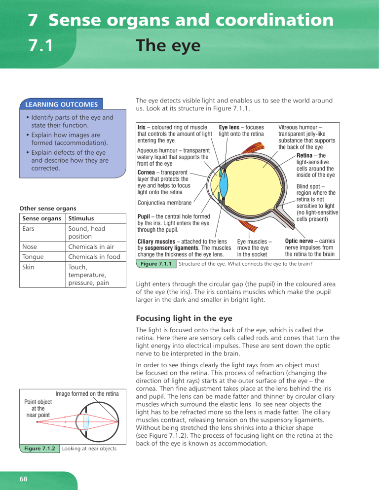
The eye detects visible light and enables us to see the world around
us. Look at its structure in Figure 7.1.1.
,ULV²FRORXUHGULQJRIPXVFOH
WKDWFRQWUROVWKHDPRXQWRIOLJKW
HQWHULQJWKHH\H
(\HOHQV²IRFXVHV
OLJKWRQWRWKHUHWLQD
$TXHRXVKXPRXU²WUDQVSDUHQW
ZDWHU\OLTXLGWKDWVXSSRUWVWKH
IURQWRIWKHH\H
&RUQHD²WUDQVSDUHQW
OD\HUWKDWSURWHFWVWKH
H\HDQGKHOSVWRIRFXV
OLJKWRQWRWKHUHWLQD
&RQMXQFWLYDPHPEUDQH
3XSLO²WKHFHQWUDOKROHIRUPHG
E\WKHLULV/LJKWHQWHUVWKHH\H
WKURXJKWKHSXSLO
(\HPXVFOHV²
&LOLDU\PXVFOHV²DWWDFKHGWRWKHOHQV
E\VXVSHQVRU\OLJDPHQWV7KHPXVFOHV PRYHWKHH\H
LQWKHVRFNHW
FKDQJHWKHWKLFNQHVVRIWKHH\HOHQV
Figure 7.1.1
9LWUHRXVKXPRXU²
WUDQVSDUHQWMHOO\OLNH
VXEVWDQFHWKDWVXSSRUWV
WKHEDFNRIWKHH\H
5HWLQD²WKH
OLJKWVHQVLWLYH
FHOOVDURXQGWKH
LQVLGHRIWKHH\H
%OLQGVSRW²
UHJLRQZKHUHWKH
UHWLQDLVQRW
VHQVLWLYHWROLJKW
QROLJKWVHQVLWLYH
FHOOVSUHVHQW
2SWLFQHUYH²FDUULHV
QHUYHLPSXOVHVIURP
WKHUHWLQDWRWKHEUDLQ
Structure of the eye. What connects the eye to the brain?
Light enters through the circular gap (the pupil) in the coloured area
of the eye (the iris). The iris contains muscles which make the pupil
larger in the dark and smaller in bright light.
Focusing light in the eye
The light is focused onto the back of the eye, which is called the
retina. Here there are sensory cells called rods and cones that turn the
light energy into electrical impulses. These are sent down the optic
nerve to be interpreted in the brain.
,PDJHIRUPHGRQWKHUHWLQD
3RLQWREMHFW
DWWKH
QHDUSRLQW
Figure 7.1.2
68
Looking at near objects
In order to see things clearly the light rays from an object must
be focused on the retina. This process of refraction (changing the
direction of light rays) starts at the outer surface of the eye – the
cornea. Then fine adjustment takes place at the lens behind the iris
and pupil. The lens can be made fatter and thinner by circular ciliary
muscles which surround the elastic lens. To see near objects the
light has to be refracted more so the lens is made fatter. The ciliary
muscles contract, releasing tension on the suspensory ligaments.
Without being stretched the lens shrinks into a thicker shape
(see Figure 7.1.2). The process of focusing light on the retina at the
back of the eye is known as accommodation.
To see distant objects, the ciliary muscles relax, pulling the suspensory
ligaments tighter and stretching the lens into a thinner shape (see
Figure 7.1.3).
,PDJHIRUPHGRQWKHUHWLQD
7RGLVWDQWSRLQWREMHFW
Defects in the eye
Short-sightedness
Some people are short-sighted. They can see near objects clearly, but
cannot focus the light from distant objects. This is because their lens
is refracting light too strongly. That makes the rays of parallel light
focus in front of the retina. To rectify this, they use corrective lenses
in spectacles that bend rays outwards before they enter the eye.
Then, if the lenses are the right strength, the light rays focus the light
precisely on the retina. Concave lenses are used to bend the light
outwards (see Figure 7.1.4). People often wear this type of spectacles
when they drive.
Figure 7.1.3
Looking at distant objects
Figure 7.1.4
How a concave lens in
spectacles helps people
who are short-sighted
Figure 7.1.5
How a convex lens in
spectacles helps people
who are long-sighted
Long-sightedness
Other people can see distant objects, but close-up objects are
blurred. Their lens cannot bend the light enough to focus the rays on
the retina. To rectify this, they wear spectacles or contact lenses with
convex lenses. These help to refract the rays more strongly towards
each other (see Figure 7.1.5). People often wear this type of glasses
for reading.
Astigmatism is a growing problem in Caribbean countries. This is
where the cornea, or lens, is not the correct shape. It can result
in blurred vision and long- or short-sightedness. It can usually be
corrected by using spectacles or contact lenses, but severe cases need
surgery.
Effects of ultraviolet or bright light
Sunlight contains ultraviolet light which can damage the front part
of the eyes, for example the cornea. Very bright light will damage the
retina at the back of the eye. Sunglasses with UV protection should
be worn in sunshine.
KEY POINTS
SUMMARY QUESTIONS
1 Draw a table to show the function of the following parts of an
eye:
a iris
b lens
c ciliary muscles
d retina
e rods and cones
f optic nerve.
2 Draw diagrams to explain how long- and short-sightedness are
rectified.
1 The main structures in the
eye are the cornea, iris,
pupil, lens, suspensory
ligaments, ciliary muscles,
retina and optic nerve.
2 People who are shortsighted need concave lenses
to help them focus light rays
on the retina, whereas longsighted people need convex
lenses.
69
7.2
Light and colour
LEARNING OUTCOMES
• Describe how white light is
separated into its constituent
colours.
• Distinguish between primary
and secondary colours.
• Discuss the effect of mixing
different primary pigments.
• Describe the effects of
lighting on the colour of
objects.
• Separate different coloured
inks by chromatography.
• Distinguish between artificial
and natural lighting, and
between transparent,
translucent and opaque
materials.
TRANSMISSION OF LIGHT
Transparent materials let light
pass straight through them.
Translucent materials let some
light pass through, but it is
scattered on the way through,
producing a blurred image.
Opaque materials do not let
light pass through them.
The visible spectrum
When a thin beam of white light passes through a glass prism the
white light is dispersed into the seven colours of the spectrum. You
have probably seen the visible spectrum in a rainbow or when the
Sun shines on a puddle with a thin film of oil on top of it. Do you
know the colours, in order?
:KLWH
OLJKW
5HG
5D\ER[
9LROHW
3ULVP
6FUHHQ
Figure 7.2.1
White light is dispersed by the glass prism into the colours of the
spectrum
White light is made up of red, orange, yellow, green, blue, indigo
and violet light. By spinning a card wheel with these colours painted
on in segments, the wheel should appear white. The different colours
have different wavelengths and change direction as they pass from
air into the glass. They are diverted in the opposite direction as they
pass out of the glass prism back into air. This changing of direction of
light travelling from one substance into another is called refraction.
Red light, with the longest wavelength is refracted least. Violet, with
the shortest wavelength in the spectrum, is refracted most.
Primary and secondary colours of light
Red, green and blue are known as the primary colours of the
spectrum. If they are combined in the right intensity, they will form
white light.
The coloured dots that make up a TV picture are formed from
combinations of red, green and blue dots. Indeed our eyes have three
types of cone cells to detect red, green and blue light.
5('
Secondary colours are made by mixing two primary colours together.
*5((1
Figure 7.2.2
70
%/8(
The primary and
secondary colours
• Red + green = yellow
• Red + blue = magenta
• Blue + green = cyan
The colour of objects
The colour an object appears depends on which parts of the visible
spectrum it absorbs and which it reflects. We see the reflected
colours. If all the colours in the visible spectrum are reflected, the
object appears white. If they are all absorbed, the object appears
black. When we mix pigments together, the colour is the common
reflected parts of the spectrum.
3DSHUFOLS
The same object can appear different colours using light filters. For
example, an object can be red in normal light, but if a filter is used
to shine blue light on it, it looks black. That is because the object
absorbs the blue end of the spectrum.
Physically separating mixtures of pigments
The technique used to separate mixtures of dyes or pigments is called
chromatography. The mixture is spotted onto some filter paper
(or special absorbent chromatography paper) using a very narrow
glass tube called a capillary tube. The paper is left standing in a little
solvent in a large beaker.
As the solvent runs up the absorbent paper, the different pigments
in the mixture are left behind at different heights. This is because
the pigments have different solubilities in the solvent. They also have
different forces of attraction with the water bonded into the paper.
Those with weak attractions to the water in the paper and high
solubilities in the solvent will be carried furthest up the paper.
Natural and artificial lighting
Lighting is very important in any building. Windows, roof-lights and
glazed doors all let natural light in. In daytime, if correctly placed,
they can let light from the Sun illuminate colours and enable the
people inside the building to perform tasks safely.
In places that are not lit up by natural light, reflective surfaces help
to bounce light around inside the building. At night, we need to
use artificial lighting. The types of light source, their fittings and
positioning are essential to the efficient functioning of a building.
KEY POINTS
1 White light is dispersed into the colours of the spectrum
because the different colours of light are refracted by
different amounts.
2 The primary colours of light (red, green and blue) form white
when they are mixed together. Two primary colours mixed
together form a secondary colour (yellow, magenta or cyan).
3 The colour an object appears depends on the colours it
reflects, as well as the colour of the light shining on it.
4 Coloured pigments can be physically separated by
chromatography.
6ROYHQWIURQW
:DWHU
WKHVROYHQW
Figure 7.2.3
2UD
QJ
H
%OXH
The ink was spotted on
the pencil line drawn
near the bottom of the
chromatography paper.
What colour pigments
made up the orange ink?
SUMMARY QUESTIONS
1 a Name the colours of the
spectrum, starting with
the shortest wavelength.
b Explain how white light is
dispersed into the colours
of the spectrum.
2 What colour is formed when
the following colours of light
are mixed?
a red, green and blue
b red and blue
c blue and yellow.
3 Write a method for an
experiment to see if an
orange felt-tipped pen is
made from a single pigment
or a mixture of pigments.
4 Look at Figure 7.2.3. Explain
how the colours were
separated.
71
7.3
LEARNING OUTCOMES
• Identify parts of the ear and
state their function.
• Explain how noises (loudness/
pitch) affect the ear.
• Describe how hearing loss is
caused by damage to the ear.
The ear
The ear has two important functions – it detects sound and helps us
to maintain our balance. Most of its structure lies within the skull.
The external ear is used to collect sounds and direct them to the inner
ear. Look at Figure 7.3.1.
7KUHHVPDOOERQHV
FDOOHGWKHRVVLFOHV
1HUYHWDNHV
PHVVDJHVWR
\RXUEUDLQ
• Describe how the ear
maintains balance.
(DUFDQDO
2XWHU
HDU
(DUGUXP
7KURDWWXEH
FRQQHFWHGWR
\RXUWKURDW
Figure 7.3.1
&RFKOHD
DVSLUDO
FRQWDLQLQJOLTXLG
DQGQHUYHFHOOV
The structure of the ear. What are the two functions of the ear?
Sound waves
Sounds are made when objects vibrate. As they vibrate, they cause
waves of disturbance in the air molecules. These sound waves spread
out from their source, getting weaker the further they travel. The
loudness of a sound when it reaches the ear is determined by the
strength of the molecular disturbance. This is called the amplitude of
the sound wave. The loudness of a sound is measured in units called
decibels (dB).
The pitch, or frequency, of a sound wave (for example high or low)
is determined by how quickly the source of the sound vibrates.
The faster the vibrations are, the higher the pitch of the sound.
We measure the frequency of a wave in units called hertz (Hz). The
human ear can usually hear sounds with a frequency in the range of
about 20 Hz to 20 000 Hz.
72
How we hear
We hear sounds when the disturbances of the air molecules travel
down the ear canal and arrive at the ear drum. The sound waves
make the ear drum vibrate. These vibrations are passed on to
the three small bones called the ossicles. Their movement causes
vibration of the inner ear drum or oval window, which in turn
disturbs liquid in the cochlea. This is detected by tiny, sensory hairs
in the cochlea. These send an electrical impulse to the brain, which
interprets the signals received into the sounds we hear.
Hearing loss
We can lose our hearing if:
• the ear canal becomes blocked with wax
• the ear drum is damaged by excessively loud noises
• the ossicles fuse together and so stop vibrating properly
• cells in the cochlea are damaged (caused by loud noises at a certain
frequency – this is why ear protectors are worn by workers in noisy
environments and why personal music players should not be turned
up too loud).
Maintaining balance
Figure 7.3.2
Excessively loud sounds can
permanently damage your
hearing
Just above the cochlea we find the semi-circular canals. Look at
Figure 7.3.3. The three canals are filled with liquid that have sensory
hairs, which detect movement in the liquid. They send their electrical
impulses to the brain, which makes the necessary adjustments to
muscles to maintain our balance.
6HPLFLUFXODUFDQDOV
$XGLWRU\QHUYH
,QQHUHDU
7REUDLQ
SUMMARY QUESTIONS
Figure 7.3.3
The semi-circular canals help us maintain our balance. What detects
movement in the liquid in the semi-circular canals?
1 Describe how sounds are
made and travel to the ear.
1 The ear enables us to hear sounds and maintain our balance.
2 Draw a flow diagram to
show the sequence of steps
when we hear a sound.
2 The vibrations of sound waves are passed along the delicate
inner structure of the ear.
3 List the causes of hearing
loss.
3 Care must be taken not to damage the sensitive structures in
your ears.
4 How does the ear help us
maintain our balance?
KEY POINTS
73
7.4
The nervous system
LEARNING OUTCOMES
• Name and describe the
function of neurones.
• Identify the major parts of the
central nervous system and
their functions.
• Explain reflex (involuntary)
actions.
• State the effects when the
central nervous system
malfunctions.
&HOOERG\
'LUHFWLRQRILPSXOVH
&16
6(16(25*$1
)DWW\VKHDWK
Your body has to cope with all sorts of changes in its environment.
These changes act as a stimulus to the body to respond in some way.
The body detects the changes using its sense organs – eyes, ears,
nose, tongue and skin.
Your sense organs contain special cells that detect changes, such as
in light, sound, smells, tastes, heat or pressure. These cells are called
sensory neurones. The neurones are linked in bundles called nerves.
There are three types of neurone, shown in Figure 7.4.1.
Voluntary actions
Here is the sequence of events when you decide how to respond to a
stimulus.
1 A stimulus detected by a sensory neurone triggers off an electrical
impulse that travels along a sensory nerve to the spinal cord.
2 The electrical impulse then travels up the spinal cord to the
brain, which coordinates what happens next.
3 The brain sends an electrical impulse back down the spinal cord
to the correct nerve which is attached from the spinal cord to a
muscle.
4 The electrical impulse leaves the spinal cord via a motor neurone
and travels down a motor nerve to the muscle.
&HOOERG\
5 When the electrical impulse arrives at a muscle, the muscle
responds by contracting.
The spinal cord and the brain make up the central nervous system
(CNS). The sequence of events for a voluntary action is shown in
Figure 7.4.2.
'LUHFWLRQRILPSXOVH
0XVFOHILEUHV
Figure 7.4.1
6HQVRU\QHXURQH
&HOOERG\
The three types of
neurones – sensory (top),
relay (middle), motor
(bottom)
5HOD\QHXURQH
6SLQDOFRUG
0RWRUQHXURQH
Figure 7.4.2
74
The sequence leading to a controlled or voluntary action
Reflex (involuntary) actions
6HQVRU\UHFHSWRULQ VNLQRIILQJHU
6WLPXOXV²
IRUH[DPSOH
WRXFKLQJD
KRWSODWH
%LFHSV
PXVFOH
FRQWUDFWV
DQG
ZLWKGUDZV
KDQG
Figure 7.4.3
6HQVRU\QHXURQH
6\QDSVH
:KLWHPDWWHU
5HOD\QHXURQH
*UH\PDWWHU
0RWRUHQGSODWH
6SLQDO
QHUYH
0RWRUQHXURQH
6SLQDOFRUG
The sequence of events in a reflex action. This is called a reflex arc.
Imagine you touch a plate that has been in a very hot oven. You drop
the plate automatically before you even feel the pain. This involuntary
action is called a reflex action.
You do not need to think about a reflex action. That’s because the
electrical impulse from the sensory neurones to the motor neurones
bypasses the brain. This helps us react more quickly to a stimulus that
might cause us harm. The action is quicker as a result of a shorter
sequence of events than in a voluntary action where we actively
decide what to do. Look at Figure 7.4.3.
The electrical impulse travels between the incoming sensory neurone
and the outgoing motor neurone via a relay neurone in the spinal
cord. Chemicals are released and received to cross the gaps at either
end of the relay neurone so the impulse can bypass the brain.
Other involuntary actions coordinated like this are your breathing,
your heart beating and your knee jerking when it is tapped. Can you
think of a reflex action in a baby?
KEY POINTS
1 Voluntary movements are
controlled by electrical
impulses sent from the brain.
2 Electrical impulses arrive at and
leave from the central nervous
system through neurones,
packaged as nerves.
3 In reflex actions the brain is
bypassed as relay neurones
transfer an electrical impulse
directly between sensory
neurones and motor neurones.
4 Damage to the nervous
system is serious as
conscious and unconscious
coordination is disrupted.
Damage to the nervous system
If electrical impulses cannot travel through the central nervous
system, you can become paralysed. That is why breaking your back
or neck is so potentially dangerous. If the break damages the spinal
cord, electrical impulses sent by the brain cannot get to muscles.
The higher up the spine the break occurs, the worse the paralysis
as more of the muscles can no longer be controlled and actions
coordinated.
Other disorders, such as motor neurone disease, affect the upper
and lower motor neurones. Degeneration of the motor neurones
leads to weakness and wasting of muscles. This causes an
increasing loss of mobility in limbs. Eventually there are difficulties
with speech, swallowing and breathing – all coordinated by the
nervous system.
SUMMARY QUESTIONS
1 Describe the function of:
a sensory neurones
b relay neurones
c motor neurones.
2 Explain the difference
between a voluntary and an
involuntary action.
3 Explain which break in the
spine, severe enough to
damage the spinal cord, is more
serious – a break near the base
of the spine or a break near the
top of the spine.
75
7.5
The endocrine system
LEARNING OUTCOMES
• Identify glands and their
location in the body.
• List hormones produced by
glands and describe their use
and effect.
• Explain how the endocrine
system works.
+\SRWKDODPXV
3LWXLWDU\
JODQG
Endocrine system
How ‘messages’
are sent around
the body
Electrical impulses
that travel along
neurones (which
make up nerves)
Hormones
(chemicals) that
travel through the
bloodstream to
target organs
Timing and effect
• Rapid response
•
• Almost immediate •
• Acts in a precise
•
area
• Effects are short
•
lasting
Slower response
Longer term
Can act over a
larger area
Effects are longer
lasting
The pituitary gland, at the base of the brain, coordinates the effects
of most of the other glands. The nearby hypothalamus is the link
between the nervous system and the endocrine system.
3DQFUHDV
The endocrine glands release (secrete) chemicals called hormones.
They are transported around the body in the blood. The hormone
molecules can be thought of as chemical messengers. Some have
general effects around the body, but others are targeted at particular
organs.
$GUHQDO
JODQG
2YDU\
LQIHPDOH
7HVWLV
LQPDOH
The main endocrine
glands. How would you
describe the position of the
pituitary gland?
LINK
For information regarding the
abuse of hormones in sports,
see 4.6 ‘Drugs’.
76
Nervous system
The endocrine system is made up of glands, located around the body.
Some glands exist as organs themselves or as parts of other organs.
Figure 7.5.1 shows where the main glands are found.
7K\URLG
JODQG
Figure 7.5.1
The endocrine system and the nervous system act together to
coordinate a variety of effects in the body. This table compares the
two systems.
Some hormones are proteins or steroids. For example, growth
hormone is a protein and testosterone is a steroid. Testosterone is
made in the testes. It stimulates the male characteristics to develop in
adolescent boys.
The table below shows some other hormones, the glands that
produce them and their effects.
Hormone
Produced by the
endocrine gland …
Effect
Adrenaline
Pancreas
Gets the body ready for
emergency action, for example
raises the pulse rate and dilates
blood vessels to muscles, lungs
and liver (ensuring muscles
have a good supply of glucose
and oxygen)
Glucagon
Pancreas
Converts glycogen back into
glucose (raises blood sugar
level)
Insulin
Pancreas
Converts glucose into glycogen
which is stored in the liver
(lowers blood sugar level)
Oestrogen
Ovary
Stimulates secondary female
characteristics
Thyroxine
Thyroid
Regulates growth
Figure 7.5.2
Taking your first sky dive will get adrenaline surging around your
body!
SUMMARY QUESTIONS
KEY POINTS
1 a Where is the hormone testosterone produced?
b Testosterone stimulates muscles to develop. How does it
get to the muscles?
c What type of chemical compound is testosterone?
1 Endocrine glands secrete
hormones into the blood.
2 a Where is the hormone adrenaline produced?
b State two effects of adrenaline on the body.
3 Sketch a diagram to show the position of the main endocrine
glands in the body.
2 The hormones act as
‘chemical messengers’,
stimulating responses
around the body.
3 The pituitary gland
coordinates the actions of
most other glands.
77
