Skeletal Muscle Structure: T-Tubules & Sarcoplasmic Reticulum
advertisement
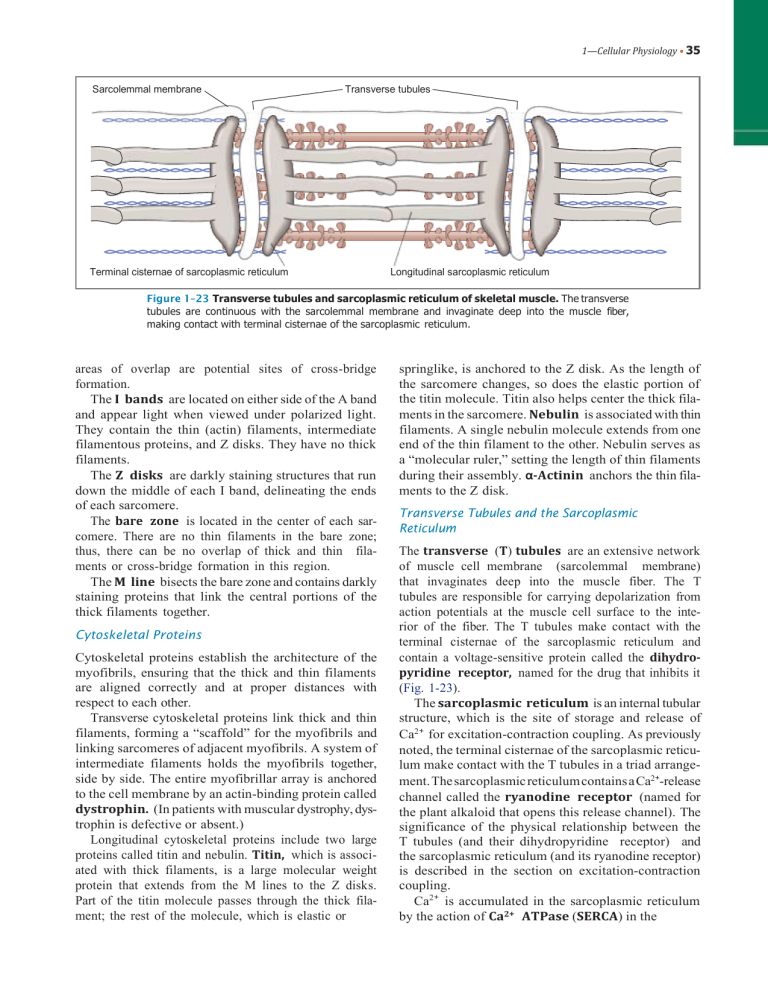
1—Cellular Physiology • 35 Sarcolemmal membrane Transverse tubules Terminal cisternae of sarcoplasmic reticulum Longitudinal sarcoplasmic reticulum Figure 1–23 Transverse tubules and sarcoplasmic reticulum of skeletal muscle. The transverse tubules are continuous with the sarcolemmal membrane and invaginate deep into the muscle fiber, making contact with terminal cisternae of the sarcoplasmic reticulum. areas of overlap are potential sites of cross-bridge formation. The I bands are located on either side of the A band and appear light when viewed under polarized light. They contain the thin (actin) filaments, intermediate filamentous proteins, and Z disks. They have no thick filaments. The Z disks are darkly staining structures that run down the middle of each I band, delineating the ends of each sarcomere. The bare zone is located in the center of each sarcomere. There are no thin filaments in the bare zone; thus, there can be no overlap of thick and thin filaments or cross-bridge formation in this region. The M line bisects the bare zone and contains darkly staining proteins that link the central portions of the thick filaments together. Cytoskeletal Proteins Cytoskeletal proteins establish the architecture of the myofibrils, ensuring that the thick and thin filaments are aligned correctly and at proper distances with respect to each other. Transverse cytoskeletal proteins link thick and thin filaments, forming a “scaffold” for the myofibrils and linking sarcomeres of adjacent myofibrils. A system of intermediate filaments holds the myofibrils together, side by side. The entire myofibrillar array is anchored to the cell membrane by an actin-binding protein called dystrophin. (In patients with muscular dystrophy, dystrophin is defective or absent.) Longitudinal cytoskeletal proteins include two large proteins called titin and nebulin. Titin, which is associated with thick filaments, is a large molecular weight protein that extends from the M lines to the Z disks. Part of the titin molecule passes through the thick filament; the rest of the molecule, which is elastic or springlike, is anchored to the Z disk. As the length of the sarcomere changes, so does the elastic portion of the titin molecule. Titin also helps center the thick filaments in the sarcomere. Nebulin is associated with thin filaments. A single nebulin molecule extends from one end of the thin filament to the other. Nebulin serves as a “molecular ruler,” setting the length of thin filaments during their assembly. α-Actinin anchors the thin filaments to the Z disk. Transverse Tubules and the Sarcoplasmic Reticulum The transverse (T) tubules are an extensive network of muscle cell membrane (sarcolemmal membrane) that invaginates deep into the muscle fiber. The T tubules are responsible for carrying depolarization from action potentials at the muscle cell surface to the interior of the fiber. The T tubules make contact with the terminal cisternae of the sarcoplasmic reticulum and contain a voltage-sensitive protein called the dihydropyridine receptor, named for the drug that inhibits it (Fig. 1-23). The sarcoplasmic reticulum is an internal tubular structure, which is the site of storage and release of Ca2+ for excitation-contraction coupling. As previously noted, the terminal cisternae of the sarcoplasmic reticulum make contact with the T tubules in a triad arrangement. The sarcoplasmic reticulum contains a Ca2+-release channel called the ryanodine receptor (named for the plant alkaloid that opens this release channel). The significance of the physical relationship between the T tubules (and their dihydropyridine receptor) and the sarcoplasmic reticulum (and its ryanodine receptor) is described in the section on excitation-contraction coupling. Ca2+ is accumulated in the sarcoplasmic reticulum by the action of Ca2+ ATPase (SERCA) in the
