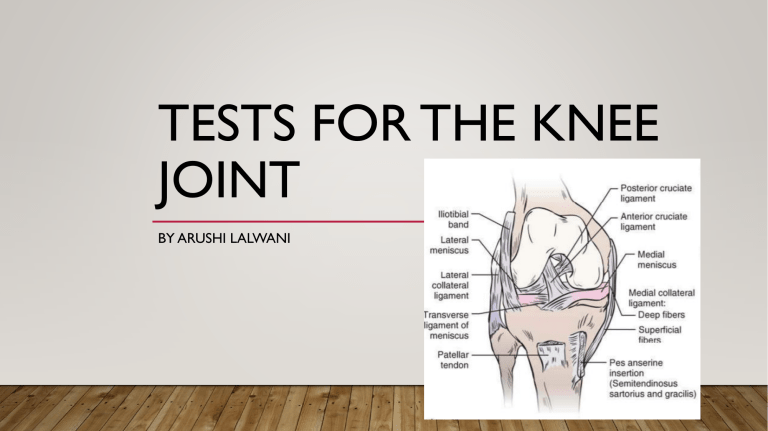
TESTS FOR THE KNEE JOINT BY ARUSHI LALWANI MENISCI • The relative tibiofemoral incongruence is improved by the addition of medial and lateral menisci, which act to convert the convex tibial plateau into concavities for the femoral condyles • These accessory joint structures play an important role in distributing weight-bearing forces, • in reducing friction between the tibia and the femur, • and in serving as shock absorbers. • The arrangement of mensical fibers allows axial loads to be dispersed in a radial direction, thereby reducing wear on the hyaline articular cartilage TEST FOR MENISCUS INJURY MC MURRAY TEST • Purpose- This test is used to determine the presence of meniscal tear within the knee • Patient position – the patient lies supine with the knee completely flexed (heel to the buttock) • Examiner Position – Standing beside the patient’s affected side • Procedure – the examiner medially rotates the tibia and extends the knee. By repeatedly changing the amount of flexion and then applying the medial rotation of the tibia followed by extension, the examiner can test the entire posterior aspect of the meniscus from the posterior horn to the middle segment. To test the medial meniscus, the examiner performs the same procedure with the knee laterally rotated. • Outcome- If there is a loose fragment of the, lateral meniscus, this action causes a snap or click that is often accompanied by pain, and is same for medial meniscus. APLEY’S TEST • Purpose- to evaluate for meniscus injury • Patient position- the patient lies in the prone position with the knee flexed to 90degree • Examiner position- the patient’s thigh is the anchored to the examining table with the examiner’s knee • Procedure- The examiner medially and laterally rotates the tibia, combined first with distraction, while noting any restriction, excessive movement or discomfort. Then the process is repeated using compression instead of distraction. • Outcome- if rotation+distraction is more painful or shows increased rotation relative to the normal side, the lesion is probably ligamentous if rotation+compression is more painful or shows decreased rotation relative to the normal side, the lesion is probably a meniscus injury. TESTS FOR PATELLOFEMORAL DYSFUNCTION CLARKE’S SIGN(PATELLAR GRIND TEST) • Purpose- to detect the presence of patellofemoral joint disorder(patellofemoral pain syndrome, chondromalacia patellae, patellofemoral degeneration) • Patient’s position- supine or long sitting with the involved knee extended • Examiner position- the examiner stands on the side to be tested • Procedure- the examiner presses down slightly proximal to the upper pole or the base of the patella with the web of the hand as the patient lies relaxed with the knee extended. The patient is then asked to contract the quadriceps muscle while the examiner pushes down. • Outcome- if the patient can complete and maintain the contraction without pain, the test is considered negative If the test causes retropatellar pain and the patient cannot hold a contraction, the test is considered positive. MC CONNELL TEST FOR CODROMALACIA PATELLA • Purpose- is done for evaluating the patellofemoral dysfunction • Patient position- the patient is sitting with the femur laterally rotated and legs hanging over the end of the table • Examiner position- sits in front of the client • Procedure(1) - therapist instructs patient to externally rotate the femur of the affected leg while performing active resisted isometric contractions of the quadriceps muscles at 0 30 60 90 and 120 degree of flexion with each contraction held for 10 seconds Therapist notes the painful degrees • Procedure(2)- Therapist passively brings the patient’s knee to full extension, resisting the heel on something so the patient relaxes the quadriceps muscle. Then therapist glides the affected patella medially and holds the patella in that position. Therapist instructs patient to perform isometric contractions at the knee ranges that were painful before. • Outcome- Pain decreases significantly after holding patella medially- patellofemoral lateral tracking problems • Pain decreases significantly after holding patella laterally- patellofemoral medial tracking problem MC CONNELL TEST LIGAMENTOUS TESTS TEST FOR ONE PLANE MEDIAL INSTABILITY MEDIAL COLLATERAL LIGAMENT • The medial collateral ligament can be divided into a superficial portion and a deep portion that are separated by a bursa • The superficial portion of the medial collateral ligament arises proximally - medial femoral epicondyle and travels distally to insert - into the medial aspect of the proximal tibia distal to the pes anserinus • The deep portion of the medial collateral ligament is continuous with the joint capsule, originates - the inferior aspect of the medial femoral condyle, and inserts - on the proximal aspect of the medial tibial plateau. It is affixed to the medial meniscus throughout its course. • The medial collateral ligament, specifically the superficial portion, is the primary restraint to excessive valgus (abduction) and lateral tibial rotation stresses at the knee. • The medial collateral ligament also plays a supportive role in resisting anterior translation of the tibia on the femur in the absence of the primary restraints against anterior tibial translation. VALGUS TEST AKA ABDUCTION STRESS TEST • Purpose- To primarily detect pain and/or laxity of the medial collateral ligament (MCL). • Patient position- Lying supine with the leg relaxed. • Clinician position- Standing on the outside of the affected leg; the patient’s lower leg is lifted and supported between the waist and the inside of the clinician’s elbow with the knee flexed to about 20 – 30 ° and the hip positioned in a degree of internal rotation and abduction. The heel of the outside hand is placed just above the lateral joint line, the inside hand is placed just below the medial joint line where the thumb can palpate the medial tibiofemoral joint line. • Action -Firm inward pressure is applied with the outside hand and outward pressure with the inside hand while rotating the body away from the end of the couch to achieve a valgus stress to the knee. The test can then be repeated with the knee in full extension. • Outcome - Positive test The reproduction of medial knee pain alone is suggestive of injury to the MCL. An intact ligament produces a normal ligamentous end-feel where firm resistance to the valgus stress is noted. Loss of this normal resistance and an increase in valgus movement (in excess of 15 ° ) suggests structural damage to the MCL indicative of a more significant injury involving other structures. In full extension stability to the joint is afforded by the cruciate ligaments and laxity in this position is likely to represent major disruption to the knee and concurrent injury to the posterior capsule, posterior cruciate ligament (PCL) and possibly the anterior cruciate ligament (ACL) should be suspected ( Malanga et al 2003 , Slocum & Larson 1968 ). A positive test in slight knee flexion incriminates the MCL and/or posterior capsule, as this position takes tension off the ACL which provides a secondary restraint to valgus in full extension. When repeated in full extension, a positive test suggests damage not only to the MCL and posterior capsule but also to one or both cruciate ligaments. The degree of MCL stress during this test can therefore be increased by adding external rotation of the tibia. LATERAL COLLATERAL LIGAMENT • The lateral collateral ligament (LCL) is located on the lateral side of the tibiofemoral joint • Attaching proximally - the lateral femoral condyle, the lateral collateral ligament then travels distally - to the fibular head (Fig. 11–16), where it joins with the tendon of the biceps femoris muscle to form the conjoined tendon. • considered to be an extracapsular ligament. • The lateral collateral ligament is primarily responsible for resisting varus stresses, and like the medial collateral ligament, limits frontal plane motion most successfully at full extension. TEST FOR ONE PLANE LATERAL INSTABILITY VARUS TEST AKA HUGHSTON’S VARUS STRESS TEST AND ADDUCTION STRESS TEST • Purpose -To primarily detect pain and/or laxity of the lateral collateral ligament (LCL). • Patient position -Lying supine towards the edge of the couch. • Clinician position- Standing on the affected side, the leg is lifted off the couch and the hip is passively abducted far enough to allow the clinician to stand in the space between the inside of the leg and the side of the couch. The patient’s lower leg is supported between the waist and the outer elbow and the hip is positioned in some degree of internal rotation. The heel of the outside hand is placed on the upper tibia just below the lateral joint line and the inside hand is placed just above the medial joint line on the lower femur. • Action -With the knee in about 20 ° of flexion and the hip internally rotated, firm pressure is applied with both hands to achieve a varus stress while rotating the body in order to increase leverage. • Outcome for positive test- Lateral knee pain or laxity on stress testing. To maximize stress on the LCL, the tibia needs to be positioned in slight flexion and as much external rotation as possible as this reduces stress on the cruciates and leaves the iliotibial band lying centrally over the lateral joint line. The test can be repeated with the knee in full extension and if instability is still detected, major disruption of the knee has occurred and injury to either the cruciates, popliteus or other lateral structures should be suspected along with injury to the LCL POSTERIOR CRUCIATE LIGAMENT • The posterior cruciate ligament attaches distally - to the posterior tibial surface, between the posterior horns of the two menisci (see Fig. 11–9) and travels - superiorly and somewhat anteriorly to attach to the lateral aspect of the medial femoral condyle • The posterior cruciate ligament, like the anterior cruciate ligament, is typically divided into bands that are named for their tibial origins: posteromedial bundle (PMB) and the anterolateral bundle (ALB). • The posterior cruciate ligament serves as the primary restraint to posterior displacement, or posterior shear, of the tibia beneath the femur. • The posterior cruciate ligament restrains approximately 90% of the posterior load directed along the tibia across the majority of the knee flexion range of motion (ROM). TEST FOR ONE PLANE POSTERIOR INSTABILITY POSTERIOR DRAWER TEST • Purpose- To detect posterior (one-plane) instability and posterior cruciate ligament (PCL) laxity. • Patient position -Supine with the hip flexed to 45 ° , the knee flexed to 90 ° and the foot placed on the couch. • Clinician position -The lower leg is stabilized by sitting on the dorsum of the forefoot. Both hands grasp around the upper tibia with thumbs placed anteriorly over the joint line with the thenar area of both hands positioned over the upper tibia. The fingers can also palpate the hamstring tendons posteriorly to ensure they are relaxed. • Action- The tibia is pushed backwards with both hands. The quality of the joint end-feel should be appreciated and a ligamentous ‘ stop ’ noted if the ligament remains intact. • Outcome for Positive test -A positive test is indicated by increased posterior excursion of the tibia and an associated loss of the normal end-feel. An increase in the slope of the infrapatellar tendon may also be noted. In a complete PCL tear, the average extent of posterior excursion of the tibia is 9.2 mm although incomplete tears will produce varied degrees of laxity • With an isolated tear of the PCL, the greatest degree of tibial excursion is noted at 90 ° flexion. Where the injury is located to the posterolateral corner, most excursion is found when the test is performed in 30 ° . In a combined posterolateral corner and PCL injury, 191 increased posterior tibial movement will be noted when the test is repeated at both 30 ° and 90 ° flexion ( Larsen & Toth 2005 ) and injury to other lateral stabilizers will be likely. Posterior sag sign Aka gravity Drawer test • Purpose- to test the posterior cruciate laxity and a useful adjunct to the posterior drawer test • Patient position- same as in the posterior drawer test • procedure- in this position the tibia “drops back”, or sags back, on the femur because of gravity if the posterior cruciate ligament is torn. Posterior tibial displacement is more noticeable when the knee is flexed 90 degree to 110 degree than when the knee is slightly flexed. • Outcome for positive test- normally the anterior tibial plateau extends 1 cm anteriorly beyond the femoral condyle when the knee is flexed to 90 degree. If this “step” is lost, this step-off test or thumb sign is considered positive. ACTIVE DRAWER TEST • Purpose is to confirm PCL insufficiency • Procedure- In the drawer test position with the foot stabilized, the patient is asked to attempt to extend the knee with the examiner holding the hip in 90 to 110 degree of flexion. The backwards sag of the tibia will be reduced with this isometric contraction of the quadriceps (bringing the tibia back into its normal position) and increased with hamstring isometric contraction. This positions the tibia in its fully displaced position and provides a good starting position for maximum anterior translation to occur when the quadriceps contract again. GODFREY’S(GRAVITY) TEST • Purpose – to test posterior cruciate ligament • Patient position- The patient lies supine with the hips and knees flexed to 90 ° and the lower legs supported by the clinician. • outcome- If posterior instability is present, a posterior ‘ sag ’ appears and additional backward pressure will increase this posterior displacement. ANTERIOR CRUCIATE LIGAMENT • The anterior cruciate ligament is attached distally - to the tibia on the lateral and anterior aspect of the medial intercondylar tibial spine (see Fig. 11–9). It extends-superiorly, laterally, and posteriorly to attach to the posteromedial aspect of the lateral femoral condyle • In addition, the anterior cruciate ligament twists inwardly (medially) as it travels proximally • The anterior cruciate ligament may also be considered to consist of two separate bundles that wrap around each other as the knee flexes. • The anteriomedial bundle (AMB) and posterolateral bundle (PLB) are named for their tibial insertions and have slightly different functions. • The anterior cruciate ligament functions as the primary restraint against anterior translation (anterior shear) of the tibia on the femur TEST FOR ONE PLANE ANTERIOR INSTABILITY ANTERIOR DRAWER TEST • Purpose To detect anterior (one-plane) instability and anterior cruciate ligament (ACL) laxity. • Patient position Lying supine with the knee flexed to 90 ° and hip flexed to 45 degree and foot placed on the couch. • Clinician position The patient’s foot is stabilized by sitting on the dorsum of the forefoot. Both hands grasp around the upper tibia with thumbs placed anteriorly over the joint line. Also, in this position the hamstring tendons can easily be palpated to ensure the muscle is completely relaxed and that voluntary or involuntary resistance to the test is avoided. • Action The tibia is drawn forwards with both hands and a comparison of the degree of anterior translation is made with the other knee. The quality of the joint end-feel should be appreciated and a ligamentous ‘ stop ’ noted if the ligament remains intact. • Outcome for positive test-Increased anterior excursion of the tibia accompanied by the loss of normal ligamentous resistance usually indicates significant injury. In a healthy knee, it is normal for approximately 6 mm of anterior tibial translation to be present ( Magee 2008 ). If the ACL is injured in combination with the medial ligament/capsule, a much greater degree of anterior translation will be present (15 mm or more). In the test position of 90 ° knee flexion, the MCL and posterior capsule provide secondary restraints to anterior tibial translation. If these structures are intact and the ACL is injured in isolation, the drawer test will be normal, leading to a false negative result. Other factors that can lead to a false negative result are hamstring spasm, a bucket-handle meniscal tear which blocks anterior translation of the tibia, or a partial ACL tear which has attached to the PCL during the healing process ( Kim & Kim 1995 ). A false positive test can occur (increased anterior translation even when the ACL is intact) if the medial coronary (meniscotibial) ligament has been disrupted ( Magee 2008 ). LACHMAN’S TEST AKA ANTERIOR DRAWER IN EXTENSION TEST, TRILLAT TEST, LACHMAN – TRILLAT TEST ,RITCHIE TEST • Purpose- To detect anterior (one-plane) instability and anterior cruciate ligament (ACL) laxity. • Patient- position Lying supine. • Clinician position -The patient’s foot is stabilized between the clinician’s thigh and the couch. The outside hand is placed over the lateral aspect of the thigh just above the knee joint and the fingers wrapped around the back of the lower thigh while counterpressure is applied anteriorly with the thumb. The inside hand is placed over the medial aspect of the leg just below the knee joint using an identical grip, with the thumb placed over the tibial tuberosity and the knee positioned in about 10 – 30 ° of flexion. • Action -With the outside hand stabilizing the femur, the lower hand firmly pulls the tibia forwards in an attempt to generate anterior translation. The quality of the joint end-feel should be appreciated and a firm ligamentous ‘ stop ’ noted in the normal knee. • Outcome for Positive test -Increased anterior excursion of the tibia on the femur with an accompanying change in the end-feel usually indicates a significant injury. The firm resistance gives way to a softer or even absent endfeel. The normal slope of the infrapatellar tendon also diminishes. MODIFICATIONS OF THE LACHMAN TEST modification s Patient position 1 Lachman test Clinician position Procedure Outcome Sitting with the leg over the Examiner sits facing edge of the table the patient and supports the foot of the test leg on the examiner’s thigh so that the patient’s knee is flexed 30 degree The examiner stabilizes the thigh with one hand and pulls the tibia forward with the other hand Abnormal forward motion is considered positive 2 Stable Lachman test Supine with knee resting on One of the examiner’s knee examiner’s hands stabilizes the femur against the examiner’s thigh, and the other hand applies an anterior stress - - 3 Supine with leg to be - - One of the GRADES OF LACHMAN TEST Grade 1 injury 3 – 6 mm Grade 2 injury 6 – 9mm Grade 3 injury 10 – 16mm Grade 4 injury 16 – 20mm THANK YOU ,
