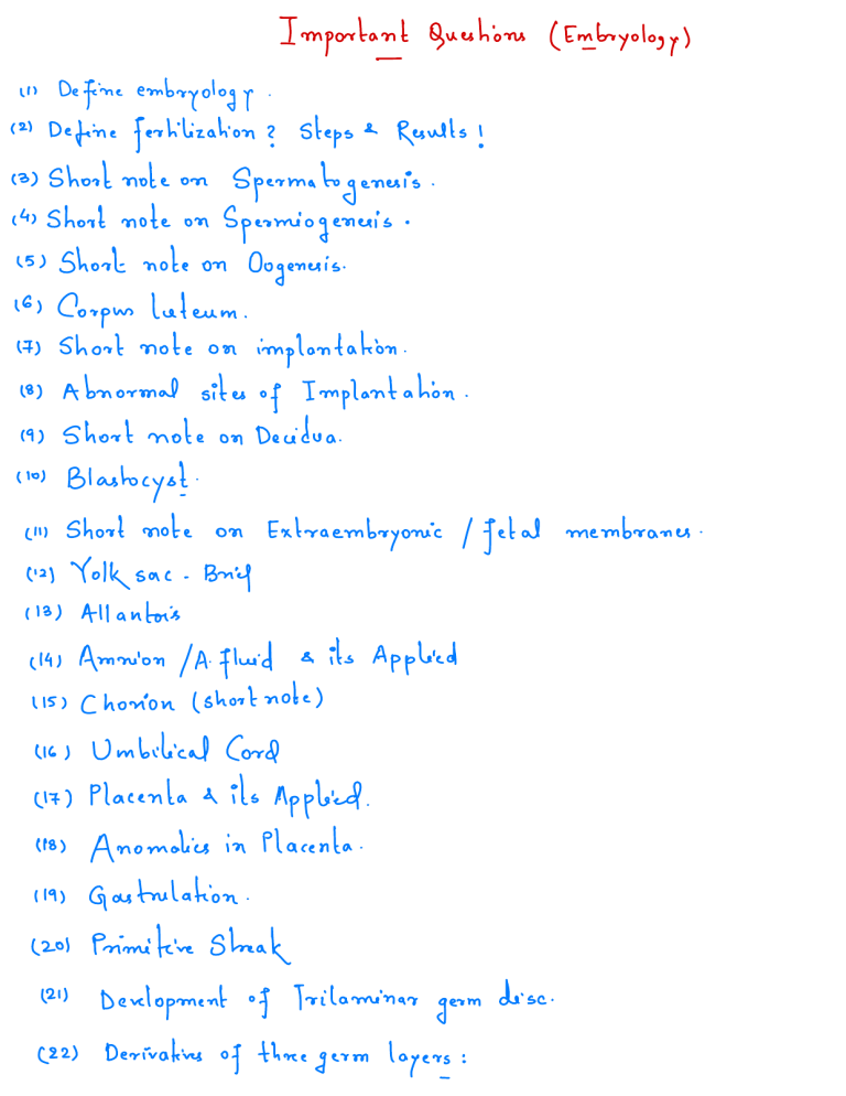
Important Define embryology in Define ( 21 Short note on (4) Short note on (5) 161 A Short note Corpus . fertilization ③ Oogenesis Blastocyst Yolk ( 121 ( 131 sac on Decidua on Extra . . . membranes embryonic / fetal Brief Allantois IA fluid Amnion 1151 Chorion ( short at ) Placenta its Applied Placenta " 8) Anomalies 1191 Gastrulation ( 201 Primitive streak in of . . . Development of Derivatives Applied note ) Umbilical Cord 4 its & . 1161 2) . . ( 141 12 " . ' short note ( in . implantation of Implantation on Short note ( lol Results ! & . Abnormal sites (9) Steps Spermiogenesis luteum short note ? Spermatogenesis on questions ( Embryology ) Tri laminar three germ germ layers : disc . . ( 231 Short note ( 241 Describe somites ( 25) Summary Notochord on . 7 ( as 1st (b) 2nd (c ) full account to g week 3rd cd , Cor) week week / summary of ② Development g 4th week Parturition Development " . Human Development from fertilization . Mednloteslmednotes.int Embryology ' '' ( Imp Que . Define embryology study g developmental Ians ) - ① . Am , ovum into divided Generally • two phones - . - Define 121 fertilization Fertilization Ans ) Steps ? in . to end 9th g 8th week g week till of union g male ° ° • ° • Penetration Penetration Fusion of • of sperm & Formation female prom udei female pronuclei Fusion og of ( a lamination n g just & fertilization ) - chromosomes 2n genetic g clean spies divisions & after zygote rg Imitation male male Determination • membranes meiotic Restoration . pellucida Zona Completion of 2nd formation • of cell Results • radiata Corona g . age birth g baby female gametes & steps ( brief ) ° . Results ! & process a a an : Embryonic Period From fer 't (b) Fetal period from beginning ( as fertilization g baby with with birth g culminating and beginning events sea oocyte of 2nd . oocyte - . Short note (3) Spermatogenesis on spermatogenesis transformed Ans , in into are stage • ° " → Two to spermatocyte 20 speomatogonia . meiotic division 2 Each which spermatocytes first → o ° spermatozoa by : Spermatogonium Each events of process a . Spermatocytes te Spermatocytes ( → n Second (n) ) meiotic division t . Spermatids ( ) spermatozoon by process spermiogenesis n • !;D Each sperm ; (4) Short note Process Ans ) - Spermiogenesis by which spermatids on . Following changes (a) ( bi ( c, (d, take place Nucleus becomes denser and ovoid to form Golgi complex centrioles cytoplasm . - in spermatid into spermatozoa . : more head tfcrosomal Cap → divides into two centrioles shed " transformed are y, one - / ::i¥ . to neck to form s o - I . form annulus (5) Short note Am ' It is by process Stages Oogenesis on which oogonia 2. oocyte ° Oocyte 20 • (n ) → Short note (6) Ans , LH , e 2nd on Corpus shrinks and later , cells g form otoyt ! star " so . luteum oocyte lo I 1 . . → Ovum ( n 2nd Ovum & ) polar body Zpnodla a yellow and . Graffam follicle under the influence g transformed into yellowish glandular . a Corpus luteum and theca internee . by surrounding polyhedral pigment develops thus called Corpus luteum If fertilization in luteal these cells . : occurs # - - - corpus called luteum persists for Corpus Secretes luteum , body . become and cells Fate in empty is vascularized vessels • . : Granada gets Oocyte division meiotic structure called o polar body a . Steps 10 1st Meiotic division to ovulation the After oocyte into Oogonium Oocytes undergo 10 ° transformed are : Oogonium enlarges to form • • . g Progesterone for 3-4 months Pregnancy initial ( under influence . 3-6 months . g Hca ) . If fertilization corpus luteum lasts - It - degenerates and days transformed into only called Corpus luteum - Ami . . og to -14 menstruation implantation Embedding of blastocyst short note Is on 6 - lo . corpus albicans . . . endometrium in Occurs : days after fertilization Stages Zona pellucida disappears Trophoblast adheres to endometrium Trophoblast differentiates into cyto and syncytio trophoblast Syncyhio trophoblast penetrates the . . : ° ° . . . . endometrium . Migration of blastocyst . site of Implantation Endometrium • Applied . into endometrium of posterior wall of uterine cavity near the fundus : If implantation in deep . it may lead to postpartum bleeding . . Abnormal sites 181 Ans The ) sites abnormal Within the uterus ca , Implantation of implantation g Near : . internal are Os : . - ( bi Outside Uterus Uterine cis implantation ) ( tubal Ampulla ( - - : common ) Infundibulum Part ( Interstitial - rare ) ciiinabdmen ( Abdominal implantation) Pouch - ciiis In g pouch ) . ( ovarian Implantation ) Short note (9) ( Recto uterine Douglas Mesentery - on Decidua . of uterus that undergoes decidua after implantation is termed Ans ) The endometrium as Decidua It . cells in bi Nuclei (c) influence of and lipid become Number of . HCG . following changes The . endometrial glycogen ( the endometrium of . : under occurs occur Cai R lez dice dual reaction in glands their rounded cytoplasm swell due cytoplasm to accumulation ( decidua . c organelles increases . cells ) of Functions • • • To To To provide ° Decidua Decidua . parietal 's embryo . remaining part of undergoes development plate component of Decidua capsular's decidua uterine cavity At the time which , of Placenta disintegrates parietals , & . delivery placenta separates and expelled out with . give , decidua , decidua to nose to and fuses . obliterates the . deep to embryo ie embryonic pole : maternal • the , embryonic pole towards the part , It surrounds the Decidua basalis with towards the part , capsular 's form decidua • . : Decidua basalis Fate ° . . cavity • suitable site for implantation provide nutrition for early embryo provide immunologically privileged site for concepts Parts ° : , ie uterine ( 101 Short note And The morula ( 4th The ( bi . . . As the separated space fluid Inner cell mass parts cell inside moral la the in cavity , called the Blastoff blastomeres . layer ) ( Embryo blast ) : Embryo blast Trophoblast Fluid appears level 9 into two Trophoblast ( Outer Fate • day after fertilization ) - cavity (a) . ( consisting fluid filled are Blastocyst of 16 blastomeres ) is transformed blastocyst shortly after it enters into uterine cavity into • on in → → the Embryo proper Embryonic part of blastocyst provides Placenta nutrition ② Meditates ( med notes . in ) . to blastomeres . Important ④ Short note Ans clues I Ans 2 - embryonic / fetal membranes All structures derived from zygote and yet not forming called Extra any part of embryo embryonic / fetal membranes : Extra on . are These ° are : ⑨ A (b) Chorion ( di fetal forms - Yotka Allantois Amniotic forms - - forms umblical Placenta if - Short note Anr : Yolk to - The car cbs a sac on . gases passages between mother Yolk embryonic disc functions of Providing sac allantois . median e for a blood fetus e to waste . to pass & . . & thus lies ventral . Yolk sac nutrition are : developing to liver Hemopoiesis until formation . nutrition , develops from blastocyst cavity is of embryo ( early stage ) formed of primordial germ Stages og Development in Placenta . . Provides - pro ④ ligament exchange Provides , og a - cel part fluid with primitive gut tube and bladder pen of urinary forms - filled sac cells . : Primayyoksac At the end of 2nd week the cavity of blastocyst becomes lined by Heuser 's membrane and becomes primary yolk - , sac . Heusen 's membrane = . Flattened cells endoderm of the derived from the embryonic disc . . Secondary Yolk (2) : sac With the - extra appearance of coelom the primary yolk . much smaller becomes flattened . (3) This Dorsal ° becomes of part Yolk 20 of his . . Yolk sac secondary part in . yolk sac " It embryo while midgut by communicate with duct Lilac cis and : vitello intestinal Thus sac incorporated ventral the sac cells become cuboidal Tertia Rem an ant o 20 is embryonic is . divided into 3 past : part Intermediate connecting part called Vitello intestinal duct Extra embryonic part called Tertiary Yolk Intra embryonic . , . His Fate ° sac : Intra embryonic and ° • . . past hindgut ) gives rise atlantic & to get diverticulum Intermediate past / Vitello intestinal duct Extra embryonic Vitelline vessels part = Atrophies supplying yolk sac ( foregut tube & = midgut , . Atrophies Disappears Celiac , sup . e detached . & Inf Mesenteric vessels . Applied If Vitello intestinal ° diverticulum ⑤ Shoot note Ans Allantois : end It ° in sac cloaca , Fate ° - The part the caudal . and passes is to dilated hindgut g allantois from . the ventral side og cloaca into the . by vascular's ed allantois vessels . : developing Allantois fibrous - from . connecting stalls The arises . becomes connected terminal which . connecting stalk folding of embryo the Meckel 's as . diverticulum a persists it . into grows it ° or , Allantois on of yolk After . sinus , fails to atrophy fistula urinary atrophies band Atlantic vessel - the bladder and is in seen continuous in arks ( median . becomes umbilical with the postnatal life umbilical vessels & allantois . as ligament ) gets attached to Placenta Congenital If • Anomalies allantois fails ④ Short note Am - It is sac ° a thin enlarges , , the • Formed by . Volume - Int ° Composition e embryonic : Urach al . . its Applied . membrane that Amniotic fluid called fistula sina.ysq.ir . embryonic embryo future forms Amniotic umbilical disc but as it cord and fetal vessels . g & . 10=30 me ele 20 et 37 - & . as fluid from maternal secreted by fetus Urine e persist : Filtration W it IA fluid the placenta lol eek , lies dorsal to the envelops fluid . • sac g se extra fluid with fetal part Amniotic tough , it fibro Amnion on filled Initially to : 350 ml = ca ' = 800 - 1000 ml Metabolites (b) Hormones ( Hey co Desquamated d) Fetal Urine & cells . HPD a fetal epithelium . Functions ° - ca , Protects (b) Permits embryo by acting symmetrical ext . as water cushion growth g Regulates body temp Forms hydrostatic bag I help in Allows during free movements 9 fetus cel embryo . . . id , ⇐ ' dilation n Cervix birth ) . Applied Amnion ° : in being ④ Short note Am : chorion in • It layer Fate ° It g gives Applied an extra embryonic membrane that and vagina . . plays a key role in envelops development the g Placenta : outer an somatopleunic to rise Villi extra layer - trophoblast and embryonic mesoderm , an inner . numerous called . : Chorionic villi 8 restructuring : chorionic week graft for a Chorion fingers like projections • As Corneas g on formed by is Repair . developing embryo Formation cis in did Amniocentesis ° used . biopsy - It's done to detect genetic disorders at . ⑥ Umbilical It Am : in Cord long a cord with which extends Formation It covered in Contents One . by umbilical Functions It ° It • It ° provides provides ! the , Cord Fetal placenta I -2cm of . connecting stalk elongates Amnion to form . . Vein g Vitello intestinal duct & Atlantic diverticulum . : passage transfer deoxygenated blood of fetus to placenta to transfer oxygenated blood from Placenta to fetus to passage . suspends fetus into Amniotic cavity Applied diameter felly Remnants • to a : Wharton 's • fetus g and . Two umbilical arteries • 50cm : umbilical cord ° length of a umbilicus from folding of embryo After . : : . stem cells Prolapse ( due to . ( cryo preserved ) companion between strangulation during delivery . fetal head & Pelvic want! ? he. , ④ Placenta Yasuda Highly Am : brief in connected to functions Exchange his transport g ciiii Transport a T to - mother ( uterus ) . by fetus intimately is . tropic Nutrients Mother to Fetus from lnlaste Products hormones hormone , , from fetus such HCG as and Retain . to mother . progesterone estrogen , , ons . Prevention of Harmful microorganisms into fetal blood storage og Glycogen Calcium and iron ( vs which gases synthesis of soma . : : in ( in . Applied disc like structure & , drugs and hormones to enter . His civil Transmission of antibodies Formation ° . , . From two Fetal in from in mother to month early fetus g Pregnancy . : sources - chorionic fetal from dos & um Maternal 4 , . Maternal in Decidua basalis . . Placenta Full term - Shape • htt • Disc - 500 = B - Thickness • shaped 600 - Diameter ° - - Maternal . Applied o : - gm 20cm 3cm Surface ④ Anomalies Ale to : a Placenta shape , - Placenta in success by Thin - Ale to Attachment g Umb tical , - is iii , Marginal Placenta Furcate Placenta ciiis Velamen tous - - Cord & blood not Conf in in vowels Elise in connected e membrane slapped Placental . : attached to reaching - . disc Margin g Umbilical vessels get divided the placenta Placenta in Overlapped by Decidua & Holes - Placenta y Small Placenta - placenta by Placenta fenestra ta µ, margin Sulcus tuna ta Diffuse Placenta in . . Peripheral = Main Placenta Consists two lobes lobes 2 > surrounded ( . : = Circumvallate iiis mole size His Multi lo bulan ( Vesicular or Bilobed ( bi discoidal in . : Hydatid form mole Am cotyledons lobes / 15-20 = Cord fails Placenta before . to reach placenta and . . to . gets attached to [ Amnion Due to at periphery . shape ] - f- Due to sites a Attachment ⑨ Meditates • g Umbilical Cord ( www.mednotes.in ) Download the App from Play store . ] ④ Gastrulation Ans : It in the process of all beginning of precursors It ° is Most . : important of formation the the event three g germ tissues embryonic morphogenesis to develop body during 3rd week of Pregnancy now ° which are . Embryo in referred to gastrula During gastrulation bi laminar embryonic tri laminar embryonic disc • layers form . . . , disc in converted into . First sign of . ④ Short note Ans : cis some It is gastrulation begins Primitive streak on important features thickened linear iis , Primitive band Epiblast that g aspect of embryonic disc Formed of proliferation and movements of median plane of embryonic disc It as cells plane week on dorsal . result a to elongates by . appears median 3rd streak . caudally in the at beginning epiblast ciii , formation : a a ( with the . the addition of cells to its caudal end . whereas proliferates cranial end its called Primitive node in A narrow depression small Primitive an of epiblast: Significance To • Primitive and cells rounded elevation a continuous with in groove Primitive called node primitive in groove form . called groove to primitive pit pit a . formed by invagination are . : cranio caudal determine of axis embryo To ° surfaces of embryo To ° determine Fate Forms • and ventral determine dorsal right . & halves left og embryo . : extra embryonic until disc embryonic mesoderm Applied Sacro coccygeal end the by ingression of 3rd y week g its cells Intrauterine into life . : I ' ' ④ teratoma Development of Ans : The cells of Tri laminar embryonic above the other • • From superficial This in The called of process . disc germ disc . differentiate into is The cells placed layers one . to deep , Tri laminar formation of these germ these are disc Ectoderm Mesoderm of embryo blast first & . Endoderm . layers cis as - ( 3 differentiate follows : into two - layers - . (as ( bi ciis superficial layer of Columnar deep layer of flattened cells cells The The cells Some the to sin The called Epiblast Hypoblast called . of epiblast migrate toward the future primitive streak ( flask shaped ) detach themselves from epiblast and slip . - underneath it ciiis cells of . these cells replace endoderm and others form Mesoderm Remaining cells the hypoblast lie above the form the newly formed endoderm cells . g Epiblast form Ectoderm . to ④ Am Derivatives : Chart = of three germ layers : ④ Short note Am It : extends cells The • solid rod is It ° Notochord on situated cells g : from primitive knot of notochord midline in to the primitive streak . . from primitive derived are disc of embryonic chordal plate pro knot the of . formation The cells • . depression called The cells from Cells Blastopore pro which at the is ° . . lat all a floor of a plate within the Canal cavity form yolk and sac to to form its lumen . form • • • supports Acts as the a Induces the axis embryo vertebral surface of embryo column ectoderm canal Thus again . notochord , the neural . . og cells which extends . form plate solid rod of embryo to a notochord a : Forms the central . . . ° Process . curved fill the in break and canal flattened in to blastopore . Functions a form notochord rat to formed again This in called definitive from primitive knot to prochondral plate in form Notochord al called amniotic the becomes proliferate tube to blastopore migrate forward notochord al canal plate Noto chordal Cells centric inward more canalized Neuro centric called neuro chordal continuous communication between This in of gets process and . the bottom toward the Noto chordal canal ° primitive knot proliferate of midline . : tube . Fate of ° It Notochord adult In the ° Applied It - in Region total of . : discs . Brief in are : - - - - - . . column of of Paraxial mesoderm extends from the notochord to form bilaterally cubical . block of mesoderm : somites are cranio caudal - cells . somites in arise . 42-44 g of gestation These follows as life in segmentation somites Formation A later . may or to caudal end undergoes called • Region longitudinal cranial It dens g of mitochondrial ④ Describe somites ° are in Intervertebral of tumor that a Cranial in sacral Thick remains pulposus remains Common Am : disappears : Chord oma - it , Apical ligament - from life Nucleus - • structure and embryonic in : 4 occipital 8 cervical direction thoracic 12 5 Lumbar 5 Sacral 8 formed during - lo coccygeal . . roth to sooth day somites Fate of . Each somite cavity ca ) ( bi (c) in in triangular center the Selene to : - me It . in og back y skin - - trunk & limbs . : Age determination determined somites . - Age g fetus by counting with subdivided into Myotome forms Mendes Dermatome forms dermis Applied section forms Axial skeleton g ° in can number be g , 3 vertebrae 9 slit small a front parts . g - like - Ribs & sternum trunk & limb muscles . on back & front ④ Development kteekwise Weeks • Beginning : Fertilization female fertilization At ° Prior to • ( as tract form It plasma over Hydrolytic to Results During first & - changes 2 meiosis I female in : the occurs • zygote a when male secondary oocyte rapidly completes Capa citation consists of sperm ° to , from cbs the uterine tube ampulla of fuse . . ferlization spermatozoa undergo genital • nuclei pro Development 7 in occurs : n membrane f hrs enzymes zona days of rep tract . several proteins of spermatozoa acne some used zygote , by . that prevents polys penny t . . from pellucida week of aero some released cortical reaction 4-5 of female in are penetrate in the removal → blastula → . Morula ( 32 - Blastocyst forms • Blastocyst • mass ° By as consists ( trophoblast ) end of week fluid develops inner cell , in morella ( mass which becomes trophoblast y - I . - . And Implantation begins . . embryo blast ) and outer placenta celled) . Glo trophoblast . syncytio trophoblast . cell Implantation : → Blastocyst usually implants . ° • posterior wall of uterus Embryonic pole of blastocyst implants first endometrium og during progestational Implants within functional layer menstrual within . . phase g cycle . lnteekz ° Embryo formation : Haft i . Bi laminar differentiates embryonic disk laminar form to yolk sac Prechondral Plate , Embryo Epiblast into Epiblast forms Amniotic cavity to • T and . and hypoblast forming , . hypoblast cells migrate . formed by fusion in site g og future Epi Hypoblast & mouth . cells , ° ° • Extra embryonic mesoderm ° embryonic The syncylio trophoblast make contact continues thymus ( upto ( 6 6 weeks ) week to occurs and in epiblast Amnion covers its the Yolk growth endometrial blood with Hematopoiesis initially sac from covers Visceral mesoderm Extra Yolk derived somatic mesoderm Extra embryonic to is . . sac . into endometrium vessels glands & Mesoderm later in 3rd trimester ) and surrounding fetal liver spleen , then bone marrow . . Intel 3 to s : Embryonic Period ( week . 3 summary ) • ° . Klee k corresponds 3 During this time process (a) ° ° ° systems begin All major organ It by Ectoderm Mesoderm with first 3 forms forms neuro also g I les take place Endoderm ectoderm & . period this are in & neural . . . the . . crest cells ( 35 pair lateral mesoderm 3- s produced Primitive streak within Paraxial mesoderm intermediate mesoderm week menstrual primary germ layers lb , Mesoderm formation develop during missed gastrulation which the Ectoderm begins to to M Epiblast . somites ) , . ⑨ Med Notes Disown load the ( med notes Med Notes app in ) from Play stone !
