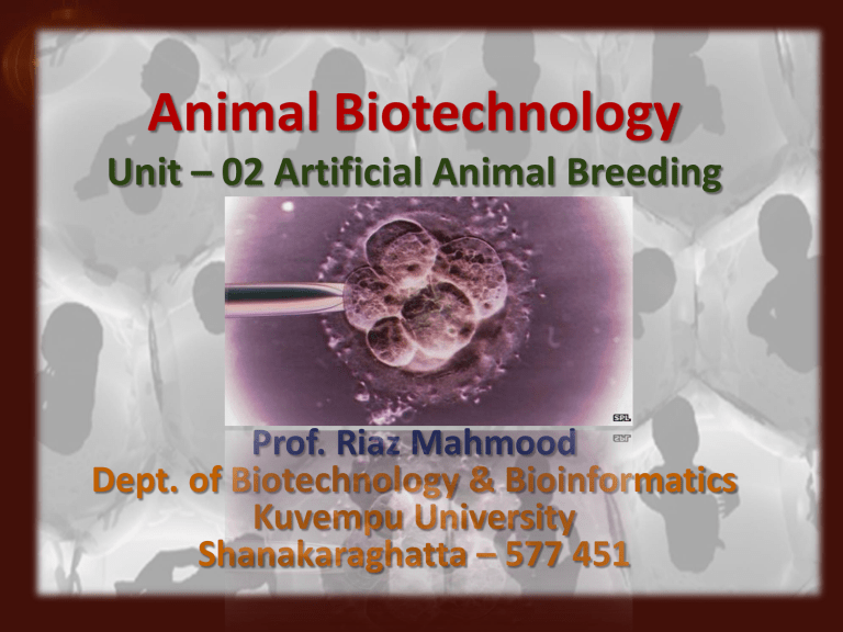
Animal Biotechnology Unit – 02 Artificial Animal Breeding Prof. Riaz Mahmood Dept. of Biotechnology & Bioinformatics Kuvempu University Shanakaraghatta – 577 451 Male Reproductive Organs Spermatogenesis. Germ cells near the outer wall of the seminiferous tubules of the testis differentiate into stem cells called spermatogonia. Spermatogonia divide by mitosis, and mature into primary spermatocytes. Each primary spermatocyte undergoes meiosis: •Meiosis I yields 2 haploid secondary spermatocytes •Meiosis II yields 4 equal-sized spermatids. The spermatids migrate toward the lumen (central opening). Sertoli cells supply nutrients for the spermatids, which mature into motile sperm in the epididymis. The ovum or the female gamete is much larger than the sperm in size. It is non - motile and laden with different types of energy rich materials like yolk, glycogen and proteins accumulated in its cytoplasm. It is enclosed by one on more egg envelops. Size of ovum varies in different animals and depends upon the amount of yolk. On the basis of amount of yolk, the ovum can be - Microlecithal (small sized egg with very small amount of yolk) - Mesolecithal (egg containing moderate amount of yolk) and - Macrolecithal (containing very large amounts of yolk). Human ovum is microlecithal. The cytoplasm is differentiated into outer transparent exoplasm or egg cortex and inner opaque endoplasm or ooplasm. Nucleus is excentric in position, so human ovum has polarity. The side of the ovum with nucleus and polar body is called animal pole and the opposite side is called vegetal pole. The line passing through the 2 poles is called primary axis or animal vegetal axis. The human ovum is surrounded by a number of egg envelopes. Vitelline membrane - A thin, inner transparent layer secreted by the ovum itself. Zona pellucida - A thick middle layer which is transparent and non - cellular. Corona radiata - An outer, thicker coat formed of radially elongated follicular cells. Between the vitelline membrane and zona pellucida, there is a narrow space called Perivitelline space. Oogenesis Fertilization occurs in the fallopian tube within 24 to 48 hours of ovulation. The initial stages of development, from fertilized ovum (zygote) to a solid mass of cells (morula), occur as the embryo passes through the fallopian tube encased within a nonadhesive protective shell (the zona pellucida). The morula enters the uterine cavity approximately two to three days after fertilization. The appearance of a fluid-filled inner cavity marks the transition from morula to blastocyst and is accompanied by cellular differentiation: the surface cells become the trophoblast (and give rise to extraembryonic structures, including the placenta) and the inner cell mass gives rise to the embryo. Within 72 hours of entering the uterine cavity, the embryo hatches from the zona, thereby exposing its outer covering of trophectoderm. The normal process by which a less specialized cell develops or matures to possess a more distinct form and function. Cell differentiation is a process in which a generic cell develops into a specific type of cell in response to specific triggers from the body or the cell itself. This is the process which allows a single celled zygote to develop into a multicellular adult organism which can contain hundreds of different types of cells. In addition to being critical to embryonic development, cell differentiation also plays a role in the function of many organisms, especially complex mammals, throughout their lives. [ Potency specifies the differentiation potential (the potential to differentiate into different cell types) of the stem cell. Totipotent: Stem cells can differentiate into embryonic and extraembryonic cell types. Such cells can construct a complete, viable, organism. These cells are produced from the fusion of an egg and sperm cell. Cells produced by the first few divisions of the fertilized egg are also totipotent. Pluiripotent: Stem cells are the descendants of totipotent cells and can differentiate into nearly all cells, i.e. cells derived from any of the three germ layers. Multipotent: Stem cells can differentiate into a number of cells, but only those of a closely related family of cells. Oligopotent: stem cells can differentiate into only a few cells, such as lymphoid or myeloid stem cells. Unipotent: Cells can produce only one cell type, their own, but have the property of self-renewal which distinguishes them from non-stem cells (e.g. muscle stem cells). Female Reproductive Tract Animal Reproduction What is Assisted Reproduction? Any directed action taken by humans to enhance reproduction in animals or “Manipulation of Reproduction” In humans: “The use of medical techniques, such as drug therapy, artificial insemination, or in vitro fertilization, to enhance fertility.” Assisted Reproduction Techniques (ART) 1) Artificial Insemination (AI) 2) Embryo Transfer (ET) 3) In vitro Fertilization (IVF) 4) Semen/embryo sexing 5) Gamete/embryo micromanipulation 6) Somatic cell nuclear transfer (SCNT) 7) Genome resource banking Five Steps Necessary for Adoption of an AR Technique 1) Technique development in a domestic animal counterpart, if available 2) Characterization of species-specific reproductive biology in a targeted non-domestic animal 3) Assessment of technique feasibility for producing offspring 4) Demonstration of adequate efficiency for applied usage 5) Application of new tool for population management First animal Biotechnology technique, has been used to obtain offspring from genetically superior males for more than 200 years. Improvements in methods to cryopreserve (freeze at -1860C in liquid nitrogen) and store semen have made AI accessible to more livestock producers. Semen from bulls is especially amenable to freezing and longterm storage. In the dairy industry, where large numbers of dairy cows are managed intensely, AI is simple, economical, and successful. However, it is more difficult to freeze and store semen from other livestock species, including horses, pigs, and poultry, than it is to freeze cattle semen. Inhibits all biological activity, esp. that which leads to cell death or deterioration Use vitrification agent to prevent crystalline formation via the conversion of cellular material to an amorphous glass-like solid Artificial Insemination Standard collection methods, • Artificial vagina • Vaginal condoms • Electro-ejaculation (under anesthesia) These can be difficult for: • Non-domestic equids, • Some great apes, • Canids • Marsupials Artificial Insemination • Wild female trapped and fertilized with sperm from zoo male, released, or vice versa. • Major application is to avoid genetic depression in fragmented populations Embryo Transfer (ET) Most important aspect is to pave the way for interspecies transfer Little is known about embryo development or feto-maternal recognition in most species Development of embryo transfer technology allows producers to obtain multiple progeny from genetically superior females. Depending on the species, fertilized embryos can be recovered from females (also called embryo donors) of superior genetic merit by surgical or non-surgical techniques. The genetically superior embryos are then transferred to females (also called embryo recipients) of lesser genetic merit. In cattle and horses, efficient techniques recover fertilized embryos without surgery, but only one or sometimes two embryos are produced during each normal reproductive cycle. In swine and sheep, embryos must be recovered by surgical techniques. To increase the number of embryos that can be recovered from genetically superior females, the embryo donor is treated with a hormone regimen to induce multiple ovulations, or superovulation. In normal course of events only one overy id so triggered by circulating hormones, but by increasing hormone dosage at the appropriate time a number of follicles can be induced to ‘ripen’ and ovulate. Donor females are frequently injected with Prostaglandin F2a (PGF2a) to induce a synchronized oestrus before treatment is started. Strating 10 days after oestrus (during luteal phase) they are injected with FSH over 4 days followed by PGF2a to induced oestrus. Osetrus should occur within 2 days and superovulated females are then mated within 24 hrs. Since the purpose of MOET is to increase the number of genetically superior progeny, the donor is usually mated by artificial insemination. The fertilized embryos are recovered after 6-8 days after insemination for cattle & 6days for sheep or goats. Gamete/Embryo Micromanipulation In vitro Fertilization (IVF) Intra-cytoplasmic sperm injection (ICSI) Sub-zonal insemination (SUZI) • Sperm placed between Zona Pelucida and vitelline membrane, can lead to polyspermy Pre-implantation genetic Diagnosis An alternative to collecting embryos from donor animals. Method to produce embryos in vitro (in the laboratory). Also called in vitro embryo production or IVP. Immature oocytes (female eggs) can be obtained from ovaries of infertile or aged females, or from regular embryo donors. Egg is picked up by a non-surgical technique that uses ultrasound and a guided needle to aspirate immature oocytes from the ovaries. The immature oocytes have been removed from the ovary, they are matured, fertilized, and cultured in vitro for up to seven days until they develop to a stage that is suitable for transfer or freezing. IVF if quite successful resulting in about 70-80% of fertilized eggs. Oocyte recovery: In the second follicular phase of the oestrus cycle, a no. of ovarian follicles (~20) grow and become filled with fluid. The fluid filled space is called the antrum and such follicles are Antral follicles (Graffian follicles) Normally only follicle ruptures releasing the egg for fertilization. Superovulation occurs when more antral follicles are stimulated to mature by injection of Gonadotropins. Pre-ovulatory follicles lies against the surface of the ovary and are quite large (about 15 mm in cattle, 8mm in sheep & Pigs). Laproscopic surgery can be used to recover oocyte from these follicles allowing them to be matured and fertilized in vitro. A number of eggs procured this way may be little costlier but more viable. In vitro Fertilization The key to widespread adoption of IVF is to bring large number of oocytes to maturation in vitro. Semen sample can be obtained from the male partner on the day of oocyte retrieval. Semen ejaculation will be done 2-5 hrs prior & will be prepared for inseminating the collected embryos. Testicular biopsy can also be done as method to extract/isolate the sperms. The follicular fluid is drawn & immediately processed for the identification of eggs, evaluation & preparation for insemination. The eggs are taken in small droplets of culture medium, each may contain 10 oocytes. Medium should be supplemented with Penicillamine or hypotaurin to facilitate the penetration of sperms in oocytes. One dose of sperm contains one million sperms per ml of medium or 100,000 – 800,000 motile sperms are needed for successful fertilization. After 24 hrs it will be evaluated for the evidence of fertilization. If no eggs are found to fertilized another technique ‘Intra-cytoplasmic sperm injection’ (ICSI) will be done. Fertilized eggs are then placed inside the uterus of Biological mother or Surrogate mother. Or the eggs can also be stored through cryopreservation. Implantation of embryos in blastocyst are suitable which can be done using a fine tube (Catheter) which is passed through the cervix. They are placed in the top part of the uterus. Typically 24 embryos can be transferred in one treatment cycle. There are complex but largely unknown factors that limit pregnancy rates. Some of the known reasons could be; Failure in egg recovery, because - Matured follicle may nor develop in treatment cycle. - Ovulation has occurred at the time of egg recovery. - Technical difficulties may prevent egg recovery. Egg may be abnormal. Insufficient semen for fertilization. Failure in fertilization even gametes are normal. Embryos may not develop sometimes. Embryo transfer into uterus may be failure. First successful Human IVF was performed in Britain. Louise Joy Brown (1978) is world’s first test tube baby In vitro Fertilization in wild animals Many attempts, few successes Requires oocyte collection, knowledge of ovarian cycles. Can use ovaries from dying animals. Pumas, tigers, cheetahs, Indian desert cats, gaur, elephants, gorillas, zebras, marmosets, minke whales, ocelots, springboks, many others Live births in only tigers, Indian desert cats, gorillas, and European mouflon Intra Cytoplasmic Sperm Injection (ICSI): It is a superior form of IVF where the sperm is injected into the egg by a micromanipulator syringe. Fertilization is outside the body using specifically selected sperm and good quality eggs. ICSI is a type of ART procedure specifically meant for poor sperm quality. This procedure is as like an IVF where a selective single sperm will be injected directly into an egg. Generally, multiple micromanipulation devices that includes microinjectors, micropipette and micromanipulator can be used to do this microscope under microscope . The mature oocyte will be hold with the help of holding pipette that hold oocyte with a gentle suction from the microinjector. On the other side, a hollow, thin glass micropipette with the sperm collected and the sperm is immobilised by cutting the tail of the sperm with the help of micropipette. Piercing of the oocyte through oolemma and then directed into the inner part of oocyte, i.e., cytoplasm, after which the sperm is released into oocyte. Intra Cytoplasmic Sperm Injection GIFT (gamete intrafallopian transfer) and ZIFT (zygote intrafallopian transfer) are modified versions of in vitro fertilization (IVF). Like IVF, these procedures involve retrieving an egg from the women, combining with sperm in a lab then transferring back to their body, but in GIFT and ZIFT the process goes more quickly. While in traditional IVF the embryos are observed and raised in a laboratory for 3 to 5 days. GIFT should be performed only when sperm level is adequate and at least one fallopian tube is open and functional. GIFT allows fertilization to take place inside the woman’s body. IN GIFT eggs are removed from the woman and placed in one of the fallopian tubes along with the sperms. In GIFT, the sperm and eggs are just mixed together before being inserted and, with luck, one of the eggs will become fertilized inside the fallopian tubes. In GIFT, fertilization actually takes place in the body rather than in a petri dish. In ZIFT, the fertilized eggs -- at this stage called zygotes -- are placed in the fallopian tubes within 24 hours. In ZIFT, the eggs are placed in the fallopian tubes rather than directly in the uterus. Zygote Intra-Fallopian Transfer (ZIFT) For some women who haven't been able to get pregnant with normal in vitro fertilization, GIFT or ZIFT may be a good idea. The processes used in GIFT and ZIFT are closer to natural conception. Since GIFT and ZIFT both require a surgical procedure that IVF does not, IVF is almost always the preferred choice in clinics. In vitro fertilization accounts for at least 98% of all assisted reproductive technology procedures performed in the U.S., while GIFT and ZIFT make up less than 2%. What is PGD ? The procedure which involves the removal of one or more blastomeres to test for mutations in a specific gene sequence or chromosomal abnormalities before transferring. Biopsy of a single cell per embryo, followed by its genetic diagnosis through different techniques (FISH, PCR, aCGH, NGS) and the subsequent transfer of those embryos classifies as genetically normal. PGD is conducted with an intention to improve the chances of a “normal” pregnancy. Handyside, Winston & Hughs (1990) used PGD for sexing embryos for preventing X linked disorder; - Used PCR for Y chromosome and transferred XX embryos. Carriers of Mutations (Autosomal dominant disorders, Autosomal recessive disorders) Female carriers of X-linked disorders HLA matching Carriers of Balanced chromosomal translocation, inversion, or other structural rearrangements Methods of Genetic Analysis : 1.PCR based techniques 2.Hybridisation based techniques; - PCR; - Array CGH 3. DNA Sequencing Techniques - NGS PGD is a procedure used along with In vitro Fertilization (IVF) in which genetic analysis is done of a single cell from an eight-cell embryo. PGD combines advances in Molecular Genetics and in ART and is conducted before the embryo is placed in the womb of the woman. Most common extra chromosomes among live births are 21, 18 & 13. Aneuploidy outcome- termination of developing fetus. Aneuploidy is most frequent cause of spontaneous abortions. WHY CONSIDER PGD? Couples with Maternal age older than 38 Family history of inherited disease. One child already affected with a genetic disease to avoid ‘Recurrent miscarriages. Procedure: 1. Monitor egg maturation in the ovary using Ultrasound & Hormone levels. 2. Collect eggs from the female in 2 steps – A. Injection of human chorionic gonadotropin (hCG) and follicle stimulating hormone (FSH) to time egg ripening B. Transvaginal aspiration using hollow needle 3. Obtain sperm from male & assess the quality 4. Combine eggs and sperm in vitro, using intracytoplasmic sperm injection (ICSI), if sperm is of low quality 5. Nurture embryo growth by incubating in medium containing various nutrients and hormones 6. For doing PGD, remove one cell called blastomere from the 6-8-cell embryo after 2-3 days (6-8 cell stage) for testing. 7. If fine on PDG, incubate until embryo is 5-6 days old (blastocyst) and transfer embryos (usually 3-6) to uterus, artifically removing zona pellucida. In skilled hands, this generally does not harm the developing embryo and this is done using a fine glass needle to puncture the zona pellucida and aspirate the cell. Blastomere Biopsy on Day 3 Genetic Analysis Removal of a single cell without breaking it or causing serious damage is technically difficult and requires skill and experience. Damage to the embryo (projected to be 0.1%) may accidentally occur during removal of the cell. It cannot detect many genetic disorders at a time and cannot guarantee that the fetus will not have an unrelated birth defect. PGD can only detect a specific genetic disease in an embryo. PGD is expensive, time and labor intensive to develop and work with single-cell diagnostic techniques Manpower cost – also very high. Need highly trained personnel High consumable cost – for PCR, FISH and CGH Embryo Splitting The most effective way of increasing pregnancy output is to produce identical twins by splitting embryos into two halves. This will double the number of pregnancy from embryo transfer. The twins are also an excellent resource of genetic studies providing a powerful approach to separating the effects of heredity & environment. It requires microscopic manipulation, minimum training & skill. It just needs few minutes to complete the process. Embryos (usually at blastocyst stage) are transferred for a few minutes into standard culture. The culture contains additional components viz., hypertonic sucrose & BSA (Bovine Serum Albumin) Embryo Splitting The medium penetrates Zona pelicuda where its high osmotic strength causes the cells contract . The BSA penetrates and adhere to the Zona pelicuda giving it a net negative charge. The embryo is then transferred to standard culture medium in a petre dish where it sinks to the bottom (due to higher density of the sucrose medium). The dish is place on the inverted microscope stage , and a fine surgical blade controlled by a micromanipulation is used to orient the embryo to bisect it. When the cells have been shrunk the blade slices between them with minimal damage to the cells. After splitting the hemispherical cell masses re-form spheres & they are simply transferred into oviducts of synchronized recipients as for normal embryos. Embryo Splitting To transfer the nucleus from either a blastomere (cells from early, and presumably undifferentiated cleavage stage embryos) or a somatic cell (fibroblast, skin, heart, nerve, or other body cell) to an enucleated oocyte (unfertilized female egg cell with the nucleus removed). This “nuclear transfer” produces multiple copies of animals that are themselves nearly identical copies of other animals (transgenic animals, genetically superior animals, or animals that produce high quantities of milk or have some other desirable trait, etc.). This process is also referred to as cloning. Somatic cell nuclear transfer has been used to clone cattle, sheep, pigs, goats, horses, mules, cats, rabbits, rats, and mice. Somatic Cell Nuclear Transfer (Animal Cloning) Through a process that is not yet understood, the nucleus from the somatic cell is reprogrammed to a pattern of gene expression suitable for directing normal development of the embryo. After further culture and development in vitro, the embryos are transferred to a recipient female and ultimately result in the birth of live offspring. The success rate for propagating animals by nuclear transfer is often less than 10 percent and depends on many factors, including Animal species, Source of the recipient ova, Cell type of the donor nuclei, Treatment of donor cells prior to nuclear transfer, The techniques used for nuclear transfer, etc. Cloning means “an exact genetic replica of an individual, clone is not an offspring but simply the copy of a given individual”. Cloning is a biological technique for copying individuals by manipulation at cellular or microscopic level. There are different procedures with three different goals comes under the animal cloning. They are; i) Embryo Cloning ii) Therapeutic Cloning iii) Adult DNA Cloning (Nuclear Transplantation Technique / Somatic Cell Nuclear Transfer) This technique produces a duplicate (replica) of an existing animal. It has been used to clone a sheep (Dolly) and other mammals. The nucleus of an embryo is removed and replaced with the nucleus from an adult animal cell. Then, the embryo is implanted in a womb of another female and allowed to develop into a new animal. This has a potential of producing a twin of an “existing” animal or human. In the normal process of reproduction the sperm nucleus fuses with egg nucleus resulting in doubling of the chromosome member in egg (2 zygote). This doubling is believed to trigger mitosis (cleavage) in zygote. In cloning, nucleus of egg is replaced with a nucleus of an adult cell (somatic cell) which contains the total chromosome compliment trick the host egg cytoplasm to initiate mitosis. Dolly – The first mammal to be cloned: The birth of “Dolly” a clone of an adult sheep was turning point in the realm of biotechnology. In 1995, Ian Wilmut and his team of researchers at Rosaline Institute in Edinburgh (Scotland) took udder cells (mammary cells) from a 6 years old female sheep – a Fin Dorset Ewe and placed it in a special solution (culture medium) that controlled its division or cell cycle. The cell was deprived of some nutrients. At the same time an unfertilized egg was obtained from another adult sheep. Its nucleus was carefully removed leaving the cytoplasm in fact the nucleus of udder cell to as taken out and transferred into the enucleated egg. Subsequently, the transferred nucleus became functional according to the new environment (egg cytoplasm) in which it had been artificially placed. This viable combination underwent cleavage like that of a normal zygote. The resulting embryo was then transplanted into the uterus of a third adult sheep (surrogate mother) for its further development. Eventually, a normal healthy little lamb – Dolly was born on July 5th , 1996 and died on 14th February 2003 due to arthritis & lung infections which is normally seen in aged sheep. ( Sheep normal life span 12 years) This new born sheep was genetically identical to the “Clone mother”, from which the nuclear DNA was taken for implantation. However, it did not have any genetic similarity with the sheep from which egg (enucleated egg or only cytoplasm) was taken or with the surrogate mother because they did not contribute any nuclear DNA during the complete process of development. The key to the success of this technique manipulation of he adult donor cell. He compelled the adult cell to undergo a kind of hibernation by starving it in a hostile salt solution. He believes that this send almost all the genes of adult cell into sleep, and later when it was introduced to an unfertilized egg cytoplasm, the complete set of genes got activated. Nuclear Transfer in Domestic Animals Applications of Animal Cloning: Clones for research purpose Propagation of desired stocks Production of pharmaceutically important compounds Preservation of the endangered animal species Success Rate (SR) of Cloning in different Animals: Dolly the sheep: 1 live birth for 277 cloned embryos. S R = 0.4%. Cloned mice: 5 live births for 613. 0.8% Cloned pigs: 5 live births for 72 cloned embryos implanted. 7%. Cloned goats: 3 live births for 85 cloned embryos implant. 3.5%. Cloned cattle: 30 live births for 496 cloned embryos implanted. 6%. Cloned cat: 1 live birth for 188 cloned embryos. 0.5%. Cloned gaur: 1 live birth for 692 cloned embryos. 0.1%. Cloned rabbits: 6 live births for 1852 cloned embryos. 0.3%. Genome resource banking is the systematic collection, storage, and redistribution of biomaterials in an organized, logistical, and secure manner. The Genome Resource Bank (GRB) is a repository of frozen biological material, including semen and embryos. Cryobanking is often used in combination with modern reproductive technologies, such as rederivation, in vitro culture, and embryo transfer. Genome cryobanks usually contain biomaterials and associated genomic information essential for progression of biomedicine, human health, and research. In that regard, appropriate genome cryobanks could provide essential biomaterials for both current and future research projects in the form of various cell types and tissues, including sperm, oocytes, embryos, embryonic or adult stem cells, induced pluripotent stem cells, and gonadal tissues. In addition to cryobanked germplasm, cryobanking of DNA, serum, blood products, and tissues from scientifically, economically, and ecologically important species has become a common practice. For revitalization of the whole organism, cryopreserved germplasm in conjunction with Assisted Reproductive Technologies, offer a powerful approach for research model management, as well as assisting in animal production for agriculture, conservation, and human reproductive medicine. Recently, many developed and developing countries have allocated substantial resources to establish genome resources banks which are responsible for safeguarding scientifically, economically, and ecologically important wild type, mutant, and transgenic plants, fish, and local livestock breeds, as well as wildlife species. Types of Genome Resource Banks Semen Banks - Spermatogonia Embryo or Oocyte Banks - Primordial Follicles; Fertilized Oocytes Tissue Graft Banks - Ovarian Tissue; Testicular Tissue Current Genome Resource Banking Facilities World wide
