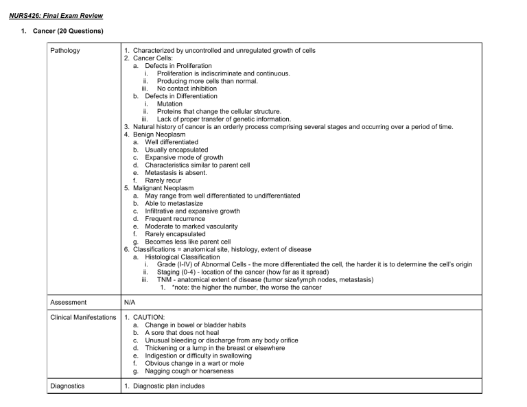
NURS426: Final Exam Review 1. Cancer (20 Questions) Pathology 1. Characterized by uncontrolled and unregulated growth of cells 2. Cancer Cells: a. Defects in Proliferation i. Proliferation is indiscriminate and continuous. ii. Producing more cells than normal. iii. No contact inhibition b. Defects in Differentiation i. Mutation ii. Proteins that change the cellular structure. iii. Lack of proper transfer of genetic information. 3. Natural history of cancer is an orderly process comprising several stages and occurring over a period of time. 4. Benign Neoplasm a. Well differentiated b. Usually encapsulated c. Expansive mode of growth d. Characteristics similar to parent cell e. Metastasis is absent. f. Rarely recur 5. Malignant Neoplasm a. May range from well differentiated to undifferentiated b. Able to metastasize c. Infiltrative and expansive growth d. Frequent recurrence e. Moderate to marked vascularity f. Rarely encapsulated g. Becomes less like parent cell 6. Classifications = anatomical site, histology, extent of disease a. Histological Classification i. Grade (I-IV) of Abnormal Cells - the more differentiated the cell, the harder it is to determine the cell’s origin ii. Staging (0-4) - location of the cancer (how far as it spread) iii. TNM - anatomical extent of disease (tumor size/lymph nodes, metastasis) 1. *note: the higher the number, the worse the cancer Assessment N/A Clinical Manifestations 1. CAUTION: a. Change in bowel or bladder habits b. A sore that does not heal c. Unusual bleeding or discharge from any body orifice d. Thickening or a lump in the breast or elsewhere e. Indigestion or difficulty in swallowing f. Obvious change in a wart or mole g. Nagging cough or hoarseness Diagnostics 1. Diagnostic plan includes a. b. c. d. Nursing Diagnosis 1. 2. 3. 4. 5. 6. 7. 8. 9. 10. 11. 12. 13. Goals Health history Identification of risk factors Physical examination Specific diagnostic studies Anticipatory Grieving Situational Low Self-Esteem Acute Pain Altered Nutrition: Less Than Body Requirements Risk for Fluid Volume Deficit Fatigue Risk for Infection Risk for Altered Oral Mucous Membranes Risk for Impaired Skin Integrity Risk for Constipation/Diarrhea Risk for Altered Sexuality Patterns Risk for Altered Family Process Fear/Anxiety 1. Collaborative effort: a. Nursing/Medical b. Spiritual c. Case management d. Social services e. Nutrition f. Physical therapy Interventions Chemotherapy 1. Response of cancer cells depend on: a. Mitotic rate b. Size of tumor c. Age of tumor Radiation 1. Primary therapy 2. Adjuvant 3. Prophylaxis/Disease control 4. Palliative a. External b. Internal Evaluations 1. 2. 3. Always make sure the IV is patent and flushing properly. Patient teaching for N/V a. Avoid eating 1-2 hours before chemo b. Take anti-emetics before treatment c. Eat small frequent meals d. Avoid anything that will increase GI upset like hot, spicy or fatty foods. Mucositis: Painful ulcerations of the mouth. a. Treated with magic mouthwash. Avoid any ETOH base mouthwashes 4. 5. 6. 7. 8. 9. Teaching Alopecia: a. Might or might not happen. Wig programs. b. Lubricate area with non-irritating lotion or cream Bone marrow suppression a. HCT may need blood products b. Know values Neutropenia a. different levels (Might need revered precautions) Def need good hand washing b. Watch your WBC and the ANC Absolute neutrophil count know values Thrombocytopenia a. Plt know values (Bleed precautions, such as using electric razor, soft bristle toothbrush, and Bowel regimen if constipation is a factor) Fatigue a. Keep up with ADLs and be as activity as they can. Pain a. Pt with cancer are going to need standing pain medications along with PRN pain meds for breakthrough pain. Prevention and Detection a. Lifestyle habits to reduce risks: i. Avoid or reduce exposure to known or suspected carcinogens. b. Cigarette smoke, excessive sun exposure i. Eat a balanced diet. ii. Exercise regularly. iii. Obtain adequate rest. c. Habits to reduce risks i. Have a regular health examination. ii. Change perceptions of stressors. iii. Know seven warning signs of cancer. iv. Practice recommended cancer screenings. v. Practice self-examination. vi. Seek medical care if cancer is suspected. 2. Renal - Acute/Chronic kidney failure, UTI, Pyelonephritis (15 Questions) Patho UTI Chronic Kidney Disease 1. Escherichia coli most common pathogen 2. Fungal and parasitic infections can cause UTIs 3. Upper tract a. Renal parenchyma, pelvis, and ureters b. Typically causes fever, chills, flank pain c. Pyelonephritis(kidney infection): inflammation of 1. Involves progressive, irreversible loss of kidney function 2. Defined as presence of a. Kidney damage 3. Pathologic abnormalities 4. Markers of damage a. Blood, urine, imaging tests b. Glomerular filtration rate (GFR) 5. <60 mL/min for 3 months or longer Acute Kidney Failure 1. Pre-renal a. Relate to cardiac output: Hemorrhage, vomiting, diarrhea, CHF, MI, renal artery thrombosis 2. Intra-renal a. Drugs, x-ray dye, lupus, toxemia of pregnancy b. ATN-acute tubular necrosis 3. Post-renal a. Bladder, prostate cancer b. Spinal cord disease Pyelonephritis 1. Inflammation of renal parenchyma and collecting system 2. Caused most commonly by bacteria 3. Fungi, protozoa, or viruses infecting kidneys can be the cause. renal parenchyma and collecting system 4. Lower tract a. Lower urinary tract b. Usually no systemic manifestations c. Bladder infection (UTI) d. Cystitis: inflammation of bladder wall 6. Leading causes of ESRD a. Glomerulonephritis b. Diabetes c. Hypertension c. Neuromuscular disorders 4. Oliguric phase: < 400 ml/ 24 hrs 5. Diuretic Phase: 1-3 litres/day / abnormal labs 6. Recovery Phase: GFR increases, BUN/Cr decrease 7. Elders: more severe disease, prognosis Assessment Subjective Assessment 1. Previous UTIs, calculi, stasis, retention, pregnancy, STDs, bladder cancer 2. Antibiotics, anticholinergics, antispasmodics 3. Urologic instrumentation 4. Urinary hygiene 5. N/V, anorexia, chills, nocturia, frequency, urgency 6. Suprapubic/lower back pain, bladder spasms, dysuria, burning on urination Objective Assessment 1. Fever 2. Hematuria, foul-smelling urine, tender, enlarged kidney 3. Leukocytosis, positive findings for bacteria, WBCs, RBCs, pyuria, ultrasound, CT scan, IVP 1. History and physical examination N/A Subjective Assessment 1. Previous UTIs, calculi, stasis, retention, pregnancy, STDs, bladder cancer 2. Antibiotics, anticholinergics, antispasmodics 3. Urologic instrumentation 4. Urinary hygiene 5. Nausea, vomiting, anorexia, chills, nocturia, frequency, urgency 6. Suprapubic or lower back pain, bladder spasms, dysuria, burning on urination Objective Assessment 1. Fever 2. Hematuria, foul-smelling urine, tender, enlarged kidney 3. Leukocytosis, positive findings for bacteria, WBCs, RBCs, pyuria, ultrasound, CT scan, IVP Clinical Manifestations Bladder storage or bladder emptying 1. Urinary frequency: abnormally frequent (> every 2 hours) 2. Urgency: sudden strong desire to void immediately 3. Incontinence: loss or leakage of urine 1. Polyuria a. Results from inability of kidneys to concentrate urine b. Occurs most often at night c. Specific gravity fixed around 1.010 2. Oliguria a. Occurs as CKD worsens 3. Anuria a. Urine output <40 mL per 24 hours Hematologic System 1. Anemia 1. Renal: Oliguria, proteinuria 2. Cardiac: Volume overload >CHF, Arrythmias 3. Resp: PE, Kussmaul respirations 4. GI: N,V,D,C 5. Heme: platelet defects (bleeding), anemia, WBCs increase 6. Neuro: Lethargy, seizures, asterixis 7. Metabolic: BUN/Creat, Lytes, Ca, PO4, bicarb 1. 2. 3. 4. 5. 6. 7. 4. Nocturia: waking up ≥2 times at night to void 5. Nocturnal enuresis: complaint of loss of urine during sleep *geriatrics. = atypical manifestations* a. Due to ↓ production of erythropoietin Mild fatigue Chills Fever Vomiting Malaise Flank pain Lower urinary tract symptoms characteristic of cystitis 8. Costovertebral tenderness usually present on affected side 9. Manifestations usually subside in a few days, even without therapy. 10.Bacteriuria and pyuria still persist. 2. From ↓ in functioning renal tubular cells 3. Bleeding tendencies a. Defect in platelet function 4. Infection a. Changes in leukocyte function b. Altered immune response and function c. Diminished inflammatory response Cardiovascular System 1. Hypertension 2. Heart failure 3. Left ventricular hypertrophy 4. Peripheral edema 5. Dysrhythmias 6. Uremic pericarditis Neurologic System 1. Expected as renal failure progresses a. Attributed to 2. ↑ nitrogenous waste products 3. Electrolyte imbalance 4. Metabolic acidosis 5. Axonal atrophy 6. Demyelination of nerve fibers 7. Restless leg syndrome 8. Muscle twitching 9. Irritability 10.Decreased ability to concentrate 11.Peripheral neuropathy Diagnostics 1. History and physical examination 2. Dipstick urinalysis a. Identify presence of nitrates, WBCs, and leukocyte esterase. 3. Urine for culture and sensitivity 4. Clean-catch sample preferred a. Specimen by catheterization or suprapubic needle aspiration more accurate b. Determine susceptibility of bacteria to antibiotics 1. History and physical examination 2. Dipstick evaluation 3. Albumin-creatinine ratio (first morning void) 4. GFR 5. Serum Levels 6. Renal ultrasound 7. Renal scan 8. CT scan 9. Renal biopsy N/A 1. History 2. Physical examination a. Palpation for CVA pain 3. Laboratory tests a. Urinalysis b. Urine for culture and sensitivity c. CBC with differential d. Blood culture (if bacteremia is suspected) 4. Ultrasound 5. IVP 6. CT scan Nursing Diagnosis 1. 2. 3. 4. Goals Educate & Hygiene Interventions Acute Pain Impaired Urinary Elimination Hyperthermia Deficient Knowledge 1. Adequate fluid intake a. Patient may think will worsen condition because of discomfort. b. Dilutes urine, making bladder less irritable c. Flushes out bacteria before they can colonize 2. Avoid caffeine, alcohol, citrus juices, chocolate, and highly spiced foods. a. Potential bladder irritants 3. Application of local heat to suprapubic or lower back may relieve discomfort. 4. Instruct patient about drug therapy and side effects. 5. Instruct patient to watch urine for changes in color and consistency and decrease in cessation of symptoms. 6. Counsel on persistence of lower tract symptoms beyond treatment; onset of flank pain or fever should be reported immediately. 7. Emphasize compliance with drug regimen. a. Take as ordered. 8. Maintain adequate fluids. 9. Regular voiding (every 3 to 4 hours) 10. Void after intercourse. 1. Excess fluid volume 2. Risk for injury 3. Imbalanced nutrition: Less than body requirements 4. Grieving 5. Risk for infection N/A 5. 6. 7. 8. N/A N/A Educate & Hygiene 1. Sodium restriction a. Diets vary from 2 to 4 g, depending on degree of edema and hypertension. b. Sodium and salt should not be equated. c. Salt substitutes should not be used because they contain potassium chloride. 2. Potassium restriction a. 2-3 g b. High-potassium foods should be avoided. 3. Acute intervention a. Daily weight b. Daily BPs c. Identify signs and symptoms of fluid overload. d. Identify signs and symptoms of hyperkalemia. e. Strict dietary adherence f. Medication education g. Motivate patients in management of their disease. 4. Drug Treatment 1. Pre-renal a. Treat dehydration, bleeding 2. Intra-renal a. Remove the drug 3. Post –renal a. Treat obstruction, BPH, tumor etc 4. Monitor a. Intake/ output (oliguric, diuretic phases) b. Cardiac function (electrolyte imbalances) c. Risk for infection (immune suppression) d. Safety (neuro changes) e. Labs f. Drug effects g. Pre-existing conditions 1. Hospitalization for patients with severe infections and complications a. Such as nausea and vomiting with dehydration 2. Signs/symptoms typically improve within 48 to 72 hours after therapy is started. 3. Drug therapy a. Antibiotics 4. Parenteral in hospital to rapidly establish high drug levels a. NSAIDs or antipyretic drugs 5. Fever 6. Discomfort a. Urinary analgesics Acute Pain Impaired Urinary Elimination Hyperthermia Deficient Knowledge Evaluations N/A N/A N/A N/A Teaching 1. Recognize individuals at risk. a. Debilitated persons b. Older adults c. Underlying diseases (HIV, diabetes) N/A N/A 1. Early treatment for cystitis to prevent ascending infection 2. Patient with structural abnormalities is at high risk 2. 3. 4. 5. d. Taking immunosuppressive drug or corticosteroids Emptying bladder regularly and completely Evacuating bowel regularly Wiping perineal area front to back Drinking adequate fluids (15 mL/lb) a. 20% fluid comes from food. 3. Stress the need for regular medical care. a. Need to continue drugs as prescribed b. Need for follow-up urine culture c. Identification of risk for recurrence or relapse d. Encourage adequate fluids a. Patho i. General Kidney Pathology 1. Kidney Function i. Regulates volume and composition of extracellular fluid ii. Produces and secretes Renin for blood pressure control iii. Erythropoietin: hormone in kidney; stimulates RBC production iv. Kidney activate Vitamin D/Calcium (bone health, muscle/heart contraction) 2. Renal Failure i. Electrolyte imbalances, fluid imbalances ii. Renin regulates BP (Too much fluid = BP↑; Too little fluid = BP↓) iii. ↓Erythropoietin = anemia iv. Altered Calcium vs. Phosphate balance 3. Aging System (“Start low, and go Slow”) i. ↓Kidney size, blood flow, nephron function, GFR, estrogen, muscle support ii. ↑Prostate size b. Assessment i. History 1. Urinary patterns 2. Weight changes / edema 3. Urinary changes a. Dysuria: pain with urination b. Polyuria: large amount of urine in a given time c. Nocturia: frequent urination at night d. Oliguria: diminished amounts of urine at a given time e. Anuria: no urine (24hr Os <100mL) 4. Incontinence 5. Itchy skin 6. Fatigue 7. Anorexia 8. N/V 9. Pain ii. iii. iv. v. Physical 1. Hypertension r/t ? 2. Skin changes / pallor r/t ? 3. Edema r/t ? 4. Anemia r/t ? 5. Bone health r/t ? 6. Change in urine: dark, bloody, concentrated, odorous *Low Calcium = High Phosphorus* Blood pressure (most cases, INCREASE in BP – kidneys loss their ability to process the fluid) Skin changes (ashy, gray) Anemia (peripheral, lung, generalized/anasarca) Bone health (muscle pain, bone pain, loss of Vitamin D) c. Clinical manifestations d. Diagnostics i. 1. Urinalysis (UA) a. Dipstick 2. Urine culture 3. Creatinine clearance a. Normal 80-135 ml/ min b. ESRD 15 ml/min 4. Glomerular filtration rate a. GFR 5. Blood tests a. Blood Urea Nitrogen(BUN) b. Creatinine(Creat) c. Electrolytes i. Potassium ii. Sodium iii. Chloride d. CBC e. Calcium, Phosphorous ii. 1. 2. 3. 4. 5. 6. KUB (x-ray; indirect image) Ultrasound CT scan (image of kidney tissue) Dye contrast studies (last resort) Biopsy (GOLD STANDARD; dangerous) Nuclear studies Urinalysis (UA) 1. Color: Hematuria, Excessive Bilirubin, Medications (Pyridium) and clarity (Cloudy urine can be a sign of an infection) 2. Odor: UTI 3. Protein: Acute or chronic renal disease (Involving the glomeruli). Check medications 4. Glucose: DM Pituitary disorder 5. Ketones: Altered Carb fat metabolism in DM, starvation, dehydration, vomiting, severe diarrhea 6. Bilirubin: Liver disorder 7. SG: low: too dilute, High dehydration 8. OSMO: Tubular dysfunction-kidney lost ability to concentrate or dilute urine 9. pH: UTI 10. RBC: Trauma, UTI, Cancer, glomerulonephritis, tuberculosis, 11. WBC: UTI or inflammation 12. Bacteria: Depending on concentration either infection or contamination of specimen 1. Asthma (5 Questions) a. Patho i. Chronic inflammatory disorder of airways ii. Causes airway hyperresponsiveness leading to wheezing, breathlessness, chest tightness, and cough iii. Triggers: 1. Allergen - seasonal, year round, exacerbated after exercise 2. Air Pollutants - heavily industrialized or densely populated areas, climatic conditions often lead to concentrated pollution in the atmosphere, especially with thermal inversions and stagnant air masses. b. Assessment i. Severe acute attack 1. Respiratory rate >30/min 2. Pulse >120/min 3. PEFR is 40% at best. c. 4. Usually seen in ED or hospitalized ii. Life-threatening asthma iii. Too dyspneic to speak iv. Perspiring profusely v. Drowsy/confused vi. Require hospital care and often admitted to ICU Clinical manifestations i. Unpredictable and variable 1. Decrease breath sounds 2. Recurrent episodes of wheezing, breathlessness, cough, and tight chest 3. May be abrupt or gradual 4. Lasts minutes to hours ii. Expiration may be prolonged. 1. Inspiration-expiration ratio of 1:2 to 1:3 or 1:4 2. Bronchospasm, edema, and mucus in bronchioles narrow the airways. 3. Air takes longer to move out. iii. An acute attack usually reveals signs of hypoxemia. 1. Restlessness iv. 2. ↑ anxiety 3. Inappropriate behavior More signs of hypoxemia 1. ↑ pulse and blood pressure 2. Pulsus paradoxus (drop in systolic BP during inspiratory cycle >10 mm Hg) d. Diagnostics i. Classification for Initial Diagnosis 1. Mild intermittent 2. Mild persistent 3. Moderate persistent 4. Severe persistent ii. Tidal volume (VT) = amount of air inhaled or exhaled during normal breathing. iii. Minute volume (MV) = total amount of air exhaled per minute. iv. Vital capacity (VC) = total volume of air that can be exhaled after inhaling as much as you can. v. Functional residual capacity (FRC) = amount of air left in lungs after exhaling normally. vi. Residual volume = amount of air left in the lungs after exhaling as much as you can. vii. Total lung capacity = total volume of the lungs when filled with as much air as possible. viii. Forced vital capacity (FVC) = amount of air exhaled forcefully and quickly after inhaling as much as you can. ix. Forced expiratory volume (FEV) = amount of air expired during the first, second, and third seconds of the FVC test. x. Forced expiratory flow (FEF) = average rate of flow during the middle half of the FVC test. xi. Peak expiratory flow rate (PEFR) = fastest rate that you can force air out of your lungs. e. Nursing Diagnoses i. Ineffective Breathing Pattern ii. Ineffective Airway Clearance iii. Deficient Knowledge iv. Anxiety v. Activity Intolerance vi. Health-Seeking Behaviors: Prevention of Asthma Attack vii. Interrupted Family Processes viii. Fatigue f. Goals i. ii. iii. iv. v. Acute intervention 1. Monitor respiratory and cardiovascular systems: Lung sounds Respiratory rate Pulse BP An important goal of nursing is to ↓ the patient’s sense of panic. 1. Stay with patient. 2. Encourage slow breathing using pursed lips for prolonged expiration. 3. Position comfortably. g. Interventions i. Bronchodilators (Short-acting) 1. Albuterol inhaler (SABA) 2. Atrovent inhaler (ipratropium) (HFA) ii. Corticosteroid (Quick-acting) 1. IV steroids iii. Bronchodilators (Long-acting) 1. Advair (Fluticasone propionate and Salmeterol) 2. Flovent (ICS) 3. Oral Prednisone 4. Singulair (monelukast) (LTRA) h. Evals i. Also monitor ABGs, pulse oximetry, and peak flow. ii. The nurse should note that louder wheezing may actually occur in the airways that are responding to the therapy as airflow in the airways increases. i. Teaching i. Avoid triggers ii. Seek medical attention for bronchospasm or when severe side effects occur. iii. Maintain good nutrition. iv. Exercise within limits of tolerance. v. Use inhaler before exercising. vi. Medication compliance. vii. Measure peak flow at least daily. viii. Compliance with Asthma Action Plan ix. Asthmatic individuals frequently do not perceive changes in their breathing. vi. 3. Musculoskeletal - OP, OA, RA, MS Assessment (10 Questions) a. Osteoporosis (OP) Patho 1. Usually bone deposition and bone reabsorption are equal so bone mass stays constant. 2. Characterized by low bone mass and structural deterioration of bone tissue leading to increased bone fragility Osteoarthritis (OA) 1. DJD 2. Degeneration of articular cartilage 3. Slow , progressive 4. Affects weight bearing joints 5. Before age 50, affects men > women 6. After age 50, affects women > men Rheumatoid Arthritis (RA) 1. Chronic, systemic 2. Inflammation of connective tissue in the synovial joints. 3. Affects all ethnic groups 4. Occurs at any time during the life span. 5. Cause – unknown 6. Etiology – thought to be autoimmune. Musculoskeletal (MS) 1. 2. 3. 4. 5. 6. Function Support Protection of internal organs Voluntary movement Blood cell production Mineral storage 3. Occurs more frequently in women but men do suffer from it. 4. Women over age 60 should be screened for it 5. Most often occurs in spine, hips and wrists 6. “silent disease” – lack of symptoms 7. First signs are back pain or spontaneous fractures 7. May be idiopathic or secondary 8. No one single cause 9. Degeneration / wearing away of cartilage 10. Body’s repairing can’t keep up with destruction 7. RF combines with IgG to form immune complexes that affect joints. Activates inflammatory response 8. See joint changes from the inflammation. 9. Genetic factors 10. May be higher incidence (have seen higher incidence with identical vs. fraternal twins) Assessment Subjective 1. Important health information 2. PMHx, MS impairment, recent trauma, pain, weakness, deformities, limitation in movement, stiffness, joint crepitation. 3. Medications 4. Anti-seizure meds, phenothiazines, corticosteroids, K depleting diuretics. 5. Surgery and other treatment 6. FHP 7. Health perceptions, Nutrition, Elimination, Activity, Sleep rest, Cognitive-perceptual, Role-relationships. Objective 1. Inspection 2. Head to toe, body build, swelling, deformity, nodules. 3. Palpation 4. Skin temp, local tenderness, swelling. 5. Motion 6. Passive and active range of motion. 7. Synovial Joint Movement Review Table 61-3 8. Muscle-strength testing 9. 0-5 Subjective 1. Important health information 2. PMHx, MS impairment, recent trauma, pain, weakness, deformities, limitation in movement, stiffness, joint crepitation. 3. Medications 4. Anti-seizure meds, phenothiazines, corticosteroids, K depleting diuretics. 5. Surgery and other treatment 6. FHP 7. Health perceptions, Nutrition, Elimination, Activity, Sleep rest, Cognitive-perceptual, Rolerelationships. Objective 1. Inspection 2. Head to toe, body build, swelling, deformity, nodules. 3. Palpation 4. Skin temp, local tenderness, swelling. 5. Motion 6. Passive and active range of motion. 7. Synovial Joint Movement Review Table 61-3 8. Muscle-strength testing 9. 0-5 Subjective 1. Important health information 2. PMHx, MS impairment, recent trauma, pain, weakness, deformities, limitation in movement, stiffness, joint crepitation. 3. Medications 4. Anti-seizure meds, phenothiazines, corticosteroids, K depleting diuretics. 5. Surgery and other treatment 6. FHP 7. Health perceptions, Nutrition, Elimination, Activity, Sleep rest, Cognitive-perceptual, Role-relationships. Objective 1. Inspection 2. Head to toe, body build, swelling, deformity, nodules. 3. Palpation 4. Skin temp, local tenderness, swelling. 5. Motion 6. Passive and active range of motion. 7. Synovial Joint Movement Review Table 61-3 8. Muscle-strength testing 9. 0-5 Subjective 1. Important health information 2. PMHx, MS impairment, recent trauma, pain, weakness, deformities, limitation in movement, stiffness, joint crepitation. 3. Medications 4. Anti-seizure meds, phenothiazines, corticosteroids, K depleting diuretics. 5. Surgery and other treatment 6. FHP 7. Health perceptions, Nutrition, Elimination, Activity, Sleep rest, Cognitive-perceptual, Rolerelationships. Objective 1. Inspection 2. Head to toe, body build, swelling, deformity, nodules. 3. Palpation 4. Skin temp, local tenderness, swelling. 5. Motion 6. Passive and active range of motion. 7. Synovial Joint Movement Review Table 61-3 8. Muscle-strength testing 9. 0-5 Clinical manifestations 1. no clinical manifestations until there is a fracture. 1. Systemic – No symptoms 2. Joints – predominant symptom is joint pain 1. Onset insidious 2. Fatigue, anorexia, weight loss, generalized stiffness. 1. Edema 2. Pain / tenderness 3. Muscle spasm 2. Many vertebral fractures are asymptomatic. 3. They may be diagnosed as an incidental finding on chest or abdominal radiographs. 4. The clinical manifestations of symptomatic vertebral fractures include pain and height loss. 3. Hips and knees (most common) 4. hands, vertebrae 5. May have crepitus, deformity 3. Articular involvement – heat, swelling, tenderness, decreased joint motion. 4. Joint symptoms – symmetrical. 5. Fingers, toes, wrists, elbows, shoulders, hips knees, ankles 4. 5. 6. 7. Diagnostics 1. Xray is not helpful 2. BMD – bone mineral density 1. 2. 3. 4. 5. Xray CT MRI Bone Scan / Imaging No lab markers for OA Labs 1. + RF; ESR, C reactive protein, ANA a. ANA (antinuclear antibody test) may be positive in clients with autoimmune disorders such as RA. 2. Synovial fluid analysis 3. Bone Scan / imaging Image 1. Xray 2. CT scan 3. MRI 4. Bone scan 5. Arthroscopy Laboratory Testing 1. Calcium 2. Phosphorus 3. Rheumatoid factor 4. Assess Autoantibodies. Assessment for connective tissue dz. 5. Erythrocyte sedimentation rate 6. Non-specific measure of inflammation 7. Uric acid 8. Levels usually increased in gout. 9. C-reactive protein 10. Used to dx inflammation, infection, malignancy Nursing Diagnoses 1. Impaired Physical Mobility 2. Imbalanced Nutrition: Less 1. 2. 3. 4. Acute Pain/Chronic Pain Impaired Physical Mobility Activity Intolerance Risk For Injury 1. 2. 3. 4. 5. 1. 2. 3. 4. 5. 6. 7. 8. 9. Pain control Rest / joint protection Heat / cold therapy Nutritional therapy / exercise Alternative therapies Drug Therapy Salicylates Tylenol NSAIDS Pain control 1. Maintain joint motion 2. Active exercise to prevent deformities 3. Nutrition therapy Drug Therapy Management 1. Salicylates 2. NSAIDS a. Cox II inhibitors Than Body Requirements 3. Risk for Poisoning 4. Deficient Knowledge Interventions 1. Proper nutrition (eating high calcium foods) 2. Calcium supplements (1000 – 1500 mg/day) 3. Vitamin D supplements (post menopausal) 4. Exercise 5. Prevention of fractures Acute Pain Impaired Physical Mobility Disturbed Body Image Self-Care Deficit Risk for Impaired Home Maintenance 6. Deficient Knowledge Deformity Ecchymosis Loss of function Crepitation 6. Medications ( Biphosphonates - fosamax, boniva) 7. take with a full glass of water, remain upright for 30 minutes after taking and take on an empty stomach 8. Fosamax – once per week oral dose 9. Boniva – once per month oral dose 10. Calcimar – nasal spray daily 11. Forteo – stimulates new bone growth – SQ daily 12. Prolia – given SQ every 6 months a. Cox II inhibitors 10. Corticosteroids 4. Hypertension (5 Questions) a. Patho i. Primary Hypertension (Essential/Idiopathic) 1. Unknown cause 2. Water & Sodium Retention (obesity/age/African Americans) ii. Secondary Hypertension 1. Specific cause (drug/endocrine b. Assessment i. Subjective Data 1. Past health history 2. Current medications 3. Functional health pattern/management ii. Objective Data 1. Elimination (kidney function) 2. Activity-Exercise, cognitive perceptual, coping = stress 3. BP, HR, Heart sounds, Lung sounds, Peripheral pulses, Peripheral edema, Carotids 4. Neurologic/Eye exam c. Clinical manifestations i. “Silent Killer” ii. Often no symptoms iii. Symptoms are often secondary to target organ disease iv. Complications 1. Target organ diseases (ex. Heart, brain, kidney, eyes) 2. CAD, LVH, HF, PVD, Nephrosclerosis, Retinal damage 3. CVD (stroke, TIAs) d. Diagnostics i. BP measurement in both arms (x2 measurements) 1. Use arm with higher reading for subsequent measurements 2. BP highest in early morning 3. Lowest at night 3. Non opioid analgesics a. Tylenol, Ultram 4. Opioid analgesics 5. Corticosteroids a. Prednisone 6. Methotrexate 7. Gold 8. Antimalarials 9. Immunosuppressants 10. Biologic therapy 11. Antibiotics a. Minocin “White coat” phenomenon 1. Repeat the BP at the end of the visit. 2. Anxiety can increase BP. iii. Echo/ECG e. Nursing Diagnoses i. Risk for decreased cardiac tissue perfusion ii. Risk for decreased cerebral tissue perfusion iii. Risk for ineffective renal perfusion iv. Ineffective self health management v. Knowledge deficit vi. Anxiety vii. Potential complication: Stroke viii. Potential complication: MI ix. Potential complication: Adverse effects from antihypertensive therapy x. Potential complication: Hypertensive crisis f. Intervention i. Lifestyle Modifications (weight, dietary sodium) ii. Moderations of alcohol consumption iii. Physical activity iv. Avoidance of tobacco products v. Stress management vi. Drug Therapy (reduce SVR, reduce volume of circulating blood) 1. Diuretics i. lasix, hydrochlorothiazide - deplete potassium, cause orthostatic hypertension ii. Spironolactone - increase in potassium 2. Direct vasodilators i. Hydralazine 3. Adrenergic inhibitors (beta-blockers) ii. i. atenolol, metoprolol - resp condition /c meds can worsen bronchospasms, ↓HR 4. Angiotensin inhibitors (ACE inhibitors) i. Lisinopril *COUGH* ii. Captopril 5. Calcium channel blockers i. Diltiazem ii. Verapamil iii. Norvasc g. Teaching i. Control blood pressure ii. Reduce CVD risk factors iii. Start with lifestyle modification iv. Identify, report, and minimize side effects: 1. Orthostatic hypotension 2. Sexual dysfunction 3. Dry mouth 4. Frequent urination 5. Diabetes Mellitus (10 Questions) a. Patho i. Normal insulin metabolism (produced by the B-cells) ii. iii. iv. v. vi. Released after food, consistently released into the bloodstream A chronic multisystem disease related to 1. Abnormal insulin production 2. Impaired insulin utilization 3. Or both Leading cause of 1. End-stage renal disease 2. Adult blindness 3. Nontraumatic lower limb amputations Major contributing factor 1. Heart disease 2. Stroke Classifications 1. Pre-diabetes 2. Type I 3. Type II 4. Gestational 5. Secondary diabetes b. Pre-diabetes Patho 1. Long-term damage already occurring 2. Heart, blood vessels 3. Usually present with no symptoms Type I Type II Gestational Secondary Diabetes 1. Most often occurs in people younger than 40 years of age 2. Occurs more frequently in younger children 3. End result of longstanding process 4. Progressive destruction of pancreatic b cells by body’s own T cells 5. Manifestations develop when pancreas can no longer produce insulin. a. Rapid onset of symptoms b. Present at ED with ketoacidosis 6. Will require exogenous insulin to sustain life 7. Diabetic ketoacidosis (DKA) a. Occurs in absence of exogenous insulin b. Life-threatening condition c. Results in metabolic acidosis 1. Usually occurs in people over 35 years of age 2. 80% to 90% of patients are overweight. 3. Prevalence increases with age. 4. Genetic basis 5. Pathophysiology a. Increase demand on pancreas b. Insulin Resistance c. Liver d. Metabolic syndrome 1. Develops during pregnancy 2. Detected at 24 to 28 weeks of gestation 3. Usually normal glucose levels at 4. 6 weeks postpartum 5. Increased risk for cesarean delivery, perinatal death, and neonatal complications 6. Increased risk for developing type 2 in 5 to 10 years 7. Therapy: First nutritional, second insulin 8. Metformin 1. Results from another medical condition a. Cushing syndrome b. Hyperthyroidism c. Pancreatitis d. Parenteral nutrition e. Cystic fibrosis f. Hemochromatosis Assessment 1. Past health history a. Viral infections b. Medications c. Recent surgery 2. Positive health history 3. Obesity 4. Weight loss 5. Thirst 6. Hunger 7. Poor healing 8. Kussmaul respirations 1. Past health history a. Viral infections b. Medications c. Recent surgery 2. Positive health history 3. Obesity 4. Weight loss 5. Thirst 6. Hunger 7. Poor healing 8. Kussmaul respirations 1. Past health history a. Viral infections b. Medications c. Recent surgery 2. Positive health history 3. Obesity 4. Weight loss 5. Thirst 6. Hunger 7. Poor healing 8. Kussmaul respirations Clinical Manifestations 1. Must watch for diabetes symptoms 2. Polyuria 3. Polyphagia 4. Polydipsia 1. Classic symptoms a. Polyuria (frequent urination) b. Polydipsia (excessive thirst) c. Polyphagia (excessive hunger) 2. Weight loss 3. Weakness 4. Fatigue 1. Nonspecific symptoms 2. Polyuria, polydipsia, polyphagia 3. Fatigue 4. Recurrent infection 5. Recurrent vaginal yeast or monilia infection 6. Prolonged wound healing 7. Visual changes Diagnostics 1. Exogenous Insulin a. Insulin from outside source b. Required for Type I DM 1. Hemoglobin A1C Test: useful in determining glycemic levels over time a. Shows the amount of glucose attached to hemoglobin molecules over RBC lifespan b. Reduces risk of retinopathy 1. Exogenous Insulin 2. SOMETIMES used for Type II a. Rapid Acting (Lispro) b. Short Acting (Regular) c. Intermediate Acting (NPH) d. Long Acting (Lantus) Nursing Diagnosis 1. Risk for Unstable Blood Glucose 2. Deficient Knowledge 3. Risk for Infection 4. Risk for Disturbed Sensory Perception 5. Powerlessness 6. Risk for Ineffective Therapeutic Regimen Management 7. Risk for Injury 1. Risk for Unstable Blood 1. Risk for Unstable Blood 1. Risk for Unstable 2. 3. 4. 5. 6. 7. Glucose Deficient Knowledge Risk for Infection Risk for Disturbed Sensory Perception Powerlessness Risk for Ineffective Therapeutic Regimen Management Risk for Injury 2. 3. 4. 5. 6. 7. Glucose Deficient Knowledge Risk for Infection Risk for Disturbed Sensory Perception Powerlessness Risk for Ineffective Therapeutic Regimen Management Risk for Injury 1. Past health history a. Viral infections b. Medications c. Recent surgery 2. Positive health history 3. Obesity 4. Weight loss 5. Thirst 6. Hunger 7. Poor healing 8. Kussmaul respirations 2. 3. 4. 5. 6. 7. Blood Glucose Deficient Knowledge Risk for Infection Risk for Disturbed Sensory Perception Powerlessness Risk for Ineffective Therapeutic Regimen Management Risk for Injury 1. Past health history a. Viral infections b. Medications c. Recent surgery 2. Positive health history 3. Obesity 4. Weight loss 5. Thirst 6. Hunger 7. Poor healing 8. Kussmaul respirations 1. Risk for Unstable Blood Glucose 2. Deficient Knowledge 3. Risk for Infection 4. Risk for Disturbed Sensory Perception 5. Powerlessness 6. Risk for Ineffective Therapeutic Regimen Management 7. Risk for Injury 8. Imbalanced Nutrition: 8. Imbalanced Nutrition: 8. Imbalanced Nutrition: 8. Imbalanced Nutrition: 8. Imbalanced Nutrition: Less Than Body Requirements 9. Risk for Deficient Fluid Volume 10. Fatigue 11. Risk for Impaired Skin Integrity Less Than Body Requirements 9. Risk for Deficient Fluid Volume 10. Fatigue 11. Risk for Impaired Skin Integrity Less Than Body Requirements 9. Risk for Deficient Fluid Volume 10. Fatigue 11. Risk for Impaired Skin Integrity Less Than Body Requirements 9. Risk for Deficient Fluid Volume 10. Fatigue 11. Risk for Impaired Skin Integrity Less Than Body Requirements 9. Risk for Deficient Fluid Volume 10. Fatigue 11. Risk for Impaired Skin Integrity Goals 1. Active patient participation 2. Few or no episodes of acute hyperglycemic emergencies or hypoglycemia 3. Maintain normal blood glucose levels. 4. Prevent or delay chronic complications. 5. Lifestyle adjustments with minimal stress 1. Active patient participation 2. Few or no episodes of acute hyperglycemic emergencies or hypoglycemia 3. Maintain normal blood glucose levels. 4. Prevent or delay chronic complications. 5. Lifestyle adjustments with minimal stress 1. Active patient participation 2. Few or no episodes of acute hyperglycemic emergencies or hypoglycemia 3. Maintain normal blood glucose levels. 4. Prevent or delay chronic complications. 5. Lifestyle adjustments with minimal stress 1. Active patient participation 2. Few or no episodes of acute hyperglycemic emergencies or hypoglycemia 3. Maintain normal blood glucose levels. 4. Prevent or delay chronic complications. 5. Lifestyle adjustments with minimal stress 1. Active patient participation 2. Few or no episodes of acute hyperglycemic emergencies or hypoglycemia 3. Maintain normal blood glucose levels. 4. Prevent or delay chronic complications. 5. Lifestyle adjustments with minimal stress Interventions - Standing Orders - Sliding Scale Order - Insulin Pump Drug Therapy 1. b-adrenergic blockers a. Mask symptoms of hypoglycemia b. Prolong hypoglycemic effects of insulin 2. Thiazide/loop diuretics a. Can potentiate hyperglycemia by inducing potassium loss “” “” 1. Insulin 2. Insulin needle sizes come in ½-5/16 inches in length, The gauges are 28, 29, 30. 3. 45-90 degree angle. 1. Treatment of a medical condition that causes abnormal blood glucose level a. Corticosteroids (Prednisone) b. Thiazides c. Phenytoin (Dilantin) d. Atypical antipsychotics (clozapine) 1. Blood sugar and A1C checks 2. Maintaining health weight 3. Regular exercise 4. Healthy diet choices 1. Blood sugar and A1C checks 2. Maintaining health weight 3. Regular exercise 4. Healthy diet choices 1. Blood sugar and A1C checks 2. Maintaining health weight 3. Regular exercise 4. Healthy diet choices 1. Blood sugar and A1C checks 2. Maintaining health weight 3. Regular exercise 4. Healthy diet choices Evals Teaching 1. Blood sugar and A1C checks 2. Maintaining health weight 3. Regular exercise 4. Healthy diet choices 5. Heart Failure (5 Questions) a. Patho i. Abnormal clinical syndrome that involves inadequate pumping and/pr filling of the heart ii. Systolic Failure 1. Inability of the heart to pump blood 2. Hallmark sign – decrease in LV ejection fraction (EF) 3. Cardiomyopathy 4. Valcular disease iii. Diastolic Failure 1. Ventricles not able to relax and fill during diastole 2. Leads to decreased CO 3. Pt has pulmonary congestion, pulmonary HTN, ventricular hypertrophy but a normal EF iv. CHF Classes 1. Class I - no limitation of physical activity i. NO: SOB, angina pain, palpitations, fatigue with usual activity 2. Class II - slight limitation. No symptoms at rest; i. YES: SOB, angina pain, palpitations, fatigue with usual activity 3. Class III - limitation with physical activity i. Comfortable at rest ii. YES: SOB, angina pain, palpitations, fatigue with usual activity 4. Class IV - inability to do activity without discomfort/symptoms i. Quality of life affected ii. YES: SOB, angina pain, palpitations, fatigue with usual activity b. Assessment i. Subjective 1. PMH, Mdx, nutrition, ROS, cardiac, resp, vascular ii. Objective 1. Full physical exam 2. Respiratory 3. O2 sat 4. Lung sounds 5. Cardiac 6. Heart rate 7. Blood pressure 8. Vascular 9. Edema 10. Cap refill c. Clinical manifestations i. Left Sided Heart Failure = most common; poor contraction in LV, back up into pulmonary tract 1. Systolic failure - low EF; LV loses pressure to eject blood out 2. Diastolic failure - inability of the ventricles to relax and fill during Diastole 3. Mixed/Both - Dilated Cardiomyopathy ii. Right Sided Heart Failure = RV fails to pump effectively *Left sided HF may lead to right sided failure (right side compensating)* 1. Venous engorgement d. Diagnostics i. Chest xray ii. Echocardiogram 1. ejection fraction decreased CO iii. 12 lead EKG 1. arrhythmias iv. BNP (b-Type natriuretic peptide) v. Electrolytes (especially K+, Na+) vi. ABG’s vii. Cardiac enzymes viii. LFT’S ix. BUN / Creat e. Nursing Diagnoses i. Impaired gas exchange ii. Decrease cardiac output iii. Excess fluid volume iv. Activity intolerance v. Risk for alterations in skin integrity f. Goals i. Decrease preload - venous return 1. Diuretics, sit up ii. Decrease afterload – work of LV 1. Dilate peripheral vessels iii. Improve gas exchange – nasal 02/ mask iv. Improve cardiac function – drugs v. Reduce anxiety g. Interventions i. Drug Therapy Positioning – High Fowler’s Get VS including O2 sat Continuous cardiac/respiratory monitoring Listen to lung sounds Supplemental O2 Diuretic - Remove the fluid a. Loop - Furosemide (Lasix) b. Thiazides - Hydrochlorothiazide( HCTZ) - Metolazone c. Potassium sparing - Spironolactone( Aldactone) 7. Ace Inhibitors - Improves systolic function a. Captopril (Capoten) b. Lisinopril 1. 2. 3. 4. 5. 6. h. Teaching i. Daily weights ii. I+O iii. Fluid restriction iv. Elevation of LE’s v. 1 – 2 gm Na diet vi. Energy conservation vii. Exercise viii. Edema management ix. Understanding symptoms and knowing what to do if they get worse. 1. Vasodilation - Increase cardiac output i. Hydralazine ii. Nitrates 2. Positive inotrope - Promotes vasodilation i. Digoxin 3. Anxiety i. Morphine *reduces preload & afterload* ii. Ativan 4. Antidysrhythmic 5. Anticoagulants 6. Antibiotics 7. Oxygen/Cardiac meds x. xi. xii. xiii. xiv. Medication compliance Understanding mediations side effects Keep all follow up appointments Diet considerations Electrolyte balance 6. Parkinson's (5 Questions) a. Patho i. Degeneration of the neurons of basal ganglia ii. characterized by 1. Slowing down in the initiation and execution of movement 2. ↑ muscle tone 3. Tremor at rest 4. Gait disturbance iii. Pathologic process of PD involves degeneration of dopamine-producing neurons in substantia nigra of the midbrain. iv. Disrupts dopamine-acetylcholine balance in basal ganglia 1. DA = neurotransmitter essential for normal functioning of the extrapyramidal motor system b. Assessment i. Observe for parkinsonian gate/shuffle c. Clinical manifestations i. Onset is gradual and insidious. ii. Classic triad of PD 1. Tremor 2. Rigidity 3. Bradykinesia: difficulty in starting, continuing or coordinating movement iii. Beginning stages may involve only mild tremor, slight limp, or ↓ arm swing. iv. Later stages may have shuffling, propulsive gait with arms flexed, and loss of postural reflexes. d. Diagnostics i. No specific tests ii. Diagnosis based solely on history and clinical features 1. Firm diagnosis can be made when at least two of three characteristics of the classic triad (tremor, rigidity, and bradykinesia) are present. iii. The ultimate confirmation of PD is a positive response to antiparkinsonian drugs. e. Nursing Diagnoses i. Ineffective Airway Clearance ii. Disturbed Thought Process iii. Impaired Verbal Communication iv. Impaired Physical Mobility v. Imbalanced Nutrition: Less Than Body Requirements vi. Impaired Swallowing vii. Risk for Injury viii. Ineffective Coping ix. Deficient Knowledge x. Other Nursing Care Plans f. Goals i. Promote safety 1. Fall precaution ii. Promote physical exercise 1. Promote independence 2. Limit the consequences from decreased mobility 3. Specific exercises to strengthen muscles involved with speaking and swallowing Well-balanced diet. 1. Aspiration precautions 2. Several small meals 3. Vit B6 supplements g. Interventions i. Enhance the release or supply of dopamine and acetylcholine 1. Levodopa 2. Levodopa with Carbidopa (sinemet) i. Monitor for dyskinesia ii. Positive effect may take several weeks to months to note ii. Antihistamine with anticholinergic properties (Benadryl) 1. Manage tremors iii. Antivirals 1. Promotes the effects of dopamine h. Evals (N/A) i. Teaching i. Safety ii. Physical exercise iii. Well-balanced diet iii. 7. Seizures (5 Questions) a. Patho i. Seizures: Uncontrolled electrical discharge of neurons in the brain that interrupt normal function. ii. Not considered epilepsy when removal of the underlying causes stop the seizures: 1. Acidosis 2. F&E imbalances 3. Hypoglycemia iii. 1. Birth - 6 months of life a. Severe birth injury b. Congenital birth defects involving CNS c. Infections d. Inborn errors of metabolism 2. Age 2 - 20 y/o a. Birth injury b. Infection c. Trauma d. Genetic factors iv. Generalized 1. Tonic-Clonic i. Tonic-muscle stiffening ii. Clonic-rhythmical jerking 2. Absence(petit mal) i. Begin on both sides of the brain ii. Lapse of awareness iii. staring 3. Atonic i. Pt loses muscle tone and goes limp 1. Age 20 - 30 y/o a. Structural lesions b. Trauma c. Brain tumor d. Vascular disease 2. Age 50 y/o + a. Cerebrovascular lesions b. Metastatic brain tumors c. 75% of seizure disorders are considered idiopathic. v. b. c. d. e. f. g. h. i. Focal 1. Focal Onset Aware (Simple partial) = Awake and aware 2. Focal Onset impaired (Complex partial) = Confused Assessment i. Accurate, comprehensive description of seizures with patient’s health history ii. EEG 1. Only a small percentage of patients with seizure disorders have abnormal findings with first test. 2. Continuous monitoring may be needed. 3. CBC, serum chemistries, liver and kidney function, UA to rule out metabolic disorders Clinical manifestations (N/A) Diagnostics i. Accurate, comprehensive description of seizures with patient’s health history ii. EEG 1. Only a small percentage of patients with seizure disorders have abnormal findings with first test. 2. Continuous monitoring may be needed. 3. CBC, serum chemistries, liver and kidney function, UA to rule out metabolic disorders Nursing Diagnoses i. Risk for Trauma or Suffocation ii. Risk for Ineffective Airway Clearance iii. Situational Low Self-Esteem iv. Deficient Knowledge v. Noncompliance Goals i. maintaining a patent airway ii. maintaining safety during an episode iii. imparting knowledge and understanding about the condition iv. monitor the patient for signs of toxicity Interventions i. Medication Therapy 1. Narrow-Spectrum AEDs - DILANTIN; used to specific seizures (ie. partial, focal, absence, myoclonic) 2. Broad-Spectrum AEDs - CLONAZEPAM; some effectiveness for a wide variety of seizures (partial/absence) ii. Acute Interventions 1. Observation and treatment of seizure 2. Clear the area for safety. 3. Put the patient on their side to maintain the airway, support head, loosen constrictive clothing, ease to floor. 4. Given IV Ativan as ordered 5. May require suctioning or oxygen after seizure 6. Assessment of level of understanding 7. Collaborative Care 8. Planning Evals (N/A) Teaching i. Medication compliance ii. Teach non-drug techniques like relaxation etc. iii. Community supports. iv. Medical alert bracelet. v. Avoid excessive alcohol intake, fatigue, loss of sleep. vi. Driving laws for patients with seizures. vii. Safety safety safety. 8. Med Calculations (5 Questions)


