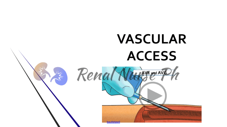
VASCULAR ACCESS AVF and AVG bautistawil OBJECTIVES Upon completion of this topic, the learner will be able to: 1. 2. 3. Describe the advantages and disadvantages of various type of vascular access Identify potential complications connected with each type of vascular access Provide adequate and appropriate education to patients and their significant others. 2 bautistawil Significant Dates in the History of the Vascular Access bautistawil AV SHUNT 1960: Scribner and Quinton developed the first permanent access. 4 bautistawil AV FISTULA 1966: Brescia and Cimino developed the internal arteriovenous (AV) fistula for repeated venipunctures for maintenance HD. bautistawil AV GRAFT 1974: Bovine carotid artery graft used for circulatory access 1975: Gore-tex® graft became commercially available for use as AV access for HD 1977: Umbilical cord vein used for AVF graft 1977: Expanded polytetrafluoroethylene (ePTFE) used as AV access for hemodialysis 6 bautistawil Preparing for Vascular Access for Hemodialysis bautistawil I. Anatomy of the vascular access for hemodialysis A. The venous system in the upper extremity includes both superficial and deep veins. It is superficial system that is most important for access creation. B. The radiocephalic AVF at the wrist is the first choice hemodialysis access and uses the forearm segment of the cephalic vein. 8 bautistawil Anatomy of the Upper Extremity Vessels 9 bautistawil II. Patient Evaluation A. Assessment, Evaluation and Preservation 1. Patiet GFR less than 30 mL/min/1.72m² (CKD Stage 4) 2. Early referral for permanent dialysis access. 3. Preservation of veins of the forearm and upper arms. 4. Recommended timeline 1. 2. AVF should be placed 6 months prior to anticipated need AVG should be inserted at least 3 to 6 weeks ahead of anticipated need 5. Nurse play crucial role in educating, explaining and reassuring patients. 10 bautistawil II. Patient Evaluation B. Evaluation Prior to Access Placement 1. Helps to optimize access survival while minimizing potential complications 2. Evaluations should include: 1. 2. History and physical examination Dupplex ultrasound of the upper extremity blood vessels 11 bautistawil III. Selection and Placement of the Vascular Access 1. Arteriovenous Fistula 2. Arteriovenous Graft 3. Central Venous Catheter A. Non Tunneled CVC B. Tunneled CVC 12 bautistawil NKF KDOQI, 2006, Clinical Practice Guidelines (CPG) Vascular access should be placed distally and in the upper extremities whenever possible. Because AVF provides the access with the longest patency rates and need for fewest interventions; options for AVF creation should be considered first, followed by prosthetic grafts, if AVF creation is not possible. Catheters should be avoided for HD and used only when the previous options are not possible, are contraindicated by the patient’s condition, or the access for hemodialysis is short term 13 bautistawil The ARTERIOVENOUS FISTULA (AVF) bautistawil Definition: A surgically created opening between an artery anastomosed to a juxtapositional (nearby) vein allowing the high pressure arterial blood to flow into the vein causing: VEIN ARTERIALIZATION a. b. c. Engorgement Enlargement Wall thickening. bautistawil Types of Anastamosis bautistawil PLACEMENT OF AVF (in order of priority) 1. 2. 3. Wrist (radial-cephalic) primary fistula An elbow (brachial-cephalic) primary fistula. An upper arm (brachial-basilic) fistula with vein transposition (surgically dissecting out and tunneling in a superficial, accessible area) bautistawil Radiocephalic Arteriovenous Fistula bautistawil Snuff-box Arteriovenous Fistula bautistawil Proximal Forearm Arteriovenous Fistula bautistawil Brachiocephalic Arteriovenous Fistula bautistawil Transposed Basilic Vein Arteriovenous Fistula bautistawil Assessment for Fistula ALLEN TEST 1. Patient clenches the fist of one hand to produce pallor in the hand 2. Clinician occludes arterial flow by compressing both radial and ulnar arteries 3. 4. Patient opens clenched fist Clinician releases pressure on the ulnar artery and counts the seconds required for color to return to the hand. More than 3 seconds indicates decreased ulnar arterial supply to the hand if the radial artery is used for the vascular access bautistawil 5. Repeat the procedure, but release pressure on radial artery this time to assess radial arterial flow to hand. 6. Repeat procedure with opposite hand. ALLEN TEST bautistawil bautistawil ADVANTAGES OF AVF 1 1. Average problem-free patency period is approximately 3 years. 2. The long-term secondary patency rate: 1. 2. 7 years for forearm fistula 3-5 years upper arm fistula 3. Lowest rate of thrombosis 4. Lower rates of infection than grafts 5. Cost of implantation and access maintenance are the lowest long-term 26 bautistawil ADVANTAGES OF AVF 2 6. Associated with increased patient survival and lower hospitalization rates. 7. Avoid potential allergic response to synthetic materials 8. Outflow veins are autogenous tissue that seal and heal after cannulation. 9. Can use buttonhole cannulation technique. 27 bautistawil DISADVANTAGES OF AVF 1. The vein may fail to enlarge or increase wall thickness (i.e., fail to mature) 2. Long maturation time 3. Some individuals, the vein may be more difficult to cannulate. 4. A thrombosed AVF may be more difficult to restore the flow. 5. Cosmetically unattractive 6. Increase cardiac output. 28 bautistawil RULE OF 6 for AVF MATURATION • The vessel should be greater than 6mm in diameter • The vessel should be less than 6 mm from surface • Flow through the vessel should exceed 600mL per min. • AVF should be expertly assessed within 6 weeks of creation for maturation 29 bautistawil ASSESSMENT OF THE FISTULA LOOK LISTEN FEEL 30 bautistawil 1. INSPECTION - Look A. Compare the access extremity with the other extremity. B. Asses the access extremity for: 1. 2. 3. 4. 5. 6. Swelling Presence of collateral veins Change in color or temperature Decreased sensation Limitation of movement Capillary refill time C. Assess the fistula itself. 31 bautistawil 2. AUSCULTATE - Listen BRUIT – continuous “whooshing” NORMAL AVF sound caused by the turbulence at the anastomosis • Note change in the sound or character of the blood flowing through the fistula. bautistawil AVF with STENOSIS 3. PALPATE - Feel To determine the blood is flowing through the fistula Thrill – Sensation that felt over the fistula a) A continuous vibration b) Result of turbulence created by the blood flow c) Pulsatile thrill may indicate stenosis bautistawil PATIENT TEACHING • Reinforce and expand pre-AVF placement education to avoid venipunctures and blood pressure measurements in targeted arm. • Elevate affected arm to decrease post-op swelling • Exercise for vessel development bautistawil PATIENT TEACHING • Instruct the ff: - How to palpate “thrill” - Avoid sleeping on access extremity • • • • bautistawil Avoid wearing anything that would tightly encircle the access extremity Wash area with soap and water before cannulation How to stop bleeding – apply localized pressure How to recognize and report s/s of infection or absence of thrill / bruit The ARTERIOVENOUS GRAFT (AVG) bautistawil ARTERIOVENOUS GRAFTS (AVG) • DEFINITON: A synthetic or, less frequently, biologic conduit implanted subcutaneously and interposed between an artery and a vein. • Needles are inserted into the graft (never into the anastomoses) to remove and return blood during hemodialysis. The average graft is 6 mm. bautistawil INDICATIONS of AVG • • Patients who do not have vasculature suitable for AVF or who have failed AVF in the location of the planned AVG. AVG is second best option for hemodialysis bautistawil TYPES OF GRAFT 1. Synthetic Grafts 2.Composite/ Polyurethane Graft 3. Biologic Graft bautistawil ANATOMIC LOCATIONS of AVG 1. 2. 3. A forearm loop graft 4. Lower extremity graft. Upper arm graft Chest wall or “necklace” prosthetic graft bautistawil FOREARM LOOP ARTERIOVENOUS GRAFT 41 bautistawil UPPER ARM ARTERIOVENOUS GRAFT 42 bautistawil THIGH ARTERIOVENOUS GRAFT 43 bautistawil AVG Surgery bautistawil bautistawil ADVANTAGES OF AVG • Larger surface area for cannulation • Easier to cannulate • Time for surgical insertion to maturation is short • • bautistawil ePTFE 1-3 weeks prior to cannulation Time allows for healing and the incorporation of the surrounding tissue ADVANTAGES OF AVG • AVG can be placed in many areas of the body • Can be placed in a variety of shape to facilitate placement and cannulation • Graft is easier to repair bautistawil DISADVANTAGES OF AVGs 1. 2. 3. 4. Higher rate of stenosis and thrombosis Higher rate of infection Higher mortality Shorter length of patency 1. Last 1-2 years before indication of failure or thrombosis 2. Long-term patency rate with treatment of stenosis and thrombosis remains at 2 years bautistawil DISADVANTAGES OF AVGs 5. Cannulation sites seal but not heal 6. Allergic response to the material 7. Steal syndrome bautistawil Patient Teaching 1. Instruct to elevate and abduct extremity and use the hand normally as much as possible 2. No venipunctures / BP taking in the access arm 3. Avoid sleeping on the access extremity 4. Wash area with soap & water before cannulation 5. Report any s/s of infection and absence of thrill / bruit bautistawil VASCULAR ACCESS COMPLICATIONS bautistawil Access Complications A. BLEEDING B. INFECTION C. VENOUS STENOSIS D.CENTRAL VENOUS STENOSIS E. THROMBOSIS F. ANEURYSM / PSEUDOANEURYSM G.STEAL SYNDROME bautistawil A. BLEEDING / INFILTRATION DEFINITION • • • Inadvertent administration of fluid into tissue surrounding the fistula. Secondary to improper cannulation technique Can occur before, during or after dialysis bautistawil A. BLEEDING / INFILTRATION SIGNS AND SYMPTOMS • Edema • Taut or stretched skin • Pain bautistawil A. BLEEDING / INFILTRATION PREVENTIONS • • • • Monitor closely for signs and symptoms of infiltration Use caution when taping needles Monitor arterial and venous pressure Proper needle removal bautistawil B. INFECTION CAUSES • • • • • Staphylococcus Aureus is the leading cause Poor patient hygiene Inadequate skin preparation Not using aseptic technique Seeding from another infected site in the body bautistawil B. INFECTION SIGNS AND SYMPTOMS • • • • bautistawil Inflammation Pain Skin break with drainage along the course of vessel Fever C. VENOUS STENOSIS DEFINITION Abnormal narrowing of the lumen of the vessel as a result of injury to the wall, causing intimal hyperplasia • • • Bruit changes to a choppy At the site of stenosis, bruit may be higher pitched Pulse will become a harsher, water hammer feel bautistawil C. VENOUS STENOSIS RELATED ABNORMALITIES 1. Reduction in BFR and potential clotting 2. 3. 4. Increase static venous pressure Access recirculation Unexplained reduction in KT/V bautistawil C. VENOUS STENOSIS CLUES INDICATING STENOSIS bautistawil 1. 2. 3. Inability to maintain BFR 4. Increase bleeding time post dialysis Increased venous pressure Difficulty cannulation or having blood squirt out C. Venous Stenosis POTENTIAL INTERVENTIONS AND TREATMENT 1. Non invasive technique to assess a fistula 2. Doppler ultrasound or fistulogram • Detect stenosis • Measure stenosis 3. Baloon angioplasty 4. Stent placement bautistawil D. CENTRAL VENOUS STENOSIS CAUSES OR CONTRIBUTING FACTORS • • • • bautistawil History of multiple central venous catheter Mechanical compression of the central venous system Arterialized high flow in the central veins Some without an identifiable cause D. CENTRAL VENOUS STENOSIS SIGNS and SYMPTOMS 1. Massive swelling in the upper extremity 2. Extensive network of collateral veins 3. Pain and discomfort during dialysis session bautistawil D. CENTRAL VENOUS STENOSIS POTENTIAL INTERVENTIONS AND TREATMENT 1. Prevention through avoidance of subclavian inserted catheters. 2. Transluminal angioplasty with possible stent placement 3. Surgical treatment is very complex and reserved for extreme situations. bautistawil E. THROMBOSIS CAUSES 1. Stenosis of main outflow vein without collateral circulation 2. Significant hypotension due to volume depletion 3. 4. 5. Hypercoagulable states Prolonged occlusive compression Supporting heavy objects bautistawil E. THROMBOSIS SIGNS and SYMPTOMS • • • • • Vein distended and does not soften when arm is elevated overhead Significant decreased intra-access blood flow Changes in quality of the bruit Difficulty or pain with cannulation Evacuation of clots bautistawil E. THROMBOSIS POTENTIAL INTERVENTIONS AND TREATMENT • • • • bautistawil Urgent referral to a surgeon Lysing the clot with a thrombolytic such as tPA Thrombectomy Anticoagulation therapy F. ANEURYSM / PSEUDOANEURYSM CAUSES • Cannulating in the same area “one-site-itis” • • Outflow stenosis / occlusion Persistent hypotension bautistawil F. ANEURYSM / PSEUDOANEURYSM SIGNS and SYMPTOMS • • • Vessel enlargement Dilatation on the weakened vessel wall Possible changes in bruit bautistawil F. ANEURYSM / PSEUDOANEURYSM POTENTIAL INTERVENTIONS AND TREATMENT • • • Assessing AVF every treatment Education of the staff Surgery based on severity bautistawil E. STEAL SYNDROME CAUSES • • Ischemia of the extremity distal to arterial anastomosis Diversion of significant volume of blood away from peripheral circulation bautistawil • Occurs more frequently in patients who: • • • • Elderly Have peripheral vascular disease Have diabetes Have history of multiple access surgeries E. STEAL SYNDROME SIGNS and SYMPTOMS • • • • • • bautistawil Pain distal to anastomosis Cold, pale hand Impaired hand movement and strength Paresthesias: numbness, tingling Poor capillary refill Progression to ulcerated, necrotic fingertips E. STEAL SYNDROME POTENTIAL INTERVENTIONS AND TREATMENT • • • • • Report any abnormal findings Surgical reperfusion of the hand using the DRIL Banding of inflow to the graft to reduce flow Severe ischemia may require urgent ligation of the access Mild ischemia may improved by wearing of a glove, exercising, and/or massaging bautistawil References • Kallenbach, J.; Review of Hemodialysis for Nurses and Dialysis Personnel, 8th Edition. Elsevier Health Sciences, 2012 • Counts, C.; Core Curriculum for Nephrology Nursing, 6th Edition. ANNA (American Nephrology Nurses Association), 2015 • Vachharajani, T.; Atlas of Dialysis Vascular Access, 2010 74 bautistawil bautistawil bautistawil
