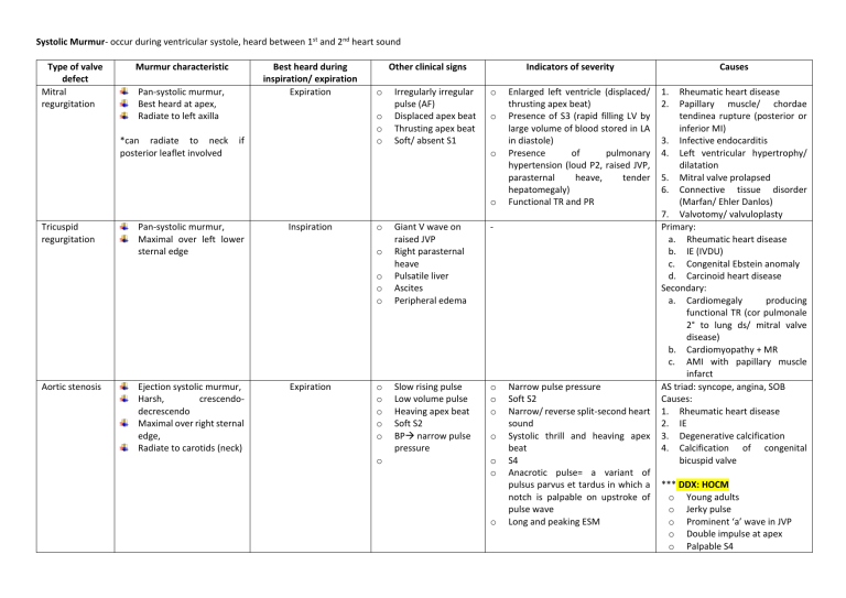
Systolic Murmur- occur during ventricular systole, heard between 1st and 2nd heart sound Type of valve defect Mitral regurgitation Murmur characteristic Best heard during inspiration/ expiration Expiration Pan-systolic murmur, Best heard at apex, Radiate to left axilla *can radiate to neck posterior leaflet involved Other clinical signs o o o o if Irregularly irregular pulse (AF) Displaced apex beat Thrusting apex beat Soft/ absent S1 Indicators of severity o o o o Tricuspid regurgitation Pan-systolic murmur, Maximal over left lower sternal edge Inspiration o o o o o Aortic stenosis Ejection systolic murmur, Harsh, crescendodecrescendo Maximal over right sternal edge, Radiate to carotids (neck) Expiration o o o o o o Giant V wave on raised JVP Right parasternal heave Pulsatile liver Ascites Peripheral edema - Slow rising pulse Low volume pulse Heaving apex beat Soft S2 BP narrow pulse pressure o o o o o o o Enlarged left ventricle (displaced/ thrusting apex beat) Presence of S3 (rapid filling LV by large volume of blood stored in LA in diastole) Presence of pulmonary hypertension (loud P2, raised JVP, parasternal heave, tender hepatomegaly) Functional TR and PR Narrow pulse pressure Soft S2 Narrow/ reverse split-second heart sound Systolic thrill and heaving apex beat S4 Anacrotic pulse= a variant of pulsus parvus et tardus in which a notch is palpable on upstroke of pulse wave Long and peaking ESM Causes 1. 2. Rheumatic heart disease Papillary muscle/ chordae tendinea rupture (posterior or inferior MI) 3. Infective endocarditis 4. Left ventricular hypertrophy/ dilatation 5. Mitral valve prolapsed 6. Connective tissue disorder (Marfan/ Ehler Danlos) 7. Valvotomy/ valvuloplasty Primary: a. Rheumatic heart disease b. IE (IVDU) c. Congenital Ebstein anomaly d. Carcinoid heart disease Secondary: a. Cardiomegaly producing functional TR (cor pulmonale 2° to lung ds/ mitral valve disease) b. Cardiomyopathy + MR c. AMI with papillary muscle infarct AS triad: syncope, angina, SOB Causes: 1. Rheumatic heart disease 2. IE 3. Degenerative calcification 4. Calcification of congenital bicuspid valve *** DDX: HOCM o Young adults o Jerky pulse o Prominent ‘a’ wave in JVP o Double impulse at apex o Palpable S4 Systolic Murmur- occur during ventricular systole, heard between 1st and 2nd heart sound o o Ventricular septal defect Pulmonary stenosis Pan-systolic murmur Over all precordium Maximal heard over left sternal edge (lower/ upper) Ejection-systolic murmur at left upper sternal edge May radiate to back No changes Inspiration Mild: displaced apex beat (LVH) Moderate: palpable thrill Severe: parasternal heave (RVH) o o o Soft P2 Normal S2 Wide splitting of S2 (ASD) Others: Functional murmur due to change in rate of blood flow/ hyperdynamic circulation: a. Fever b. Anaemia c. Exercise d. Hyperthyroidism Systolic thrill at left lower sternal edge Family history of sudden death at young age Systolic Murmur- occur during ventricular systole, heard between 1st and 2nd heart sound Systolic Murmur- occur during ventricular systole, heard between 1st and 2nd heart sound Diastolic Murmur- occur during cardiac diastole, heard after 2nd heart sound Type of valve defect Mitral stenosis Aortic regurgitation Characteristic of murmur Mid-diastolic murmur, Low pitched Best heard left lateral position Early diastolic murmur, High pitched Maximal at left upper sternal edge Best heard leaning forward Inspiration/ Expiration Expiration o o Expiration o o o o Other clinical signs Indicators of severity Loud S1 (occurs when leaftlets are mobile, slammed shut during ventricular systole) Opening snap (d/t opening of stenosed mitral valve pliable leaftlets) o Collapsing pulse (waterhammer pulse) Corrigan’s sign- visible carotid pulsation due to wide pulse pressure Quincke sign- nail bed capillary pulsation Displaced apex beat + thrusting o o o o o Others: o De Musset’s sign- rhythmic bobbing of head in synchrony with heartbeat o Mueller’s sign: uvular pulsation in time with HR o Duroziez’s sign- to and fro murmur when femoral artery compressed by stethoscope o Traube’s sign- systolic pistol-shots over femoral artery o Hill’s sign (SBP in LL> UL) o o o o o Narrow distance between opening snap and S2 Longer MDM Pulmonary HPT Right-sided heart failure Wide pulse pressure Signs of LVF Displaced apex beat Long EDM Austin Flint murmur (MDM with no opening snap) Presence of S3 Hill’s sign Causes Congenital: congenital parachute valve Acquired: - Rheumatic heart disease - Calcification of mitral annulus & leaflets - CTD (RA, SLE) - Malignant carcinoid Intrinsic valvular disease: - Congenital bicuspid valve - IE - Rheumatic heart disease - RA Aortic root disease: - Calcific AS - Aortic dissection - Syphilis - Seronegative spondyloarthropathy - Marfan syndrome Position: 45°, expose chest and neck General: - Body built - Sign of respiratory distress - Syndromic facies Hands: - Finger clubbing - Stigmata of IE: splinter haemorrhage, Osler’s node, Janeway lesion - Peripheral cyanosis - Radial scar* - Arterial pulse: a. Rate b. Rhythm c. Volume d. Character e. Any radio-radial delay f. Collapsing pulse Face: Eye: pallor, jaundice Mouth: oral hygiene, high-arched palate, dental caries, central cyanosis Neck: JVP Chest Inspection: - Any midline sternotomy scar - Visible heartbeat Palpation: - Apex beat + characteristic - Palpable thrill - Parasternal heave Auscultation: listen for murmur + manouever Description of murmur 1. Timing- Systolic/ diastolic 2. Character/ pitch 3. Maximally heard at which site 4. Any radiation 5. Intensity (grading) 6. Effects of posture + respiration Continue with 1. Listen to lung bases for basal crepitations 2. Peripheral edema- sacral+ leg Complete with - Abdominal examination: Palpate liver- enlargement, tenderness, pulsation Palpate spleen- if suspect IE - Measure blood pressure - Presence and equality of peripheral pulses


