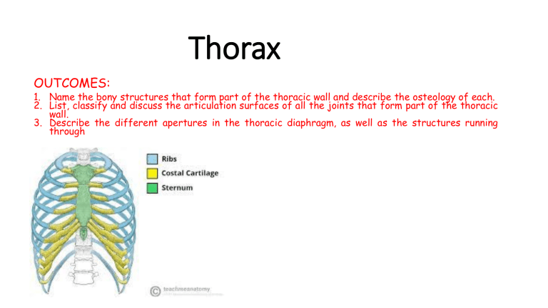
Thorax OUTCOMES: 1. Name the bony structures that form part of the thoracic wall and describe the osteology of each. 2. List, classify and discuss the articulation surfaces of all the joints that form part of the thoracic wall. 3. Describe the different apertures in the thoracic diaphragm, as well as the structures running through Thoracic cage Sternum Ribs R1-R12 Costal cartilage Sternum Manubrium sterni: sternoclavicular joint Body Xiphisternum = xiphoid proc. Sternal angle Typical rib (R3-R10): head (1/2) head has 2 facets (demifacets) for joints with adjacent vertebral bodies Typical rib (R3-R10): head (2/2) an intermediate crest for attachment with intervertebral disc Typical rib (R3-R10) neck, tubercle (joint with transverse proc.), angle, costal cartilage costal groove Typical rib (R3-R10) R3-7 joint to sternal body R8-10 attach to the costal cartilage of the rib above ( → costal arch) The first rib (R1) flattened, costal cartilage to sternal manubrium The first rib (R1): inferior surface contact pleura - smooth The first rib (R1): superior surface The second rib (R2) costal cartilage has 2 facets 2nd rib The 11th and 12th ribs (R11, R12) end in small cap of cartilage: floating rib (R11, R12): head


