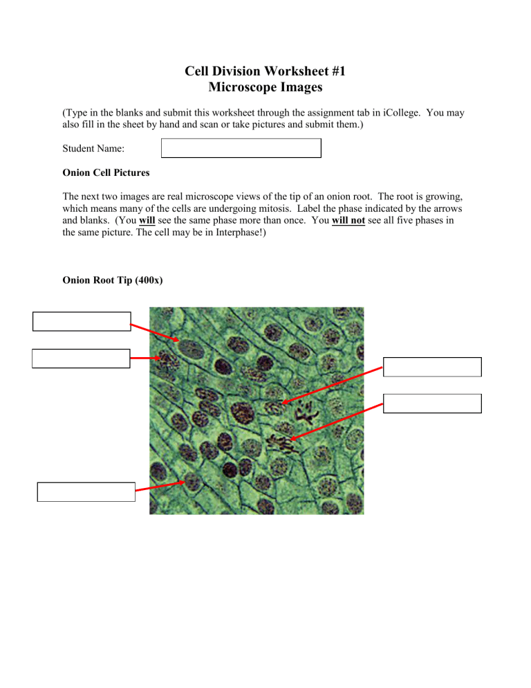
Cell Division Worksheet #1 Microscope Images (Type in the blanks and submit this worksheet through the assignment tab in iCollege. You may also fill in the sheet by hand and scan or take pictures and submit them.) Student Name: Onion Cell Pictures The next two images are real microscope views of the tip of an onion root. The root is growing, which means many of the cells are undergoing mitosis. Label the phase indicated by the arrows and blanks. (You will see the same phase more than once. You will not see all five phases in the same picture. The cell may be in Interphase!) Onion Root Tip (400x) Onion Root Tip (400x) Fish Cell Pictures The next two images are real microscope views of a developing fish embryo. The embryo is growing, which means many of the cells are undergoing mitosis. Label the phase indicated by the arrows and blanks. (You will see the same phase more than once. You will not see all five phases in the same picture. The cell may be in Interphase!) Fish Embryo (400X) Fish Embryo (400X)
