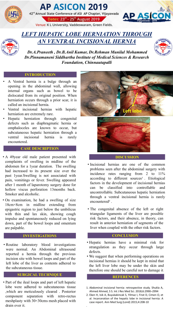
LEFT HEPATIC LOBE HERNIATION THROUGH AN VENTRAL INCISIONAL HERNIA Dr.A.Praneeth , Dr.B.Anil Kumar, Dr.Rehman Munilal Mohammed Dr.Pinnamaneni Siddhartha Institute of Medical Sciences & Research Foundation, Chinnautapalli INTRODUCTION • A Ventral hernia is a bulge through an opening in the abdominal wall, allowing internal organs such as bowel to be dislocated from its original position. When herniation occurs through a prior scar, it is called an incisional hernia. • Ventral incisional hernias with hepatic herniation are extremely rare. • Hepatic herniation through congenital defects such as diaphragmatic hernia or omphaloceles are known to occur, but subcutaneous hepatic herniation through a ventral incisional hernia is rarely encountered. Left lobe CASE DESCRIPTION • A 49year old male patient presented with complaints of swelling in midline of the abdomen for a 1year duration. The swelling had increased to its present size over the past 1year.Swelling is not associated with pain, vomitings or fever. Swelling appeared after 1 month of laparotomy surgery done for hollow viscus perforation 13months back. Smoker and alcoholic. • On examination, he had a swelling of size 18cm×8cm in midline extending from epigastric region to just below the umbilicus with thin and lax skin, showing cough impulse and spontaneously reduced on lying down, part of the bowel loops and omentum are palpable. DISCUSSION • Incisional hernias are one of the common problems seen after the abdominal surgery with incidence rates ranging from 2 to 11% according to different sources¹[. Etiological factors in the development of incisional hernias can be classified into controllable and uncontrollable. Subcutaneous hepatic herniation through a ventral incisional hernia is rarely encountered² . • The congenital absence of the left or right triangular ligaments of the liver are possible risk factors, and their absence, in theory, can result in anterior herniation of segments of the liver when coupled with the other risk factors. INVESTIGATIONS CONCLUSION • Routine laboratory blood investigations were normal. An Abdominal ultrasound reported a hernia through the previous incision site with bowel loops and part of the left lobe of the liver as contents adhered to the subcutaneous tissue. • Hepatic hernias have a minimal risk for strangulation as they occur through large defects. • We suggest that when performing operations on incisional hernias it should be kept in mind that the left liver lobe may be under the skin and therefore one should be careful not to damage it. SURGICAL TECHNIQUE • Part of the ileal loops and part of left hepatic lobe were adhered to subcutaneous tissue ,which are meticulously cleared . Posterior component separation with retro-rectus meshplasty with 30×30cms mesh placed with drain over it. REFERENCES 1. Abdominal incisional hernia: retrospective study. Shukla A, Ahmed Ahmed, S S. Int J Res Med Sci. 2018;6:2990–2994 2. Eken H, Isik A, Buyukakincak S, Yilmaz I, Firat D, Cimen O, et al. Incarceration of the hepatic lobe in incisional hernia: A case report. Ann Med Surg (Lond) 2015;4:208-10 .
