International Journal of Trend in Scientific Research and Development (IJTSRD)
Volume 5 Issue 1, November-December 2020 Available Online: www.ijtsrd.com e-ISSN: 2456 – 6470
Taxonomic Identification of Micro-Organisms Growing on and
Cause Biodeterioration of Cultural Heritage of Agra and
Mathura Region
Dr. Anuradha Chauhan1, Ravindra Goswami2, Dr. Seema Bhadauria2
1Department
of Botany, SBS (PG) College, Umedpur, Etah, Uttar Pradesh, India
of Botany, RBS College, Agra, Uttar Pradesh, India
2Department
ABSTRACT
Fungi and bacteria have been known to degrade dye particles and other
coloring agents. However in the present investigation fungal enzymes
deteriorate monument walls and make them ugly. The various historical
monuments have been made cracked by fungal and bacterial enzymes like
lipase, cellulose, ligninase, pectinase etc. These are secreted by their cell wall.
Fungi and bacteria release these enzymes and in presence of moisture and
suitable temperature and environmental conditions degrade and break the
walls of monuments by deteriorating their rocks and calcium particles. Also
various magnesium particles are broken by fungal and bacterial enzymes by
growth of fungi in long period of time. The fungi and bacteria have the ability
to grow fast also and they continue their growth in historical monuments.
How to cite this paper: Dr. Anuradha
Chauhan | Ravindra Goswami | Dr. Seema
Bhadauria "Taxonomic Identification of
Micro-Organisms Growing on and Cause
Biodeterioration of Cultural Heritage of
Agra and Mathura
Region" Published in
International Journal
of Trend in Scientific
Research
and
Development (ijtsrd),
ISSN:
2456-6470,
IJTSRD38238
Volume-5 | Issue-1,
December 2020, pp.1293-1295, URL:
www.ijtsrd.com/papers/ijtsrd38238.pdf
Copyright © 2020 by author (s) and
International Journal of Trend in Scientific
Research and Development Journal. This
is an Open Access article distributed
under the terms of
the
Creative
Commons Attribution
License
(CC
BY
4.0)
(http://creativecommons.org/licenses/by/4.0)
INTRODUCTION
We can see many fungi and bacteria of different colors
growing in monumental walls. Hence our historical
monuments get deteriorated. No care is taken by spraying
antifungals or antibacterials. Hence the monuments have lost
their beauty. Many fungi like Aspergillus niger, Cladosporium
spahaerospermum, Trichoderma harzianum, Albugo candida,
Aspergillus flavus, Aspergillus fumigatus etc. have their
dominance on the walls of historical monuments. Bacteria
like Streptococcus, Streptobacillus, Vibrio, Clostridium etc.
also cut the beauty of monumental walls.[10]
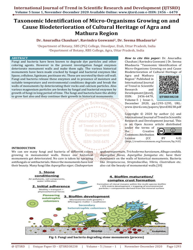
Fig-1: Fungi and bacteria degrading stone monuments (process)
@ IJTSRD
|
Unique Paper ID – IJTSRD38238
|
Volume – 5 | Issue – 1
|
November-December 2020
Page 1293
International Journal of Trend in Scientific Research and Development (IJTSRD) @ www.ijtsrd.com eISSN: 2456-6470
We took the different colored fungi and bacteria by forceps
in petriplates filled with Potato Dextrose Agar, Sabouraud’s
dextrose agar or blood agar medium and brought them to
the laboratory for identification and dominance. [1]
OBSERVATION AND DISCUSSION
Table -1 fungal species identified from isolation from
historical monuments in Agra
S. No.
Fungal species
Colony count
1.
Aspergillus niger
10
2.
Aspergillus flavus
8
3.
Aspergillus fumigatus
7
4.
Candida albicans
7
5.
Fusarium oxysporum
6
6.
Rhizopus nigricans
9
7.
Cladosporium sphaerospermum
6
8.
Alternaria Solani
5
9.
Geotrichum indicum
4
10. Trichoderma harzianum
7
10 fungal species were obtained from Agra monuments on
an average.[8,9] Out of these Aspergillus niger was the most
dominant. This means the air borne spores of fungi were
maximum of those of Aspergillus niger followed by Rhizopus
nigricans.[2]
Fig.3: Mixed bacterial colonies in petriplates
Fig.4: the Taj Mahal losing its sheen in Agra
Table -2: Bacterial species identified from isolation
from historical monuments in Agra
S. No.
Fungal species
Colony count
1.
Clostridium botulinum
5
2.
Bacillus spp.
8
3.
Streptococcus spp.
12
4.
Streptobacillus spp.
5
5.
Salmonella typhus
6
In case of bacteria Streptococcus spp had maximum colonies.
Fig.2: Mixed fungal colonies in petriplate
Fig.5: Krishna Janmabhoomi in Mathura
Among all these species, Aspergillus niger was the most
dominant, followed by Rhizopus nigricans. The fungi were
taken from different areas and by forceps put in petriplates.
Then the number of colonies was counted. This shows that
these fungi degrade the wall of historical monuments. The
wall of monuments is made of calcium, magnesium basically
cement. Fungal enzymes are so strong that they dissolve the
wall in years. As care is not taken fungal enzymes keep
deteriorating walls of monuments. [3]
@ IJTSRD
|
Unique Paper ID – IJTSRD38238
|
Table-2: Fungal species isolated from historical
monuments in Mathura
S. No.
Fungal species
Colony count
1.
Aspergillus niger
10
2.
Aspergillus flavus
6
3.
Aspergillus fumigatus
8
4.
Candida albicans
6
5.
Fusarium oxysporum
7
6.
Rhizopus nigricans
8
7.
Cladosporium sphaerospermum
5
8.
Alternaria Solani
3
9.
Geotrichum indicum
6
10. Trichoderma harzianum
9
11. Colletotrichum albicans
7
12. Trichothecium roseum
8
Volume – 5 | Issue – 1
|
November-December 2020
Page 1294
International Journal of Trend in Scientific Research and Development (IJTSRD) @ www.ijtsrd.com eISSN: 2456-6470
In case of monuments in Mathura on an average the colony count of Aspergillus niger was again the highest. This was followed
by Trichoderma harzianum. Hence we can say that Aspergillus niger spores were again widespread in the air of Mathura.[4]
Table-3: bacterial species obtained from monuments of Mathura
S. No.
Bacteria
Colony count
1.
Streptococcus spp.
9
2.
Streptobacillus spp.
6
3.
Salmonella typhus
7
Here again Streptococcus spp. had maximum colony count.
Fig.6: fungal identification and sensitivity
CONCLUSION
Strict measures should be taken to prevent fungi from
degrading historical monumental walls:
1. Regular antifungal sprays like Bordeaux mixture and
antibacterial sprays
2. Washing and cleaning of monumental walls
3. Strict checking of monuments regularly
4. Painting and cementing wherever degradation has
occurred[5]
The precious historical walls are fun for tourists and
economy for India. They require to be preserved. There are
fungal and bacterial airborne spores which attach
themselves to the walls of historical monuments and
degrade them. Regular checking and preservation of
monuments is necessary as they are economically useful
tourist spots.[6,7]
REFERENCES
[1] Pepe O, Sannino L, Palomba S, Anastasio M, Blaiotta G,
Villani F, et al. Heterotrophic microorganisms in
deteriorated medieval wall paintings in southern
Italian churches. Microbiological research. 2010;
165(1):21–32. pmid:18534834
[2]
[3]
Cuezva S, Fernandez-Cortes A, Porca E, Pasic L, Jurado
V, Hernandez-Marine M, et al. The biogeochemical
role of Actinobacteria in Altamira Cave, Spain. FEMS
Microbiol Ecol. 2012; 81(1):281–90. pmid:22500975
Lan W, Li H, Wang WD, Katayama Y, Gu JD. Microbial
community analysis of fresh and old microbial
biofilms on Bayon temple sandstone of Angkor Thom,
@ IJTSRD
|
Unique Paper ID – IJTSRD38238
|
Cambodia. Microb
pmid:20593173
Ecol.
2010;
60(1):105–15.
[4]
Saiz-Jimenez C, Miller AZ, Martin-Sanchez PM,
Hernandez-Marine M. Uncovering the origin of the
black stains in Lascaux Cave in France. Environ
Microbiol. 2012; 14(12):3220–31. pmid:23106913
[5]
Gaylarde P, Gaylarde C. Deterioration of siliceous
stone monuments in Latin America: Microorganisms
and mechanisms. Corros Rev. 2004; 22(5–6):395–
415.
[6]
Gaylarde CC, Ortega-Morales BO, Bartolo-Perez P.
Biogenic black crusts on buildings in unpolluted
environments. Curr Microbiol. 2007; 54(2):162–6.
pmid:17211538
[7]
Saiz-jimenez C. Deposition of Airborne Organic
Pollutants on Historic Buildings. Atmos Environ BUrb. 1993; 27(1):77–85.
[8]
Flores M, Lorenzo J, Gomez-Alarcon G. Algae and
bacteria on historic monuments at Alcala de Henares,
Spain. Int Biodeter Biodegr. 1997; 40(2–4):241–6.
[9]
Scheerer S, Ortega-Morales O, Gaylarde C. Chapter 5
Microbial Deterioration of Stone Monuments—an
Updated Overview. 2009; 66:97–139.
[10]
Saiz-jimenez C. Microbial Melanins in Stone
Monuments. Sci Total Environ. 1995; 167:273–86.
[11]
Tiano P, Accolla P, Tomaselli L. Phototrophic
Biodeteriogens on Lithoid Surfaces—an Ecological
Study. Microbial Ecol. 1995; 29(3):299–309.
Volume – 5 | Issue – 1
|
November-December 2020
Page 1295


