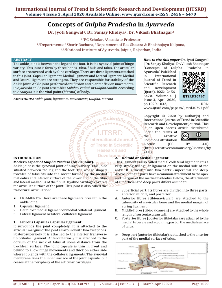
International Journal of Trend in Scientific Research and Development (IJTSRD) Volume 4 Issue 3, April 2020 Available Online: www.ijtsrd.com e-ISSN: 2456 – 6470 Concepts of Gulpha Pradesha in Ayurveda Dr. Jyoti Gangwal1, Dr. Sanjay Kholiya2, Dr. Vikash Bhatnagar3 1,2PG Scholar, 3Associate Professor, 1,3Department of Sharir Rachana, 2Department of Ras Shastra & Bhaishajaya Kalpana, 1,2,3National Institute of Ayurveda, Jaipur, Rajasthan, India How to cite this paper: Dr. Jyoti Gangwal | Dr. Sanjay Kholiya | Dr. Vikash Bhatnagar "Concepts of Gulpha Pradesha in Ayurveda" Published in International Journal of Trend in Scientific Research and Development (ijtsrd), ISSN: 24566470, Volume-4 | IJTSRD30797 Issue-3, April 2020, pp.1029-1032, URL: www.ijtsrd.com/papers/ijtsrd30797.pdf ABSTRACT The ankle joint is between the leg and the foot. It is the synovial joint of hinge variety. This joint is form by three bones- tibia, fibula and talus. The articular surface are covered with hyaline cartilage. There are three ligaments attached to this joint- Capsular ligament, Medial ligament and Lateral ligament. Medial and lateral ligament are strongest. They are responsible for stability of the Ankle Joint. Ankle joint performs dorsiflexion and plantar flexion movements. In Ayurveda ankle joint resembles Gulpha Pradesh or Gulpha Sandhi. According to Acharyas it is the vital point (Marma) of body. KEYWORDS: Ankle joint, ligaments, movements, Gulpha, Marma Copyright © 2020 by author(s) and International Journal of Trend in Scientific Research and Development Journal. This is an Open Access article distributed under the terms of the Creative Commons Attribution License (CC BY 4.0) (http://creativecommons.org/licenses/by /4.0) INTRODUCTION Modern aspect of Gulpha Pradesh (Ankle joint)Ankle joint is the synovial joint of hinge variety. This joint situated between the leg and the foot. The wedge shaped trochlea of talus fits into the socket formed by the medial malleolus and inferior surface of the lower end of the tibia and lateral malleolus of the fibula. Hyaline cartilage covered the articular surface of the joint. This joint is also called the “talocrural articulation”. 1. 2. 3. LIGAMENTS- There are three ligaments present in the ankle joint. Capsular ligament. Deltoid or medial ligament or medial collateral ligament. Lateral ligament or lateral collateral ligament. 1. Fibrous Capsule/ Capsular ligament It surrounds the joint completely. It is attached to the articular margins of the joint all around with two exceptions. Posterosuperiorly it is attached to the inferior transverse tibiofibular ligament. Anteroinferiorly it is attached to the dorsum of the neck of talus at some distance from the trochlear surface. The joint capsule is thin in front and behind to allow hinge movements and thick on either side where it blends with the collateral ligaments. The synovial membrane lines the inner surface of the joint capsule, but ceases at the periphery of the articular cartilages. @ IJTSRD | Unique Paper ID – IJTSRD30797 | 2. Deltoid or Medial Ligament This ligament is also called medial collateral ligament. It is a very strong triangular ligament on the medial side of the ankle. It is divided into two parts: -superficial and deep. Above, both the parts have a common attachment to the apex and margins of the medial malleolus. Below, the attachment of superficial and deep parts differs as under: • Superficial part: Its fibres are divided into three parts: anterior, middle, and posterior. A. Anterior fibres (tibionavicular) are attached to the tuberosity of navicular bone and the medial margin of spring ligament. B. Middle fibres (tibiocalcanean) are attached to the whole length of sustentaculum tali. C. Posterior fibres (posterior tibiotalar) are attached to the medial tubercle and adjoining part of the medial surface of talus. • Deep part (anterior tibiotalar) is attached to the anterior part of the medial surface of talus. Volume – 4 | Issue – 3 | March-April 2020 Page 1029 International Journal of Trend in Scientific Research and Development (IJTSRD) @ www.ijtsrd.com eISSN: 2456-6470 3. Lateral Ligament The lateral ligament also called lateral collateral ligament. It consists of three parts: -Anterior talofibular, posterior talofibular, and calcaneofibular. 1. Anterior talofibular ligament is a weak flat band, which extends forward and medially from the anterior margin of lateral malleolus, to the neck of talus just in front of the fibular facet. 2. Posterior talofibular ligament is a strong band, which extends backward and medially from the posterior margin of the lateral malleolus to the posterior tubercle of the talus. 3. Calcaneofibular ligament is a long rounded cord, which runs downward and backward from the notch on the lower border of lateral malleolus to the tubercle on the lateral surface of the calcaneum. Movements - The following movements take place at the ankle joint: 1. Dorsiflexion 2. Plantar flexion MOVEMENTS PRINCIPAL MUSCLES Dorsiflexion Tibialis anterior muscle Planter Flexion 1. Gastrocne mius muscle 2. Soleus muscle ACCESSORY MUSCLES 1. Extensor Digitorum longus muscle 2. Extensor hallucis longus muscle 3. Peroneus tertius muscle 1. Plantaris muscle 2. Tibialis posterior muscle 3. Flexor Hallucis Longus muscle 4. Flexor Digitorum Longus muscle Arterial SupplyIt is by the malleolar branches of anterior tibial, posterior tibial and peroneal arteries. Stability of the Ankle Joint- Stability of this joint depends on some factors, mention as below Close interlocking of its articular surfaces. Deepening of tibiofibular socket posteriorly by the inferior transverse tibiofibular ligament. Strong, medial, and lateral collateral ligaments. Tendons (four in front and five behind) crossing the ankle joint. Relations of the Ankle Joint (transverse section of the Ankle Joint)Anterior: Anteriorly from medial to lateral side the ankle joint is related to the following structures: 1. Tibialis anterior. 2. Extensor Hallucis longus. 3. Anterior tibial Artery. 4. Deep peroneal Nerve. 5. Extensor Digitorum longus. 6. Peroneus tertius. Posterior: Posteriorly from medial to lateral side the ankle joint is related to the following structures: 1. Tibialis posterior 2. Flexion Digitorum longus. 3. Posterior tibial Artery. 4. Posterior tibial Nerve. 5. Flexor Hallucis longus. Nerve SupplyBranches of deep peroneal and tibial nerves. Clinical correlation 1. Ankle sprains :The excessive stretching and tearing of ligaments of the ankle joint is called the ankle sprain. The ankle sprains are normally caused by the falls from height or twists of ankle. When the plantar-flexed foot is excessively inverted, the anterior and posterior talofibular and calcaneofibular ligaments are stretched and torn. The anterior talofibular ligament is most commonly torn. 2. Dislocation of the ankle:The dislocations of ankle joint are rare because it is a very stable joint due to tibiofibular mortise. However, whenever dislocation occurs it is always accompanied by the fracture of one of the malleoli. 3. Pott’s fracture or Dupuytren fractureIt occurs when the foot is caught in the deep hole and everted forcefully. In this condition, the following sequence of events takes place:1. Oblique fracture of the lateral malleolus due to internal rotation of the tibia. @ IJTSRD | Unique Paper ID – IJTSRD30797 | Volume – 4 | Issue – 3 | March-April 2020 Page 1030 International Journal of Trend in Scientific Research and Development (IJTSRD) @ www.ijtsrd.com eISSN: 2456-6470 2. 3. Transverse fracture of the medial malleolus due to pull by deltoid ligament. Fracture of the posterior margin of the lower end of tibia because it is carried forward. These stages are also termed first, second, and third degree of Pott’s fracture, respectively. The third degree of Potts fracture is also called trimalleolar fracture. lohitaakshaani vitapan cheti, etenetarasakthi vyaakhyaataam | (su.sha. 6/6) Kshipra, Talahridaya, Kurcha, Kurchashira, Gulpha, Indrabasti, Janu, Ani, Urvi, Lohitaksha and Vitapa-one each. The same are in the other leg. • Sandhi marma- 20, gulph- 2 Jānukūrparasīmantādhipatigulphamaṇibandhakukundarā var takr̥ kā ṭikāścēti sandhimarmāṇi | (su.sha. 6/7, a.hr. sha. 5/44) Janu, Kurpara, Simanta, Adhipati, Gulpha, Manibandha, Kukundara, Avarta and Krikatika are Sandhi Marmas. Rujākaramarma- 8, gulpha- 2 gulphau dvau manibandhau dvau dve dve koorchashiraansi ch | (su.sha.6/14) Gulpha (two), Manibandha (two), Kurchashira (२ + २)these eight Rujakara Marmas, cause pain. Ayurvedic aspect of Gulpha PradeshNiruktiयोपारोपतकोशांशःकपम ु ः- गुफः, पुं, (गल + “क लग ल यां फगयोच ।” उणां । ५ । २६ अमरकोशः-गुफ पुं। | पादथी | समानाथक:घु"टका,गुफ 2।6।72।1।2 वाचपयम ्- '''गुफ'''¦ पु॰ गुल--फक् &न॰ अयोच। पादथौ। Pramana (measurement) of Gulpha Chaturdashangulparanihani Padgulfjangha (Su.su.35 / 12) 14 finger circumference ...... | Total no. of Sandhi, Snayu, Peshi, Jaal, Sanghata,Simanta, Marma in Gulpha Pradesha Sandhi 210, in branches – 68, Jānugulphavaṅkṣaṇēṣvēkaikaḥ (Su.sha .5 / 28) • Sandhi prakāra tēṣāmaṅgulimaṇibandhagulphajānukūrparēṣu kōrāḥ sandhayaḥ| (su.Śā. 5/32) • Types of sandhi - These joints are of eight types- Kora, Ulukhala, Samudga, Pratara, TunnaSevani, Vayasatunda, Mandala and Shankhavarta. Of them, Kora joints are found in fingers, wrists, ankles, knees and elbows. • Kora sandhi (Condyloid) -4 blades Khalla, sadansa, cakrakōra, paraspara (pratyakṣa śārīra) Snāyu – 900, śākhāgata snāyu -600 tala, kūrca, gulpha – 30 | (su.Śā.5/35) Pēśiyā- 500, śākhāgata pēśiyā – 400 daśa gulphatalayōḥ, gulphajānvantarē vinśatiḥ | (su.Śā.5/46) Jāla-16, gulpha- 4-4 pratyēkaṁ catvāri; tāni maṇibandhagulphasanśritāni | (su.Śā. 5/12) Saṅghāta-14, gulpha 1-1 (su.Śā. 5/16) seemant-14, gulph 1-1 (su.sha.5/17) Marma- 107, According to shadang - shaakhaagat marm44, sakthigat marm-11 • Tatr sakthimarmaani kshipr-talahrday-koorchkoorchashiro-gulfendrabasti jaanvaanyoorva- @ IJTSRD | Unique Paper ID – IJTSRD30797 | Pramana of marma Vid'dhyaṅguladvayamitaṁ maṇibandhagulphaṁ | (su.sha. 6/29 , a.hr.sha. 4/60) Manibandha and Gulpha are two fingers each (gulph -2 angula) lakshan of asthi saar purush paarshnigulphajaanu......parvasthoola: | (ch.vi. 8/107) Signs of long life age boy naatyupachitau naatyapachitau gulphau | (ch.sha. 8/51 Neither full of muscle nor covered with less muscle, that Gulpha or ankle is superior. naanaatmaj vikaar- gulphagrahashch | (ch.su. 20/11 Sparśagamya ariṣṭa – Gulphajānuvaṅkṣaṇa………….. Srastāni vyastāni cyutāni sthānēbhyaḥ | (ca.I. 3/5) If we touches the the patient’s Gulf, Janu, Vankshana etc. and it feels relaxed or separated from the place of joint then that man will die soon. Treatment of Gridhrasi roga Antarākaṇḍarāgulphaṁ sirā bastyagnikarma ca| gr̥ dhrası̄ṣu prayuñjīta || (ca.Ci. 28/101) In Gridhrasi disease,siravedhana between the tendon and the gulpha should be done,and anuvasana and niruha basti given and cauterization between the tendon and the gulpha should be done. Niruha basti kē yōgya rōgī Sphigjānujaṅghōrugulphapārṣṇi…………parvāsthiśūla….| (Ca.Si. 2/16) symptoms of antrayama Aṅ gulı̄gulphajaṭ harahr̥ dvakṣ ōgalasanś ritaḥ | snā yupratā namanilō yadā ̕̕kṣ ipati vē gavā n | (su.Ni.1/54) The vitiated vayu (air) is established in the fingers, gulpha, abdomen, heart, thorax and throat and attacks the siras and snayus of the extremities. Symptoms of trauma on marma Gulphasandhēradha ubhayata: Kūrcaśirō nāma, tatra rujāśōphau| (su.Śā. 6/25) On both sides of the gulpha, there is a marma called Kurchashira, on which trauma causes pain and edema. Volume – 4 | Issue – 3 | March-April 2020 Page 1031 International Journal of Trend in Scientific Research and Development (IJTSRD) @ www.ijtsrd.com eISSN: 2456-6470 Pādajaṅghayō : Sandhā nē gulphō nā ma, tatra ruja: Stabdhapā datā khan̄ jatā vā | (su.Śā.6/25) There is a gulpha marma in the joints of the foot and the legs on which trauma causes pain and aggravation or stigma. Symptoms of sandhi vidhha marma vastu shookairivaakeernam roodhe ch kunikhanjata | balacheshtaakshay: shosh: parvashophashch sandhije || (a.hr.sha. 4/51) The place of trauma appears to be full of spikes and produced Kunita or khanjata and the force and effort is destroyed, atrophy and swelling in the joints. Purvaroopa of vata rakta Shyaavaarunamandalotpattishchaakasmaat paanipaadatalaanguligulphamanibandhaprabhrtishu | (su.chi. 5/4) In reference to siravedhanaKroshatukashirsha Khañjapaṅgulavātavēdanāsu jaṅghāyāṁ gulphasyōpari caturaṅgulē | (su.Śā. 8/17) Caturaṅgulē ūrdhvaṁ gulphasya sakthyartau, tathā krōṣṭukaśīrṣakē | (a.Hra.Sū. 27/16) Treatment of apachi Ā gulphakarṇāt sumitasya jantōstasyāṣṭabhāgaṁ khuḍakādvibhajya | ghōṇarjuvēdhaḥ | (su.Ci. 18/26) From the ear (ankle) of the gulpha, measure it thoroughly and make a straight incision similar to the nostril except for the location of the eighth section (Indrabasti marma). Treatment of shlipada ś lı̄padē ̕nilajē bhiṣak | kr̥ tvā gulphō pari sirā ṁ vidhyēttu caturaṅgulē | (su.Ci. 19/54) Gulphasyādhaḥ sirāṁ vidhyēcchlīpadē pittasambhavē | (su.Ci. 19/55) Reference– [1] Ashtanga Hridaya english translation by Prof. K. R. Shrikantha Murthy, vol. 1 Sutra sthana and Sharir sthana, Krishanadas academy, Varanasi, edition 1994. @ IJTSRD | Unique Paper ID – IJTSRD30797 | [2] Charaka samhita English translation by Prof. Priyavrat sharma vol. 1 Sutra Sthana to Indriya Sthana, Chaukhambha orientalia, Varanasi. [3] Charaka samhita with Vidyotini hindi commentary by Pt. Kashinath shastri & Dr. Gorakhanatha Chaturvedi, Part-1&2, Published by Chaukhambha Bharati Academy Varanasi, 22nd Edition 1996. [4] Clinical Anatomy by Regions, Richard S. Snell, 8th Edition, Published by Wolters Kluwer (India) Pvt. Ltd, New Delhi. [5] Clinically Oriented Anatomy, Keith L. Moore, Arthur F. Dalley, Anne M.R. Agur, 6th Edition, Published by Lippincott Williams & Wilkins. [6] Grants method of anatomy, John V. Basmajian and Charles E. Slonecker, 11th edition, Williams and Wilkins. [7] Gray’s Anatomy, 38th edition, Peter L. Williams (Chairman editorial board), London, UK; churchill Livingstone, British Library. [8] Human Anatomy, Chaurasia B. D., Vol. II, 4th edition, New Delhi ; Satish Kumar Jain for CBS publications and distributors, Darya Ganj, 2004, [9] Shabdakalpadrum; Vol (1-5) by Raja Radha Kanta Deva; Choukhamba Sanskrit Series; Varanasi. [10] Sushruta Samhita English translation by Prof. K. R. Shrikantha Murthy, vol.2 Sharir Sthana, Chaukhambha orientalia, Varanasi. [11] Sushruta Samhita English translation by Prof. K. R. Shrikantha Murthy, vol.1 Sutra Sthana and nidana Sthana, Chaukhambha orientalia, Varanasi. [12] Sushruta Samhita of Sushruta with Ayurveda Tatva Sandipika hindi commentary by Kaviraja Ambika Dutta Shastri, Chaukhambha Sanskrit Sansthan Varanasi, Part 1-2 9th edition 1995. [13] Pratyaksha sharir, Sanskrit translation by Acharya Gannatha Sen, vol. 1. [14] https://en.wikipedia.org/wiki/Ankle Volume – 4 | Issue – 3 | March-April 2020 Page 1032
