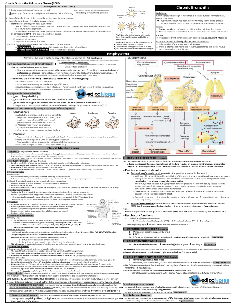
Chronic Obstructive Pulmonary Disease (COPD) Pathogenesis of COPD : (IHLL) Inflammation & fibrosis of the bronchial wall, 2- Hypertrophy of the submucosal glands and hypersecretion of mucus 1- 34- Chronic Bronchitis Both lead to obstruction of airflow & cause : mismatching of ventilation & perfusion. Definition : Chronic hronic producCve cough of more than 3 months’ duraCon for more than 2 consecutive years . Typically,the cough has been present for many years, with a gradual increase in acute exacerbations that produce frankly purulent sputum. Loss of alveolar tissue decreases the surface area for gas exchange. Loss of elastic fibers leads to airway collapse. Normally the elas c fibers have 2 func ons: 1- Recoil of elastic fibers that were stretched during inspiration provides the force needed to move air out of the lung during expiration. 2- Elastic fibers are attached to the airways,providing radial traction to hold airways open during expiration In persons with COPD : the loss of elastic fibers causes: 1- Predisposes edisposes to airway collapse. 2- Increases air trapping. 3- Impairs the expiratory flow rate. The term COPD encompasses two types of obstructive airway disease: • Emphysema, with enlargement of air spaces and destruction of lung tissue. • Chronic obstructive bronchitis, with obstruction of airways. Types : 1- Simple bronchitis Chronic bronchitis tis without airflow obstruction. 2- Chronic hronic obstructive bronchitis chronic bronchitis with airflow obstruction. (Top) Normal bronchial airway with elastic fibers that provide traction and hold the airway open. (Bottom)) Obstruction of the airway caused by : (A)) hypertrophy of the bronchial wall, (B)) inflammation and hypersecretion of mucus (C) loss of elastic fibers ibers that hold the airway open Causes : • It's associated with chronic irritation from smoking & recurrent infections. infection In chronic bronchitis, airway obstruction is caused by : 1- Inflammation nflammation of the major and small airways. 2- There is edema and hyperplasia of submucosal glands and excess mucus excretion into the bronchial tree. Emphysema Normally, the lung is p protected by antiprotease enzymes as : α1-antitrypsin Two recognized causes of emphysema : 1- Increased elastase production: In smokers in whom COPD develops : - Inadequate antiprotease production and release - Excess protease production to neutralize • Cigarette smoke stimulate movement of inflammatory cells into the lungs lungs,, resulting in increased release of proteinases as: elastase ( se serine elastase from neutrophils / metalloelastase from alveolar macrophages ) that digests elastin resulting in breakdown of elastin and other alveolar wall components. components 2- Inherited deficiency of α1--proteinase inhibitor (α α1 – antitrypsin): antitrypsin) • Accounts for 1% of all cases of COPD. • More ore common in young persons before age of 40 years . • Smoking & repeated respiratory tract infections, decrease α1-antitrypsin antitrypsin levels • Human α1-antitrypsin is available ailable for replacement therapy. risk for emphysema . Emphysema is characterized by : 1- Loss of lung elasticity 2- Destruction estruction of the alveolar walls and capillary beds. 3- Abnormal bnormal enlargement of the air spaces distal to the terminal bronchioles, Enlargement of the air spaces leads to hyperinflation of the lungs produces an increase in (TLC). There are two commonly recognized types of emphysema: 1- Centriacinar : • Affects the bronchioles in the central part of the respiratory lobule ( terminal bronchioles (TB) & respiratory bronchioles (RB) ), with initial preservation of the alveolar ducts and sacs. • Most ost common type of emphysema emphysema. Seen een predominantly in male smokers smokers. • Centriacinar entriacinar changes in upper parts of the lung. 2- Panacinar • Produces roduces initial involvement of the peripheral alveoli Later extends to involve the more central bronchioles. • More ore common in persons with α1-antitrypsin deficiency. Also lso found in smokers in association with centrilobular emphysema. • Panacinar changes are seen in lower parts of the lung Clinical Manifestations 1. Dyspnea : In Emphysemaa & chronic bronch bronchitis - Persons with emphysema have marked dyspnea and struggle to maintain normal blood gas levels with increased ventilatory effort (overventilate) , including prominent use of the accessory muscles. leads to - The seated position,which stabilizes chest structures and allows for maximum chest expansion and use of accessory muscles, is preferred. 2. Productive chough : In Chronic Bronchitis The lung’s reduced ability to retract ((flaccid lung)) can lead to obstructive lung disease, because : - Cough is productive of thick, purulent sputum owing to ongoing local inflammation & infection. reduced elastic recoil (increased compliance) of the lung requires an increase in intrathoracic pressure for - Sputum viscosity is increased largely as a result of presence of free DNA (of high molecular weight and highly viscous) from lysed cells. expiration, resulting in compression of the intrathoracic airways massive increase in flow resistance. - Hemoptysis with increased inflammation ation and mucosal injury. - Cough is much less effective owing to 1- narrow airway caliber & 2- greater volume and viscosity of secretions. • Positive pressure in alveoli : 1- Reduced elastic recoil: 3. Breath sounds : 1-- Reduced lung’s elastic recoil generates the positive pressure in the alveoli : - With loss of lung elasticity and hyperinflation of the lungs greater reater intrathoracic pressure is necessary for expiration because compliance and resistance are increased This causes compression of the bronchioles so, airway pressure increases further. further - The he airways often collapse during expiration because pressure in surrounding lung tissues exceeds airway pressure Air becomes trapped in lungs, producing an increase in the anteroposterior anteropos dimensions of the chest, the so-called barrel chest. - Elastic recoil can be raised by increasing the inspiratory volume leading to a shift in the resting position toward inspiration (barrel barrel chest). chest In Emphysema : - Decreased in intensity of breathing sound reflecting decreased airflow. - Wheezes, when present , are of diminished intensity - Crackles & rhonchi,, in superimposed processes as infection In Chronic Bronchitis : Persistent airway narrowing & mucus obstruction : - Produce localized or diffuse wheezing ( responsive to bronchodilators). - Prolonged expiratory time . - Inspiratory & expiratory coarse crackles : mucus production + defective mucociliary clearance excessive secretions in the airways. 4. Cardiac examination : - Tachycardia as in chronic bronchitis, especially with exacerbations of bronchitis or hypoxemia. - Pulmonary hypertension If hypoxemia mia is significant and chronic,,, Cardiac examination may reveal : • Prominent pulmonary valve closure (increased P2, pulmonary component of the 2nd heart sound) or • Elevated jugular venous pressure AND peripheral edema resulting from Rt heart failure. - Pursed-lip breathing, increases the resistance to the outflow of air, increasing airway pressure. 5. Imaging : - 2-- External compression produces positive pressure in the alveoli by contraction of expiratory muscles, BUT this will also compress the bronchioles thus bring a massive increase in flow resistance. resistance Hyperinflation with 1- fla8ened hemidiaphragms 22- anteroposterior chest diameter. Parenchymal destruction produces : attenuated peripheral vascular markings. Pulmonary hypertension produces : proximal pulmonary artery dilation . Cardiac size may be increased, suggesting right heart volume overload overload. Cystic or bullous changes. THUS : Maximal expiratory flow rate (V max) is a function of the ratio between elastic recoil (K) and resistance (RL) . • 6. Pulmonary function tests : In Emphysema : • The loss of elastic recoil in lung tissue supporting the airways results in increased dynamic compression of airways, (especially during forced expiration) all flow rates are reduced:With With premature airway collapse, FEV1, FVC, FEV1/FVC (FEV1% ratio) . • "Expiratory flow-volume volume curve" shows substantial limitation in flow flow. In Chronic Bronchitis : • Diffuse airway obstruction is demonstrated as a global reduction in expiratory flows & volumes. FEV1, FVC, FEV1/FVC (FEV1%) • expiratory flow-volume volume curve" shows substantial limitation in flow flow. preventing airway collapse by Respiratory function : - If tidal volume(TV) remains constant : 1- functional residual capacity (FRC) - V Vital capacity (VC) is 2 2- residual volume (RV) 3- dead space. because of the reduced expiratory volume. 2- Bronchial obstruction : leads to . - Increased RV and FRC, reflecting air trapped in the lung as a result of : a- Diffuse iffuse airway obstruction (chronic bronchitis) b- Early airway closure caused by loss of elastic recoil (emphysema) - TLC is increased substantial amount of this increase comes from gas trapped in poorly lung units, including bullae. 1maximum breathing capacity (V˙ max) 2FEV1 . 3- differing ventilation of various alveoli results in abnormal distribution resulting in : Hypoxemia. Hypoxemia 3- Loss of alveolar wall : leads to 1- diminished diffusion area abnormal diffusion of gases resulting in : Hypoxemia . 7. Arterial blood gases : In Emphysema : (Emphysema is a disease of alveolar wall destruction) - The loss of the alveolar capillaries creates: areas of high ventilation relative to perfusion. - They may able to maintain nearly normal PO2 and PCO2 levels despite advanced disease - Hypercapnia, respiratory acidosis, and a compensatory metabolic alkalosis are common in severe disease. In Chronic Bronchitis : - In contrast to persons with emphysema, those with chronic obstructive bronchitis are unable to maintain normal blood gases by increasing their breathing effort " Ventilation Ventilation-perfusion mismatching " is common in chronic bronchitis. - Hypoxemia (arterial arterial PO2 levels fall below 55 mm Hg) at rest tends to be more profound than in emphysema causes reflex vasoconstriction of the pulmonary vessels persons with chronic obstructive bronchitis develop pulmonary hypertension and, eventually, right-sided sided heart failure with peripheral edema ((i.e., cor pulmonale). - Hypercapnia, Cyanosis, respiratory acidosis, and a compensatory metabolic alkalosis alkalosis. 8. Polycythemia : Chronic hypoxemia (especially in chronic bronchitis) is associated with erythropoietin-mediated erythropoietin increase in hematocrit. The hypoxia of underventilated alveoli leads to pulmonary hypertension, vasoconstriction, increased pulmonary vascular resistance, an increased right ventricular load (cor pulmonale). pulmonale) 4- Loss of pulmonary capillaries : leads to 1- increase in functional dead space 2- increased pulmonary artery pressure and vascular resistance with development of " cor pulmonale" • The fluid retention and peripheral oedema is due to failure of excretion of sodium and water by the hypoxic kidney rather than Rt. heart failure. • With severe fluid overload, tricuspid incompetence may develop with : elevated jugular venous pressure (JVP) / ascites / upper abdominal discomfort due to liver swelling. The mnemonics “pink puffer” and “blue bloater” used to differentiate the clinical manifestations of : Emphysema & Chronic obstructive bronchitis ( In practice, differentiation between the t two types is often difficult ) A major difference between the pink puffers & the blue bloaters is the respiratory responsiveness to the hypoxic stimuli. stimuli • Chronic obstructive bronchitis is characterized by excessive bronchial secretions and airway obstruction that causes mismatching of ventilation & perfusion Thus, persons with chronic bronchitis are unable to compensate by increasing their ventilation; instead, hypoxemia & cyanosis develop. (These are blue bloaters, or nonfighters ). • Pulmonary emphysema : there iis a proportionate loss of ventilation & perfusion perfusi area in the lung. - These persons are pink puffers, ffers, or fighters able to overventilate and thus maintain mai relatively normal blood gas levels until late in the disease. Centrilobular emphysema : • In centrilobular emphysema a distribution abnormality also develops,, because of differing resistances resist in different bronchioles resulting in hypoxemia.. • Patients with centrilobular emphysema are called “blue bloaters” . Panlobu Panlobular emphysema : • In pa panlobular emphysema , an enlargementt of the ffunctional dead space forces them to breathe more m deeply. • Patie Patients with panlobular emphysema are called alled are called “pink puffers”. Investigations 1- Lung function tests: - Show evidence of airflow limitation : FEV1: FVC ratio is reduced /and and PEFR is low. In many patients the airflow limitation is partly reversible (usually a change in FEV1 of <15%), and it can be difficult to distinguish between COPD and asthma. - Lung volumes : be normal or increased; - Carbon monoxide gas transfer factor: is low when significant emphysema is present. 2345- Chest X-ray : see above in clinical manifestations High-resolution CT scans : when the plain chest XX-ray is normal. Haemoglobin level and PCV : can be elevated as a result of persistent hypoxaemia (2ry ( polycythaemia). Blood gases : - At rest normal - On exercise patients desaturate . - In more advanced cases 1- resting hypoxaemia and 2- hypercapnia. 6- Sputum examination : - Strep. pneumoniae and H. influenzae are the only common organisms to produce acute exacerbations. - Occasionally, Moraxella catarrhalis may cause infective exacerbations. 7- Electrocardiogram: is often normal normal, BUT In advanced pulmonary hypertensio tension : - P wave is tall (P pulmonale) . - Right bundle branch block (RBBB) - Rt. ventricular hypertrophy. 8- Echocardiogram : is useful to assess cardiac function where there is disproportionate dyspnoea. 9- α1-Antitrypsin : levels & genotype are worth measuring in in: 1- premature disease or 2- lifelong non-smokers. Acute exacerbations of COPD Presence of hypercarbia (PCO2 >45 mmHg) has important implications for treatment (discussed below). - Exacerbations are episodes of increased dyspnea and cough and change in the amount and character of sputum. - They may or may not be accompanied by other signs of illness, including fever, myalgias, and sore throat. throat - Presence of cyanosis, peripheral oedema or alteration in consciousness indicates the need for referral to hospital. 1- Oxygen : ( Inn patients with an exacerbation of severe COPD, high concentrations of oxygen may cause respiratory depression & worsening acidosis) • The aim is maintaining : PaO2 > 60 mmHg or SaO2 between 88% & 92% without worsening acidosis. 2- Bronchodilator s : • Nebulised short-acting β2-agonists agonists (salbutamol) combined with anticholinergic agent (ipratropium). • These may be administered separately or together. 3- Glucocorticoids : • 1- Reduce length of stay 2- hasten recovery 3- reduce chance of subsequent exacerbaCon or relapse for a period of up to 6 m. m • The GOLD guidelines recommend : oral prednisolone (30 mg ) for a period of 2 weeks. 4- Antibiotics : • Patients with COPD are frequently colonized with potential respiratory pathogenss (s. pneumoniae, H. influenzae, M. catarrhalis). • Indications: Currently recommended ded for patients reporting increase in sputum purulence, sputum volume or breathlessness. • Regimens : Aminopenicillin minopenicillin or a macrolide macrolide. / Co-amoxiclav if β-lactamase-producing producing organisms susceptible. 5- Mechanical Ventilatory Support : A) Noninvasive positive-pressure pressure ventilation (NIPPV) : • Indications: If, despite above measures, patient remains tachypnoeic, hypercapnic ,acidotic (PaCO2 > 45 mmHg / pH < 7.35) • Its use is associated with reduced requirements for mechanical ventilation and reduced mortality B) Invasive Mechanical Ventilation : • Indications: 1- Severe evere respiratory distress despite initial therapy, 2- life-threatening threatening hypoxemia, 3- severe hypercarbia and/or acidosis, 4- markedly impaired mental status 55- respiratory arrest 6- hemodynamic instability Pharmacotherapy Reducing exposure to noxious particles & gases • Complete cessation of smoking is accompanied by 1- improvement in lung function 2- deceleraCon in the rate of FEV1 decline . • Pharmacologic approaches: 1- Bupropion 2- Nicotine replacement therapy as: gum, transdermal patch, inhaler 3- varenicline,, nicotinic acid receptor agonist/antagonist. - Bronchodilator therapy is central to the management of breathlessness. - Compound bronchodilators, a selective β2 2 agonist and an anCmuscarinic agent, are used. - Oral bronchodilator therapy may be used in patients who cannot use inhaled devices efficiently. - Significant improvements in breathlessness may be reported, despite despite minimal changes in FEV1 reflecting improvements in lung emptying that reduce dynamic hyperinflation . Bronchodilators β-Adrenergic agonists Antimuscarinic drugs Theophyllines Phosphodiesterase inhibitors • In mild COPD : Short-acting β2-agonists agonists Salbutamol or Terbutaline . Formoterol or salmeterol . • In moderate & severe COPD : long-acting acting β2 agonists • In mild COPD : Short-acting Ipratropium pratropium bromide (improves symptoms and produces acute improvement in FEV1 ). • In moderate & severe COPD : long-acting acting Tiotropium bromide (improve improve symptoms and reduce exacerbations BUT does not affect the decline in FEV1 ). ) • Theophylline preparations: improve breathlessness and quality of life, BUT their use is limited by : 1- side-effects 2- unpredictable metabolism 3- drug interactions. • Roflumilast is an inhibitor with anti-inflammatory inflammatory properties. It is used as an adjunct to bronchodilators for the maintenanc maintenancee treatment of COPD patients. Inhaled Corticosteroids (ICS) Corticosteroids Combination of : - Corticosteroid with - Long-acting β2 agonist may protect against lung function decline BUT does not improve overall mortality. Oxygen therapy • Indications : - Recommended in patients with severe disease (FEV1 < 50%) who report two or more exacerbaLons requiring anLbioLcs or oral steroids per year . • Effects : - Reduce educe the frequency and severity of exacerbations. exacerbations - Regular use is associated with th a small improvement in FEV1 (but ICS do not alter the natural history of the FEV1 declin decline). Oral Corticosteroids Prednisolone 30 mg daily should be given for 2 weeks • Indications : - Useful during exacerbations ( BUT maintenance therapy contributes to osteoporosis "other side effects" should be avoided ) . • effects : - If there is objective evidence of a substantial degree of improvement in airflow limitaLon (FEV1 increase >15%), prednisolone should be discontinued and replaced by : inhaled corticosteroids (beclometasone 40 μgg twice daily adjusted according to response) • long-term term domiciliary oxygen therapy (LTOT) will benefit patients who have: Arterial blood gases measured in clinically stable patients on optimal medical therapy on at least two occasions 3 weeks apart : 1- PaO2 < 55 mmHg irrespecCve of PaCO2 and FEV1 < 1.5 L 2- PaO2 55–60 mmHg plus secondary polycythaemia, pulmonary hypertension, peripheral oedema or nocturnal hypoxaemia 3- Carboxyhaemoglobin of <3% 3% (i.e. paLents who have stopped smoking). Long-term term oxygen therapy (LTOT) Use : at least 15 hrs/day at 2–4 2 L/min Aim : to achieve PaO2 O2 > 60 mmHg or SaO2 > 90 % Other measures 1234567- • A fall in pulmonary artery pressure : was achieved if oxygen was given for 15 hours daily, • Substantial improvement in mortality: was only achieve if oxygen was given for 19 hours daily. Vaccination: 1- Influenza vaccine (annually) 2- Polyvalent pneumococcal vaccin vaccine (single dose) Mucolytic therapy : 4-week week trial of Carbocysteine (Reduce educe sputum viscosity + can reduce the number of acute exacerbations) . Diuretic therapy: This is necessary for all oedematous patients. α1-Antitrypsin Antitrypsin replacement: replacement Weekly or monthly infusions of α1-antitrypsin antitrypsin have been recommended for : 1-paLents paLents with serum levels below 310 mg/L & 2-abnormal 2 abnormal lung function. Heart failure : should be treated. treate Secondary polycythaemia : requires venesection if the PCV is >55% Sensation ensation of breathlessness: breathlessness an be reduced by either promethazine or dihydrocodeine Although opiates are the most effective treatment for intractable breathlessness they depress ventilation and carry the risk of respiratory failure. Nonpharmacologic Therapies Pulmonary rehabilitation • • Exercise should be encouraged at all stages Multidisciplinary programmes that incorporate: 1- Physical training 2- Disease isease education and nutrition nutritional counselling reduce symptoms 3- Improve health status and enhance confidence. 1- Bullectomy : - Patients in whom large bullae compress surrounding normal lung tissue, who otherwise have minimal airflow limitation and a lack of generalised emphysema, Surgical intervention - Predominantly ominantly upper lobe emphysema - with preserved gas transfer - No o evidence of pulmonary hypertension, peripheral emphysematous lung tissue is resected Lung Transplantation tation considered for bullectomy. 2- Lung volume reduction surgery (LVRS) : Patients with : benefit from lung volume reduction surgery (LVRS), in which: with the aim of reducing hyperinflation and decreasing the work of breathing. • Candidates for lung transplantation: 11 <65 years 2- have severe disability despite espite m maximal medical therapy 3- free of comorbid orbid cconditions ( liver, renal diseases). • In contrast to LVRS, the anatomic ic distribution distrib of emphysema AND presence of pulmona pulmonary hypertension are not contraindications ations to lu lung transplantation.
