Carotid Pressure Is a Better Predictor of Coronary
advertisement
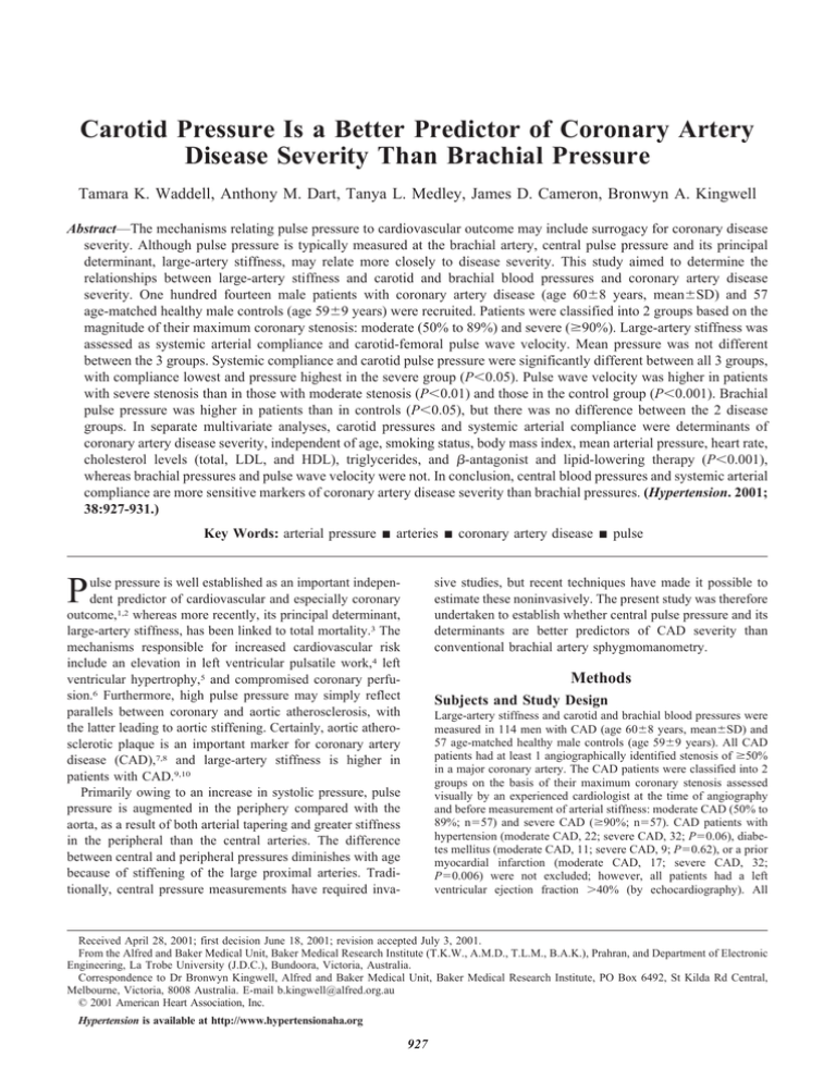
Carotid Pressure Is a Better Predictor of Coronary Artery Disease Severity Than Brachial Pressure Tamara K. Waddell, Anthony M. Dart, Tanya L. Medley, James D. Cameron, Bronwyn A. Kingwell Abstract—The mechanisms relating pulse pressure to cardiovascular outcome may include surrogacy for coronary disease severity. Although pulse pressure is typically measured at the brachial artery, central pulse pressure and its principal determinant, large-artery stiffness, may relate more closely to disease severity. This study aimed to determine the relationships between large-artery stiffness and carotid and brachial blood pressures and coronary artery disease severity. One hundred fourteen male patients with coronary artery disease (age 60⫾8 years, mean⫾SD) and 57 age-matched healthy male controls (age 59⫾9 years) were recruited. Patients were classified into 2 groups based on the magnitude of their maximum coronary stenosis: moderate (50% to 89%) and severe (ⱖ90%). Large-artery stiffness was assessed as systemic arterial compliance and carotid-femoral pulse wave velocity. Mean pressure was not different between the 3 groups. Systemic compliance and carotid pulse pressure were significantly different between all 3 groups, with compliance lowest and pressure highest in the severe group (P⬍0.05). Pulse wave velocity was higher in patients with severe stenosis than in those with moderate stenosis (P⬍0.01) and those in the control group (P⬍0.001). Brachial pulse pressure was higher in patients than in controls (P⬍0.05), but there was no difference between the 2 disease groups. In separate multivariate analyses, carotid pressures and systemic arterial compliance were determinants of coronary artery disease severity, independent of age, smoking status, body mass index, mean arterial pressure, heart rate, cholesterol levels (total, LDL, and HDL), triglycerides, and -antagonist and lipid-lowering therapy (P⬍0.001), whereas brachial pressures and pulse wave velocity were not. In conclusion, central blood pressures and systemic arterial compliance are more sensitive markers of coronary artery disease severity than brachial pressures. (Hypertension. 2001; 38:927-931.) Key Words: arterial pressure 䡲 arteries 䡲 coronary artery disease 䡲 pulse P sive studies, but recent techniques have made it possible to estimate these noninvasively. The present study was therefore undertaken to establish whether central pulse pressure and its determinants are better predictors of CAD severity than conventional brachial artery sphygmomanometry. ulse pressure is well established as an important independent predictor of cardiovascular and especially coronary outcome,1,2 whereas more recently, its principal determinant, large-artery stiffness, has been linked to total mortality.3 The mechanisms responsible for increased cardiovascular risk include an elevation in left ventricular pulsatile work,4 left ventricular hypertrophy,5 and compromised coronary perfusion.6 Furthermore, high pulse pressure may simply reflect parallels between coronary and aortic atherosclerosis, with the latter leading to aortic stiffening. Certainly, aortic atherosclerotic plaque is an important marker for coronary artery disease (CAD),7,8 and large-artery stiffness is higher in patients with CAD.9,10 Primarily owing to an increase in systolic pressure, pulse pressure is augmented in the periphery compared with the aorta, as a result of both arterial tapering and greater stiffness in the peripheral than the central arteries. The difference between central and peripheral pressures diminishes with age because of stiffening of the large proximal arteries. Traditionally, central pressure measurements have required inva- Methods Subjects and Study Design Large-artery stiffness and carotid and brachial blood pressures were measured in 114 men with CAD (age 60⫾8 years, mean⫾SD) and 57 age-matched healthy male controls (age 59⫾9 years). All CAD patients had at least 1 angiographically identified stenosis of ⱖ50% in a major coronary artery. The CAD patients were classified into 2 groups on the basis of their maximum coronary stenosis assessed visually by an experienced cardiologist at the time of angiography and before measurement of arterial stiffness: moderate CAD (50% to 89%; n⫽57) and severe CAD (ⱖ90%; n⫽57). CAD patients with hypertension (moderate CAD, 22; severe CAD, 32; P⫽0.06), diabetes mellitus (moderate CAD, 11; severe CAD, 9; P⫽0.62), or a prior myocardial infarction (moderate CAD, 17; severe CAD, 32; P⫽0.006) were not excluded; however, all patients had a left ventricular ejection fraction ⬎40% (by echocardiography). All Received April 28, 2001; first decision June 18, 2001; revision accepted July 3, 2001. From the Alfred and Baker Medical Unit, Baker Medical Research Institute (T.K.W., A.M.D., T.L.M., B.A.K.), Prahran, and Department of Electronic Engineering, La Trobe University (J.D.C.), Bundoora, Victoria, Australia. Correspondence to Dr Bronwyn Kingwell, Alfred and Baker Medical Unit, Baker Medical Research Institute, PO Box 6492, St Kilda Rd Central, Melbourne, Victoria, 8008 Australia. E-mail b.kingwell@alfred.org.au © 2001 American Heart Association, Inc. Hypertension is available at http://www.hypertensionaha.org 927 928 Hypertension October 2001 -blocking agents and nitrates were discontinued for 24 hours before the study. Control subjects were healthy, normotensive nonsmokers with normal plasma lipid levels, were not involved in regular physical activity, and were not taking cardiovascular medication. All subjects gave informed consent for their participation in the study, which was approved by the Ethics Committee of The Alfred Healthcare Group and performed in accordance with both the institutional guidelines and the Declaration of Helsinki (1989) of the World Medical Association. Resting Blood Pressure Resting brachial arterial blood pressure and heart rate were measured at 3-minute intervals with a Dinamap vital signs monitor (1846 SX, Critikon), with subjects remaining undisturbed in the supine position for 10 minutes. The mean of 3 values was taken to represent resting levels. Systemic Arterial Compliance Systemic arterial compliance (SAC) was determined by calculations based on the area method of Liu et al,11 as described previously.12,13 Briefly, SAC was derived from measurements of aortic flow velocity with continuous-wave Doppler velocimetry (Multi-Dopplex MD1, Huntleigh Technology) and simultaneous right carotid pressure, measured by applanation tonometry (SPT-301, Millar Instruments). Brachial arterial blood pressure was measured simultaneously by a Dinamap vital signs monitor (1846 SX, Critikon) to permit calibration of the carotid arterial pressure contour with brachial mean and diastolic blood pressure and to derive carotid systolic blood pressure.12,14 Aortic volume flow was obtained by multiplication of the mean velocity flow by the left ventricular outflow tract (LVOT) area, measured by 2-dimensional echocardiography (Hewlett-Packard Sonos 1500). Pulse Wave Velocity Pulse wave velocity (PWV) was measured by simultaneous recordings of arterial pressure waves from the carotid to the femoral artery by applanation tonometry (SPT-301, Millar Instruments), as described previously.13,15 Biochemical Analyses Fasting venous blood samples were drawn for analyses of lipids. Total, LDL, and HDL cholesterol and triglyceride levels were determined enzymatically with a Cobas-BIO centrifugal analyzer (Roche Diagnostic Systems).16 Statistical Analyses Group comparisons for all variables were by 1-way ANOVA, with a least significant difference post hoc test used to compare individual means. Stepwise multiple regression analysis was performed to examine determinants of maximum coronary stenosis in CAD patients, whereas categorical variables were compared with a 2 test. Receiver operating characteristic curves were used to examine the relative predictive value of central pressures and SAC for CAD severity class. All data were analyzed with SPSS for Windows version 9.0.1. (SPSS Inc). Unless otherwise stated, group results are presented as mean⫾SEM, and statistical significance was deemed to have been achieved at P⬍0.05. Results The 3 study groups were similar in age, LVOT area, and cardiac output, whereas body mass index was higher and total cholesterol lower in the group with severe CAD than in controls (Table). Total triglycerides were higher and HDL and LDL cholesterol levels lower in CAD patients than in controls, but there was no difference between the 2 CAD groups (Table). The number of diseased coronary vessels (single/double/triple: moderate CAD, 36/17/4; severe CAD, 26/23/8; P⫽0.20) and previous revascularization procedures (angioplasty: moderate CAD, Figure 1. Resting brachial and carotid blood pressures in controls (n⫽57), moderate CAD group (n⫽57), and severe CAD group (n⫽57). SBP indicates systolic blood pressure; DBP, diastolic blood pressure; MAP, mean arterial pressure; and PP, pulse pressure. Data presented as mean⫾SEM. *P⬍0.05, **P⬍0.01, ***P⬍0.001. 13; severe CAD, 10; P⫽0.48; bypass grafts: moderate CAD, 4; severe CAD, 7; P⫽0.34) were also similar between the groups with moderate and severe CAD. Compared with the moderate CAD group, more patients in the group with severe CAD were taking -antagonists (moderate CAD, 23; severe CAD, 41; P⫽0.001) and lipid-lowering therapy (moderate CAD, 33; severe CAD, 43; P⫽0.07), but there was no difference in use of any other medications (ACE inhibitors: moderate CAD, 17; severe CAD, 17; P⫽0.84; calcium antagonists: moderate CAD, 12; severe CAD, 21; P⫽0.10; nitrates: moderate CAD, 18; severe CAD, 25; P⫽0.25; and diuretics: moderate CAD, 7; severe CAD, 3, P⫽0.32). There were more ex-smokers (moderate CAD, 26; severe CAD, 40; P⫽0.01) and fewer nonsmokers (moderate CAD, 27; severe CAD, 14; P⫽0.03) in the severe CAD group, whereas the number of current smokers was similar between the 2 CAD populations (moderate CAD 4; severe CAD 3; P⫽0.68). Blood Pressure Brachial mean and diastolic blood pressures (Figure 1) and heart rate (Table) did not differ between the 3 groups. The average difference between brachial and carotid pulse pressure was significantly higher in controls than in the group with severe CAD (P⫽0.02) and tended to be higher in the group with moderate CAD than in the group with severe CAD (controls, 8.0⫾1.1 mm Hg; moderate CAD, 6.5⫾1.8 mm Hg; and severe CAD, 2.6⫾1.8 mm Hg; P⫽0.08). Brachial systolic pressure was higher in the group with severe CAD than in controls, but other comparisons were not significant (controls, 124⫾2 mm Hg; moderate CAD, 128⫾2 mm Hg; and severe CAD, 134⫾3 mm Hg) (Figure 1). Brachial pulse pressure was more discriminating, with differences evident between controls and CAD patients (controls, 46⫾1 mm Hg; moderate CAD, 52⫾2 mm Hg; and Waddell et al Carotid Blood Pressure and CAD Severity 929 Group Characteristics Variable Controls Age, y Body mass index, kg 䡠 m⫺2 LVOT area, cm2 Moderate CAD Severe CAD 59⫾1 60⫾1 61⫾1 26.5⫾0.5 27.7⫾0.5 28.9⫾0.6† 3.6⫾0.1 3.5⫾0.1 3.6⫾0.1 Heart rate, bpm 59⫾1 60⫾1 59⫾1 Cardiac output, AFU 1.5⫾0.2 1.1⫾0.1 1.1⫾0.1 Total cholesterol, mmol 䡠 L⫺1 5.1⫾0.1 4.8⫾0.1 4.6⫾0.1* HDL cholesterol, mmol 䡠 L⫺1 1.4⫾0.1 1.1⫾0.1† 1.0⫾0.1‡ ⫺1 LDL cholesterol, mmol 䡠 L 3.2⫾0.1 2.7⫾0.1† 2.6⫾0.1† Total triglycerides, mmol 䡠 L⫺1 1.2⫾0.1 2.0⫾0.2‡ 1.8⫾0.1‡ AFU indicates arbitrary flow units (dimensionally equivalent to L 䡠 min⫺1). Values are mean⫾SEM. *P⬍0.05, †P⬍0.01, ‡P⬍0.001 significant difference from controls. severe CAD 56⫾2 mm Hg); however, brachial pulse pressure was not different between the 2 CAD groups (Figure 1). In contrast to brachial pressures, carotid systolic blood pressure was significantly higher in the group with severe CAD than in either the moderate CAD group or the controls (controls, 116⫾2 mm Hg; moderate CAD, 122⫾3 mm Hg; and severe CAD, 132⫾3 mm Hg) (Figure 1). Carotid pulse pressure was the best discriminator, with highly significant differences between all groups (controls, 38⫾1 mm Hg; moderate CAD, 45⫾2 mm Hg; and severe CAD, 53⫾3 mm Hg) (Figure 1). Brachial and carotid pressures were also examined as determinants of the magnitude of the maximum coronary stenosis in CAD patients, with systolic and pulse pressure examined in separate multivariate analysis that incorporated age, smoking status, body mass index, mean arterial pressure, heart rate, cholesterol (total, LDL, and HDL) levels, triglycerides, and -antagonist and lipid-lowering therapy. Carotid systolic pressure (r⫽0.47; P⬍0.001) and carotid pulse pressure (r⫽0.45; P⬍0.001) were both significant independent determinants of CAD severity, but brachial pressures were not. Arterial Stiffness Like central pulse pressure, SAC was able to discriminate between all groups, with values highest in the control group and lowest in the patients with severe CAD (controls, 0.62⫾0.03 mL/mm Hg; moderate CAD, 0.51⫾0.03 mL/ mm Hg; and severe CAD, 0.41⫾0.03 mL/mm Hg) (Figure 2a). Carotid-femoral PWV was higher in the severe CAD group than in the moderate CAD and control groups but was unable to discriminate the control group from those with only moderate CAD (controls, 9.3⫾0.2 m/s; moderate CAD, 10.2⫾0.3 m/s; and severe CAD, 11.5⫾0.5 m/s) (Figure 2b). SAC and carotid-femoral PWV were examined as determinants of the magnitude of the maximum coronary stenosis in CAD patients in multivariate analyses that incorporated age, smoking status, body mass index, mean arterial pressure, heart rate, cholesterol (total, LDL, and HDL) levels, triglycerides, and -antagonist and lipid-lowering therapy. Although SAC was an independent predictor of disease severity (r⫽0.47, P⬍0.001), PWV was not. Furthermore, receiver operating characteristic curves indicated that SAC was most Figure 2. SAC (a) and carotid-femoral (C-F) PWV (b) in controls (n⫽57), moderate CAD group (n⫽57), and severe CAD group (n⫽57). Data presented as mean⫾SEM. *P⬍0.05, **P⬍0.01, ***P⬍0.001. strongly associated with CAD severity class (receiver operating characteristic curve area⫽0.64, SE⫽0.05; P⫽0.01), followed closely by carotid pulse pressure (area⫽0.62, SE⫽0.05; P⫽0.03) and carotid systolic blood pressure (area⫽0.61, SE⫽0.05; P⫽0.05). Discussion The major finding of this study was that the large arteries are stiffer in patients with more severe CAD and that carotid pressures and SAC were more sensitive markers of CAD severity than brachial pressures. These relationships were evident despite the limitations involved in estimation of carotid pressure and the relatively advanced age of the study patients, which would minimize pressure differences between the central and peripheral circulations. The current data emphasize the importance of the central arterial circulation as a surrogate marker of CAD severity, assessed by the magnitude of the maximum coronary stenosis. The relationship between CAD severity with largeartery stiffness and central pulse pressure is likely to be due to parallels between coronary and aortic atherosclerosis, with the latter leading to aortic stiffening. Indeed, aortic atherosclerosis detected by transesophageal echocardiography is related to the presence of CAD,7,8 and it is well known that CAD patients have stiffer aortas.9,10 More detailed analyses in nonhuman primates17–19 have demonstrated an association between elasticity and severity of atherosclerosis in the aorta. At the cellular level, atherosclerosis is associated with smooth muscle cell prolifera- 930 Hypertension October 2001 tion and deposition of lipids,20 whereas aortic stiffening may also be related to nutritional deficiency arising from reduced blood flow in the vasa vasorum.21 The severity of CAD and large-artery stiffness have not previously been related; however, there is a report linking elasticity of the abdominal aorta to the number of diseased coronary arteries in patients after myocardial infarction.22 The postinfarction status of the patients in this previous analysis, however, precluded assessment of relationships with maximum coronary stenosis. Large-artery stiffening and pulse-pressure elevation may also convey risk beyond that associated with CAD severity. Animal data indicate that cardiac ejection into a stiffened aorta increases cardiac work4 and reduces coronary perfusion.6 In humans, this relationship has been substantiated by the observation that ischemic threshold during a standard exercise stress test is lower in patients with stiffer aortas, independent of disease severity.23 These data indicate that the risk of myocardial ischemia in CAD patients is related not only to the severity of coronary stenoses but also to aortic stiffness, which increases in parallel with CAD severity. Although the ability of largeartery stiffness and carotid versus brachial pressure to assess this hemodynamic risk was not examined in the present study, the proximity of the large vessels to the left ventricle again makes it likely that central pressures would be more informative in this regard. The observed differences in aortic stiffness between the 2 CAD groups and control subjects were not attributable to differences in age, mean arterial pressure, cardiac output, heart rate, or aortic size. Furthermore, limiting the study to male subjects avoided the confounding effects of gender on large-artery stiffening. The greater number of exsmokers in the severe CAD group is likely to have contributed to arterial stiffening; however, both carotid pressures and SAC were related to disease severity independently of smoking status. Consistent with the presence of coronary disease, HDL cholesterol was lower and triglyceride levels were higher in the CAD groups than in controls. Total and LDL cholesterol were lower in the CAD groups, presumably owing to the effects of treatment. However, carotid pressures and SAC were related to CAD severity independently of lipid levels and lipid-lowering therapy. The only other medication difference was a greater prevalence of -antagonist therapy in the severe compared with the moderate CAD group. -Antagonists were discontinued 24 hours before the study, and all relationships were independent of -antagonist therapy. In addition, heart rate was not different between groups. Finally, SAC was more sensitive in discriminating CAD severity than carotid-femoral PWV. This difference in sensitivity may relate to the fact that directly measured carotid-femoral PWV does not incorporate a measure of proximal arterial stiffness. This is because at the time the pressure is measured in the carotid artery, the wave moving toward the femoral artery will have traversed the same distance along the aorta. Thus, the length of aorta actually assessed is the carotid to manubrium sternum distance subtracted from the manubrium sternum to femoral distance. In summary, the present study demonstrated that carotid systolic and pulse pressures were more sensitive markers of CAD severity than brachial pressure. This likely reflects the greater aortic stiffness in patients with more severe CAD, which suggests a correlation between the severity of coronary and aortic atherosclerosis. In view of the known experimental effects of increased aortic stiffness on blood flow through critically stenosed vessels, these findings suggest an important role for aortic stiffness in modulating the onset of myocardial ischemia. Furthermore, they suggest that a reduction in large-artery stiffness, and thus central pressure, may be an important therapeutic goal in the management of angina in patients with CAD. Acknowledgments Drs Kingwell and Dart are National Health and Medical Research Council Research Fellows. This work was supported by grants from VicHealth and The National Heart Foundation of Australia and the Alfred Hospital, Melbourne, Australia. We thank Melissa Formosa, Elizabeth Dewar, and Karen Murchie for their technical expertise and assistance with this study. References 1. Benetos A, Safar M, Rudnichi A, Smulyan H, Richard JL, Ducimetiere P, Guize L. Pulse pressure: a predictor of long-term cardiovascular mortality in a French male population. Hypertension. 1997;30:1410 –1415. 2. Madhavan S, Ooi WL, Cohen H, Alderman MH. Relation of pulse pressure and blood pressure reduction to the incidence of myocardial infarction. Hypertension. 1994;23:395– 401. 3. Laurent S, Boutouyrie P, Asmar R, Gautier I, Laloux B, Guize L, Ducimetiere P, Benetos A. Aortic stiffness is an independent predictor of all-cause mortality in hypertensive patients. J Hypertens. 2000; 18:S20. Abstract. 4. Kelly RP, Tunin R, Kass DA. Effect of reduced aortic compliance on cardiac efficiency and contractile function of in situ canine left ventricle. Circ Res. 1992;71:490 –502. 5. Boutouyrie P, Laurent S, Girerd X, Benetos A, Lacolley P, Abergel E, Safar M. Common carotid artery stiffness and patterns of left ventricular hypertrophy in hypertensive patients. Hypertension. 1995;25: 651– 659. 6. Watanabe H, Ohtsuka S, Kakihana M, Sugishita Y. Decreased aortic compliance aggravates subendocardial ischaemia in dogs with stenosed coronary artery. Cardiovasc Res. 1992;26:1212–1218. 7. Khoury Z, Gottlieb S, Stern S, Keren A. Frequency and distribution of atherosclerotic plaques in the thoracic aorta as determined by transesophageal echocardiography in patients with coronary artery disease. Am J Cardiol. 1997;79:23–27. 8. Fazio GP, Redberg RF, Winslow T, Schiller NB. Transesophageal echocardiographically detected atherosclerotic aortic plaque is a marker for coronary artery disease. J Am Coll Cardiol. 1993;21: 144 –150. 9. Gatzka CD, Cameron JD, Kingwell BA, Dart AM. Relation between coronary artery disease, aortic stiffness, and left ventricular structure in a population sample. Hypertension. 1998;32:575–578. 10. Cameron JD, Jennings GL, Dart AM. Systemic arterial compliance is decreased in newly-diagnosed patients with coronary heart disease: implications for prediction of risk. J Cardiovasc Risk. 1996;3: 495–500. 11. Liu Z, Brin KP, Yin FCP. Estimation of total arterial compliance: an improved method and evaluation of current methods. Am J Physiol. 1986;251:H588 –H600. 12. Cameron JD, Dart AM. Exercise training increases total systemic arterial compliance in humans. Am J Physiol. 1994;266:H693–H701. 13. Rajkumar C, Kingwell BA, Cameron JD, Waddell TK, Mehra R, Christophidis N, Komesaroff PA, McGrath B, Jennings GL, Sudhir K, Dart AM. Hormonal therapy increases arterial compliance in postmenopausal women. J Am Coll Cardiol. 1997;30:350 –356. Waddell et al 14. Chen CH, Ting CT, Nussbacher A, Nevo E, Kass DA, Pak P, Wang SP, Chang MS, Yin FCP. Validation of carotid artery tonometry as a means of estimating augmentation index of ascending aortic pressure. Hypertension. 1996;27:168 –175. 15. Vaitkevicius PV, Fleg JL, Engel JH, O’Conner FC, Wright JG, Lakatta LE, Yin FCP, Lakatta EG. Effects of age and aerobic capacity on arterial stiffness in healthy adults. Circulation. 1993;88: 1456 –1462. 16. Kingwell BA, Sherrard B, Jennings GL, Dart AM. Four weeks of cycle training increases basal production of nitric oxide from the forearm. Am J Physiol. 1997;272:H1070 –H1077. 17. Farrar DJ, Bond MG, Riley WA, Sawyer JK. Anatomic correlates of aortic pulse wave velocity and carotid artery elasticity during atherosclerosis progression and regression in monkeys. Circulation. 1991; 83:1754 –1763. 18. Farrar DJ, Bond MG, Sawyer JK, Green HD. Pulse wave velocity and morphological changes associated with early atherosclerosis progression in the aortas of cynomolgus monkeys. Cardiovasc Res. 1984; 18:107–118. Carotid Blood Pressure and CAD Severity 931 19. Farrar DJ, Green HD, Wagner WD, Bond MG. Reduction in pulse wave velocity and improvement of aortic distensibility accompanying regression of atherosclerosis in the rhesus monkey. Circ Res. 1980; 47:425– 432. 20. Ross R. The pathogenesis of atherosclerosis: an update. N Engl J Med. 1986;314:488 –500. 21. Stefanadis C, Vlachpoulos C, Karayannacos P, Boudoulas H, Stratos C, Filippides T, Agapitos M, Toutouzas P. Effect of vasa vasorum flow on structure and function of the aorta in experimental animals. Circulation. 1995;91:2669 –2678. 22. Hirai T, Sasayama S, Kawasaki T, Yagi S. Stiffness of systemic arteries in patients with myocardial infarction: a noninvasive method to predict severity of coronary atherosclerosis. Circulation. 1989;80: 78 – 86. 23. Kingwell BA, Medley TL, Waddell TK, Cameron JD, Jennings GL, Dart AM. Large artery compliance predicts time to ischemia during treadmill testing in coronary artery disease patients. Circulation. 2000;102(suppl II):II-782. Abstract.
