Phonetic Feature Encoding in Human
Superior Temporal Gyrus
Nima Mesgarani,1* Connie Cheung,1 Keith Johnson,2 Edward F. Chang1†
During speech perception, linguistic elements such as consonants and vowels are extracted from
a complex acoustic speech signal. The superior temporal gyrus (STG) participates in high-order
auditory processing of speech, but how it encodes phonetic information is poorly understood. We used
high-density direct cortical surface recordings in humans while they listened to natural, continuous
speech to reveal the STG representation of the entire English phonetic inventory. At single electrodes,
we found response selectivity to distinct phonetic features. Encoding of acoustic properties was
mediated by a distributed population response. Phonetic features could be directly related to tuning for
spectrotemporal acoustic cues, some of which were encoded in a nonlinear fashion or by integration of
multiple cues. These findings demonstrate the acoustic-phonetic representation of speech in human STG.
P
honemes—and the distinctive features composing them—are hypothesized to be the
smallest contrastive units that change a word’s
1
Department of Neurological Surgery, Department of Physiology, and Center for Integrative Neuroscience, University of
California, San Francisco, CA 94143, USA. 2Department of
Linguistics, University of California, Berkeley, CA 94720, USA.
*Present address: Department of Electrical Engineering,
Columbia University, New York, NY 10027, USA.
†Corresponding author. E-mail: changed@neurosurg.ucsf.edu
meaning (e.g., /b/ and /d/ as in bad versus dad)
(1). The superior temporal gyrus (Brodmann area
22, STG) has a key role in acoustic-phonetic processing because it responds to speech over other
sounds (2) and focal electrical stimulation there
selectively interrupts speech discrimination (3).
These findings raise fundamental questions about
the representation of speech sounds, such as
whether local neural encoding is specific for phonemes, acoustic-phonetic features, or low-level
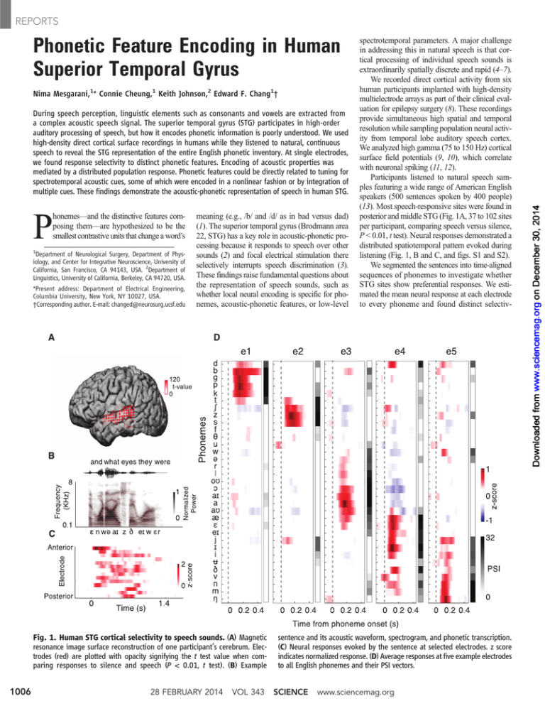
Fig. 1. Human STG cortical selectivity to speech sounds. (A) Magnetic
resonance image surface reconstruction of one participant’s cerebrum. Electrodes (red) are plotted with opacity signifying the t test value when comparing responses to silence and speech (P < 0.01, t test). (B) Example
1006
28 FEBRUARY 2014
VOL 343
spectrotemporal parameters. A major challenge
in addressing this in natural speech is that cortical processing of individual speech sounds is
extraordinarily spatially discrete and rapid (4–7).
We recorded direct cortical activity from six
human participants implanted with high-density
multielectrode arrays as part of their clinical evaluation for epilepsy surgery (8). These recordings
provide simultaneous high spatial and temporal
resolution while sampling population neural activity from temporal lobe auditory speech cortex.
We analyzed high gamma (75 to 150 Hz) cortical
surface field potentials (9, 10), which correlate
with neuronal spiking (11, 12).
Participants listened to natural speech samples featuring a wide range of American English
speakers (500 sentences spoken by 400 people)
(13). Most speech-responsive sites were found in
posterior and middle STG (Fig. 1A, 37 to 102 sites
per participant, comparing speech versus silence,
P < 0.01, t test). Neural responses demonstrated a
distributed spatiotemporal pattern evoked during
listening (Fig. 1, B and C, and figs. S1 and S2).
We segmented the sentences into time-aligned
sequences of phonemes to investigate whether
STG sites show preferential responses. We estimated the mean neural response at each electrode
to every phoneme and found distinct selectiv-
sentence and its acoustic waveform, spectrogram, and phonetic transcription.
(C) Neural responses evoked by the sentence at selected electrodes. z score
indicates normalized response. (D) Average responses at five example electrodes
to all English phonemes and their PSI vectors.
SCIENCE
www.sciencemag.org
Downloaded from www.sciencemag.org on December 30, 2014
REPORTS
REPORTS
ity. For example, electrode e1 (Fig. 1D) showed
large evoked responses to plosive phonemes /p/,
/t/, /k/, /b/, /d/, and /g/. Electrode e2 showed
selective responses to sibilant fricatives: /s/, /ʃ/,
and /z/. The next two electrodes showed selective responses to subsets of vowels: low-back
(electrode e3, e.g., /a/ and /aʊ/), high-front vowels
and glides (electrode e4, e.g., /i/ and /j/). Last,
neural activity recorded at electrode e5 was selective for nasals (/n/, /m/, and /ŋ/).
To quantify selectivity at single electrodes, we
derived a metric indicating the number of phonemes with cortical responses statistically distinguishable from the response to a particular
phoneme. The phoneme selectivity index (PSI)
is a dimension of 33 English phonemes; PSI = 0
is nonselective and PSI = 32 is extremely selective (Wilcox rank-sum test, P < 0.01, Fig. 1D;
methods shown in fig. S3). We determined an
optimal analysis time window of 50 ms, centered
150 ms after the phoneme onset by using a phoneme separability analysis (f-statistic, fig. S4A).
The average PSI over all phonemes summarizes
an electrode’s overall selectivity. The average PSI
was highly correlated to a site’s response magnitude to speech over silence (r = 0.77, P < 0.001,
t test; fig. S5A) and the degree to which the
response could be predicted with a linear spectrotemporal receptive field [STRF, r = 0.88, P <
0.001, t test; fig. S5B (14)]. Therefore, the ma-
Fig. 2. Hierarchical clustering of single-electrode and population
responses. (A) PSI vectors of selective electrodes across all participants. Rows
correspond to phonemes, and columns correspond to electrodes. (B) Clustering across population PSIs (rows). (C) Clustering across single electrodes (columns). (D) Alternative PSI vectors using rows now corresponding to phonetic
www.sciencemag.org
SCIENCE
jority of speech-responsive sites in STG are selective to specific phoneme groups.
To investigate the organization of selectivity
across the neural population, we constructed an
array containing PSI vectors for electrodes across
all participants (Fig. 2A). In this array, each column
corresponds to a single electrode, and each row
corresponds to a single phoneme. Most STG electrodes are selective not to individual but to specific groups of phonemes. To determine selectivity
patterns across electrodes and phonemes, we
used unsupervised hierarchical clustering analyses. Clustering across rows revealed groupings of
phonemes on the basis of similarity of PSI values
in the population response (Fig. 2B). Clustering
features, not phonemes. (E) Weighted average STRFs of main electrode clusters. (F) Average acoustic spectrograms for phonemes in each population cluster. Correlation between average STRFs and average spectrograms: r = 0.67,
P < 0.01, t test. (r = 0.50, 0.78, 0.55, 0.86, 0.86, and 0.47 for plosives, fricatives,
vowels, and nasals, respectively; P < 0.01, t test).
VOL 343
28 FEBRUARY 2014
1007
REPORTS
Fig. 3. Neural encoding of vowels. (A) Formant frequencies, F1 and F2, for
English vowels (F2-F1, dashed line, first principal component). (B) F1 and F2
partial correlations for each electrode’s response (**P < 0.01, t test). Dots (elecacross columns revealed single electrodes with
similar PSI patterns (Fig. 2C). These two analyses revealed complementary local- and globallevel organizational selectivity patterns. We also
replotted the array by using 14 phonetic features
defined in linguistics to contrast distinctive articulatory and acoustic properties (Fig. 2D; phonemefeature mapping provided in fig. S7) (1, 15).
The first tier of the single-electrode hierarchy
analysis (Fig. 2C) divides STG sites into two distinct groups: obstruent- and sonorant-selective electrodes. The obstruent-selective group is divided
into two subgroups: plosive and fricative electrodes (similar to electrodes e1 and e2 in Fig. 1D)
(16). Among plosive electrodes (blue), some were
responsive to all plosives, whereas others were
selective to place of articulation (dorsal /g/ and /k/
versus coronal /d/ and /t/ versus labial /p/ and /b/,
labeled in Fig. 2D) and voicing (separating voiced
/b/, /d/, and /g/ from unvoiced /p/, /t/, and /k/;
labeled voiced in Fig. 2D). Fricative-selective
electrodes (purple) showed weak, overlapping selectivity to coronal plosives (/d/ and /t/). Sonorantselective cortical sites, in contrast, were partitioned
into four partially overlapping groups: low-back
vowels (red), low-front vowels (orange), high-front
vowels (green), and nasals (magenta) (labeled in
Fig. 2D, similar to e3 to e5 in Fig. 1D).
Both clustering schemes (Fig. 2, B and C) revealed similar phoneme grouping based on shared
phonetic features, suggesting that a substantial portion of the population-based organization can be
accounted for by local tuning to features at single electrodes (similarity of average PSI values
for the local and population subgroups of both
clustering analyses is shown in fig. S8; overall
r = 0.73, P < 0.001). Furthermore, selectivity is
organized primarily by manner of articulation distinctions and secondarily by place of articulation,
corresponding to the degree and the location of
constriction in the vocal tract, respectively (16).
This systematic organization of speech sounds is
consistent with auditory perceptual models positing that distinctions are most affected by manner
contrasts (17, 18) compared with other feature
hierarchies (articulatory or gestural theories) (19).
1008
trodes) are color-coded by their cluster membership. (C) Neural population decoding of fundamental and formant frequencies. Error bars indicate SEM. (D)
Multidimensional scaling (MDS) of acoustic and neural space (***P < 0.001, t test).
We next determined what spectrotemporal
tuning properties accounted for phonetic feature
selectivity. We first determined the weighted average STRFs of the six main electrode clusters
identified above, weighting them proportionally by their degree of selectivity (average PSI). These
STRFs show well-defined spectrotemporal tuning
(Fig. 2E) highly similar to average acoustic spectrograms of phonemes in corresponding population clusters (Fig. 2F; average correlation = 0.67,
P < 0.01, t test). For example, the first STRF in
Fig. 2E shows tuning for broadband excitation
followed by inhibition, similar to the acoustic spectrogram of plosives. The second STRF is tuned to
a high frequency, which is a defining feature of
sibilant fricatives. STRFs of vowel electrodes show
tuning for characteristic formants that define lowback, low-front, and high-front vowels. Last, STRF
of nasal-selective electrodes is tuned primarily to
low acoustic frequencies generated from heavy
voicing and damping of higher frequencies (16).
The average spectrogram analysis requires a priori
phonemic segmentation of speech but is modelindependent. The STRF analysis assumes a linear
relationship between spectrograms and neural responses but is estimated without segmentation.
Despite these differing assumptions, the strong
match between these confirms that phonetic feature selectivity results from tuning to signature
spectrotemporal cues.
We have thus far focused on local feature selectivity to discrete phonetic feature categories.
We next wanted to address the encoding of continuous acoustic parameters that specify phonemes
within vowel, plosive, and fricative groups. For
vowels, we measured fundamental (F0) and formant (F1 to F4) frequencies (16). The first two
formants (F1 and F2) play a major perceptual role
in distinguishing different English vowels (16),
despite tremendous variability within and across
vowels (Fig. 3A) (20). The optimal projection of
vowels in formant space was the difference of F2
and F1 (first principal component, dashed line,
Fig. 3A), which is consistent with vowel perceptual studies (21, 22). By using partial correlation
analysis, we quantified the relationship between
28 FEBRUARY 2014
VOL 343
SCIENCE
electrode response amplitudes and F0 to F4. On
average, we observed no correlation between the
sensitivity of an electrode to F0 with its sensitivity to F1 or F2. However, sensitivity to F1 and
F2 was negatively correlated across all vowelselective sites (Fig. 3B; r = –0.49, P < 0.01, t test),
meaning that single STG sites show an integrated
response to both F1 and F2. Furthermore, electrodes selective to low-back and high-front vowels
(labeled in Fig. 2D) showed an opposite differential tuning to formants, thereby maximizing vowel
discriminability in the neural domain. This complex sound encoding matches the optimal projection
in Fig. 3A, suggesting a specialized higher-order
encoding of acoustic formant parameters (23, 24)
and contrasts with studies of speech sounds in nonhuman species (25, 26).
To examine population representation of vowel
parameters, we used linear regression to decode
F0 to F4 from neural responses. To ensure unbiased estimation, we first removed correlations
between F0 to F4 by using linear prediction and
decoded the residuals. Relatively high decoding
accuracies are shown in Fig. 3C (P < 0.001, t test),
suggesting fundamental and formant variability
is well represented in population STG responses
(interaction between decoder weights with electrode STRFs shown in fig. S9). By using multidimensional scaling, we found that the relational
organization between vowel centroids in the acoustic domain is well preserved in neural space (Fig.
3D; r = 0.88, P < 0.001).
For plosives, we measured three perceptually
important acoustic cues (fig. S10): voice-onset
time (VOT), which distinguishes voiced (/b/, /d/,
and /g/) from unvoiced plosives (/p/, /t/, and /k/);
spectral peak (differentiating labials /p/ and / b /
versus coronal /t/ and /d/ versus dorsal /k/ and /g/);
and F2 of the following vowel (16). These acoustic parameters could be decoded from population
STG responses (Fig. 4A; P < 0.001, t test). VOTs
in particular are temporal cues that are perceived
categorically, which suggests a nonlinear encoding (27). Figure 4B shows neural responses for
three example electrodes plotted for all plosive
instances (total of 1200), aligned to their release
www.sciencemag.org
REPORTS
Fig. 4. Neural encoding of plosive and fricative
phonemes. (A) Prediction accuracy of plosive and
fricative acoustic parameters from neural population responses. Error bars indicate SEM. (B) Response
of three example electrodes to all plosive phonemes
sorted by VOT. (C) Nonlinearity of VOT-response
transformation and (D) distributions of nonlinearity
for all plosive-selective electrodes identified in Fig.
2D. Voiced plosive-selective electrodes are shown
in pink, and the rest in gray. (E) Partial correlation
values between response of electrodes and acoustic parameters shared between plosives and fricatives
(**P < 0.01, t test). Dots (electrodes) are color-coded
by their cluster grouping from Fig. 2C.
time and sorted by VOT. The first electrode responds to all plosives with same approximate
latency and amplitude, irrespective of VOT. The
second electrode responds only to plosive phonemes with short VOT (voiced), and the third
electrode responds primarily to plosives with long
VOT (unvoiced).
To examine the nonlinear relationship between
VOT and response amplitude for voiced-plosive
electrodes (labeled voiced in Fig. 2D) compared
with plosive electrodes with no sensitivity to
voicing feature (labeled coronal, labial and dorsal
in Fig. 2D), we fitted a linear and exponential
function to VOT-response pairs (fig. S11B). The
difference between these two fits specifies the
nonlinearity of this transformation, shown for all
plosive electrodes in Fig. 4C. Voiced-plosive electrodes (pink) all show strong nonlinear bias for
short VOTs compared with all other plosive electrodes (gray). We quantified the degree and direction of this nonlinear bias for these two groups
of plosive electrodes by measuring the average
second-derivative of the curves in Fig. 4C. This
measure maps electrodes with nonlinear preference for short VOTs (e.g., electrode e2 in Fig. 4B)
to negative values and electrodes with nonlinear
preference for long VOTs (e.g., electrode e3 in
Fig. 4B) to positive values. The distribution of this
measure for voiced-plosive electrodes (Fig. 4D,
red distribution) shows significantly greater nonlinear bias compared with the remaining plosive
electrodes (Fig. 4D, gray distribution) (P < 0.001,
Wilcox rank-sum test). This suggests a specialized mechanism for spatially distributed, nonlinear rate encoding of VOT and contrasts with
previously described temporal encoding mechanisms (26, 28).
Plosive
Fricative
Vowel LB
Vowel LF
Vowel HF
We performed a similar analysis for fricatives,
measuring duration, which aids the distinction between voiced (/z/ and /v/) and unvoiced fricatives
(/s/, /ʃ/, /q/, /f/); spectral peak, which differentiates
/f/ and /v/ versus coronal /s/ and /z/ versus dorsal /ʃ/;
and F2 of the following vowel (16) (fig. S12).
These parameters can be decoded reliably from
population responses (Fig. 4A; P < 0.001, t test).
Because plosives and fricatives can be subspecified by using similar acoustic parameters, we
determined whether the response of electrodes to
these parameters depends on their phonetic category (i.e., fricative or plosive). We compared the
partial correlation values of neural responses with
spectral peak, duration, and F2 onset of fricative
and plosive phonemes (Fig. 4E), where each point
corresponds to an electrode color-coded by its cluster grouping in Fig. 2D. High correlation values
(r = 0.70, 0.87, and 0.79; P < 0.001; t test) suggest that electrodes respond to these acoustic parameters independent of their phonetic context.
The similarity of responses to these isolated acoustic parameters suggests that electrode selectivity
to a specific phonetic features (shown with colors
in Fig. 4E) emerges from combined tuning to multiple acoustic parameters that define phonetic contrasts (24, 25).
We have characterized the STG representation
of the entire American English phonetic inventory.
We used direct cortical recordings with high spatial
and temporal resolution to determine how selectivity for phonetic features is correlated to acoustic spectrotemporal receptive field properties in
STG. We found evidence for both spatially local
and distributed selectivity to perceptually relevant
aspects of speech sounds, which together appear to
give rise to our internal representation of a phoneme.
www.sciencemag.org
SCIENCE
VOL 343
We found selectivity for some higher-order
acoustic parameters, such as examples of nonlinear, spatial encoding of VOT, which could have
important implications for the categorical representation of this temporal cue. Furthermore, we
observed a joint differential encoding of F1 and
F2 at single cortical sites, suggesting evidence of
spectral integration previously speculated in theories of combination-sensitive neurons for vowels
(23–25, 29).
Our results are consistent with previous singleunit recordings in human STG, which have not
demonstrated invariant, local selectivity to single
phonemes (30, 31). Instead, our findings suggest
a multidimensional feature space for encoding the
acoustic parameters of speech sounds (25). Phonetic features defined by distinct acoustic cues for
manner of articulation were the strongest determinants of selectivity, whereas place-of-articulation
cues were less discriminable. This might explain
some patterns of perceptual confusability between
phonemes (32) and is consistent with feature hierarchies organized around acoustic cues (17),
where phoneme similarity space in STG is driven
more by auditory-acoustic properties than articulatory ones (33). A featural representation has greater
universality across languages, minimizes the need
for precise unit boundaries, and can account for
coarticulation and temporal overlap over phonemebased models for speech perception (17).
References and Notes
1. N. Chomsky, M. Halle, The Sound Pattern of English
(Harper and Row, New York, 1968).
2. J. R. Binder et al., Cereb. Cortex 10, 512–528 (2000).
3. D. Boatman, C. Hall, M. H. Goldstein, R. Lesser,
B. Gordon, Cortex 33, 83–98 (1997).
4. E. F. Chang et al., Nat. Neurosci. 13, 1428–1432 (2010).
28 FEBRUARY 2014
1009
REPORTS
5. E. Formisano, F. De Martino, M. Bonte, R. Goebel, Science
322, 970–973 (2008).
6. J. Obleser, A. M. Leaver, J. Vanmeter, J. P. Rauschecker,
Front. Psychol. 1, 232 (2010).
7. M. Steinschneider et al., Cereb. Cortex 21, 2332–2347
(2011).
8. Materials and methods are available as supplementary
materials on Science Online.
9. N. E. Crone, D. Boatman, B. Gordon, L. Hao, Clin. Neurophysiol.
112, 565–582 (2001).
10. E. Edwards et al., J. Neurophysiol. 102, 377–386 (2009).
11. M. Steinschneider, Y. I. Fishman, J. C. Arezzo, Cereb. Cortex
18, 610–625 (2008).
12. S. Ray, J. H. R. Maunsell, PLOS Biol. 9, e1000610 (2011).
13. J. S. Garofolo, TIMIT: Acoustic-Phonetic Continuous
Speech Corpus (Linguistic Data Consortium, Philadelphia,
1993).
14. F. E. Theunissen et al., Network 12, 289–316 (2001).
15. M. Halle, K. Stevens, in Music, Language, Speech, and
Brain, J. Sundberg, L. Nord, R. Carlson, Eds. (Wenner-Gren
International Symposium Series vol. 59, Macmillan,
Basingstoke, UK, 1991).
16. P. Ladefoged, K. Johnson, A Course in Phonetics
(Cengage Learning, Stamford, CT, 2010).
17. K. N. Stevens, J. Acoust. Soc. Am. 111, 1872–1891
(2002).
18. G. Clements, Phonol. Yearb. 2, 225–252 (1985).
19. C. A. Fowler, J. Phonetics 14, 3–28 (1986).
20. G. E. Peterson, H. L. Barney, J. Acoust. Soc. Am. 24, 175
(1952).
21. J. D. Miller, J. Acoust. Soc. Am. 85, 2114–2134 (1989).
22. A. K. Syrdal, H. S. Gopal, J. Acoust. Soc. Am. 79,
1086–1100 (1986).
23. H. M. Sussman, Brain Lang. 28, 12–23 (1986).
24. I. Nelken, Curr. Opin. Neurobiol. 18, 413–417 (2008).
25. N. Mesgarani, S. V. David, J. B. Fritz, S. A. Shamma,
J. Acoust. Soc. Am. 123, 899–909 (2008).
26. C. T. Engineer et al., Nat. Neurosci. 11, 603–608 (2008).
27. L. Lisker, A. S. Abramson, Lang. Speech 10, 1–28
(1967).
28. M. Steinschneider et al., Cereb. Cortex 15, 170–186
(2005).
29. G. Chechik, I. Nelken, Proc. Natl. Acad. Sci. U.S.A. 109,
18968–18973 (2012).
30. A. M. Chan et al., Cereb. Cortex, published online 16 May
2013 (10.1093/cercor/bht127).
31. O. Creutzfeldt, G. Ojemann, E. Lettich, Exp. Brain Res.
77, 451–475 (1989).
Detection of a Recurrent DNAJB1-PRKACA
Chimeric Transcript in Fibrolamellar
Hepatocellular Carcinoma
Joshua N. Honeyman,1,2* Elana P. Simon,1,3* Nicolas Robine,4* Rachel Chiaroni-Clarke,1
David G. Darcy,1,2 Irene Isabel P. Lim,1,2 Caroline E. Gleason,1 Jennifer M. Murphy,1,2
Brad R. Rosenberg,5 Lydia Teegan,1 Constantin N. Takacs,1 Sergio Botero,1
Rachel Belote,1 Soren Germer,4 Anne-Katrin Emde,4 Vladimir Vacic,4 Umesh Bhanot,6
Michael P. LaQuaglia,2 Sanford M. Simon1†
Fibrolamellar hepatocellular carcinoma (FL-HCC) is a rare liver tumor affecting adolescents and
young adults with no history of primary liver disease or cirrhosis. We identified a chimeric
transcript that is expressed in FL-HCC but not in adjacent normal liver and that arises as the
result of a ~400-kilobase deletion on chromosome 19. The chimeric RNA is predicted to code
for a protein containing the amino-terminal domain of DNAJB1, a homolog of the molecular
chaperone DNAJ, fused in frame with PRKACA, the catalytic domain of protein kinase A.
Immunoprecipitation and Western blot analyses confirmed that the chimeric protein is expressed
in tumor tissue, and a cell culture assay indicated that it retains kinase activity. Evidence
supporting the presence of the DNAJB1-PRKACA chimeric transcript in 100% of the FL-HCCs
examined (15/15) suggests that this genetic alteration contributes to tumor pathogenesis.
ibrolamellar hepatocellular carcinoma
(FL-HCC) is a rare liver tumor that was
first described in 1956 and that historically
has been considered a variant of hepatocellular
carcinoma (1, 2). It is histologically characterized
by well-differentiated neoplastic hepatocytes and
F
1
Laboratory of Cellular Biophysics, Rockefeller University, 1230
York Avenue, New York, NY 10065, USA. 2Division of Pediatric
Surgery, Department of Surgery, Memorial Sloan-Kettering
Cancer Center, 1275 York Avenue, New York, NY 10065, USA.
3
The Dalton School, 108 East 89th Street, New York, NY 10128,
USA. 4New York Genome Center, 101 Avenue of the Americas,
New York, NY 10013, USA. 5Whitehead Presidential Fellows
Program, The Rockefeller University, 1230 York Avenue, New
York, NY 10065, USA. 6Pathology Core Facility Memorial SloanKettering Cancer Center, 1275 York Avenue, New York, NY
10065, USA.
*These authors contributed equally to this work.
†Corresponding author. E-mail: simon@rockefeller.edu
1010
thick fibrous bands in a noncirrhotic background
(3, 4). FL-HCC has a clinical phenotype distinct
from conventional hepatocellular carcinoma and
usually occurs in adolescents and young adults.
Patients have normal levels of alpha fetoprotein
without underlying liver disease or history of viral hepatitis (3–6). Little is known of its molecular pathogenesis. FL-HCC tumors do not
respond well to chemotherapy (7, 8), and surgical
resection remains the mainstay of therapy, with
overall survival reported to be 30 to 45% at
5 years (1, 6, 8, 9).
To investigate the molecular basis of FL-HCC,
we performed whole-transcriptome and wholegenome sequencing of paired tumor and adjacent
normal liver samples. To determine whether there
were tumor-specific fusion transcripts among the
coding RNA, we ran the program FusionCatcher
28 FEBRUARY 2014
VOL 343
SCIENCE
32. G. A. Miller, P. E. Nicely, J. Acoust. Soc. Am. 27, 338
(1955).
33. A. M. Liberman, Speech: A Special Code (MIT Press,
Cambridge, MA, 1996).
Acknowledgments: We thank A. Ren for technical help with
data collection and preprocessing. S. Shamma, C. Espy-Wilson,
E. Cibelli, K. Bouchard, and I. Garner provided helpful
comments on the manuscript. E.F.C. was funded by NIH
grants R01-DC012379, R00-NS065120, and DP2-OD00862
and the Ester A. and Joseph Klingenstein Foundation. E.F.C.,
C.C., and N.M. collected the data. N.M. and C.C. performed the
analysis. N.M. and E.F.C. wrote the manuscript. K.J. provided
phonetic consultation. E.F.C. supervised the project.
Supplementary Materials
www.sciencemag.org/content/343/6174/1006/suppl/DC1
Materials and Methods
Figs. S1 to S12
Reference (34)
16 September 2013; accepted 17 January 2014
Published online 30 January 2014;
10.1126/science.1245994
(10) on RNA sequencing (RNA-Seq) data from
29 samples, including primary tumors, metastases,
recurrences, and matched normal tissue samples,
derived from a total of 11 patients (table S1).
There was only one recurrent candidate chimeric
transcript detected in every tumor sample. This
candidate transcript is predicted to result from
the in-frame fusion of exon 1 from the DNAJB1
gene, which encodes a member of the heat
shock 40 protein family, with exons 2 to 10 from
PRKACA, the gene encoding the adenosine 3′,5′monophosphate (cAMP)–dependent protein kinase A (PKA) catalytic subunit alpha. This fusion
transcript was not detected in any of the available
paired normal tissue samples (n = 9). This fusion
is not found in the COSMIC database (11) and
has not previously been reported in the literature.
To further characterize the candidate fusion
transcript, we directly examined those RNA-Seq
reads that mapped to PRKACA and DNAJB1. We
examined PRKACA transcript levels with DESeq2
(12) and found that they were increased relative
to normal in tumors from all nine patients tested
[P value adjusted for multiple testing (pAdj) < 10−12,
range three- to eightfold]. To determine whether
the increased expression was attributable to a
specific isoform of PRKACA, we quantified reads
mapping to different exons and evaluated differential expression using DEXSeq (13). In all nine
patients, there was an increase in the expression
of exons 2 to 10 of PRKACA in the tumor relative to exon 1 and relative to the expression in normal tissue (Fig. 1A, left). This exon expression
pattern does not correspond to a known isoform
of PRKACA. Rather, it reflects an increase in
PRKACA transcripts lacking the first exon, which
encodes the domain that engages the regulatory
subunits of PKA. All reads mapping to PRKACA
in normal tissue were either contained within
exons or bridged the junctions between adjacent
exons at annotated splicing sites (Fig. 1B, left,
blue). All tumor samples additionally had reads
mapping from the start of the second exon of
www.sciencemag.org
Phonetic Feature Encoding in Human Superior Temporal Gyrus
Nima Mesgarani et al.
Science 343, 1006 (2014);
DOI: 10.1126/science.1245994
If you wish to distribute this article to others, you can order high-quality copies for your
colleagues, clients, or customers by clicking here.
Permission to republish or repurpose articles or portions of articles can be obtained by
following the guidelines here.
The following resources related to this article are available online at
www.sciencemag.org (this information is current as of December 30, 2014 ):
Updated information and services, including high-resolution figures, can be found in the online
version of this article at:
http://www.sciencemag.org/content/343/6174/1006.full.html
Supporting Online Material can be found at:
http://www.sciencemag.org/content/suppl/2014/01/29/science.1245994.DC1.html
A list of selected additional articles on the Science Web sites related to this article can be
found at:
http://www.sciencemag.org/content/343/6174/1006.full.html#related
This article cites 28 articles, 9 of which can be accessed free:
http://www.sciencemag.org/content/343/6174/1006.full.html#ref-list-1
This article has been cited by 5 articles hosted by HighWire Press; see:
http://www.sciencemag.org/content/343/6174/1006.full.html#related-urls
This article appears in the following subject collections:
Neuroscience
http://www.sciencemag.org/cgi/collection/neuroscience
Science (print ISSN 0036-8075; online ISSN 1095-9203) is published weekly, except the last week in December, by the
American Association for the Advancement of Science, 1200 New York Avenue NW, Washington, DC 20005. Copyright
2014 by the American Association for the Advancement of Science; all rights reserved. The title Science is a
registered trademark of AAAS.
Downloaded from www.sciencemag.org on December 30, 2014
This copy is for your personal, non-commercial use only.


