Comparative cephalometric study of Class II division 1 malocclusion
advertisement
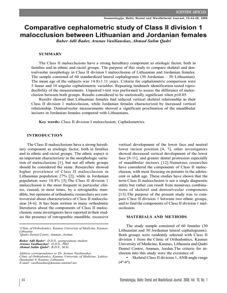
SCIENTIFIC ARTICLES Stomatologija, Baltic Dental and Maxillofacial Journal, 10:44-48, 2008 Comparative cephalometric study of Class II division 1 malocclusion between Lithuanian and Jordanian females Baher Adli Bader, Arunas Vasiliauskas, Ahmad Salim Qadri SUMMARY The Class II malocclusions have a strong hereditary component as etiologic factor, both in families and in ethnic and racial groups. The purpose of this study to compare skeletal and dentoalveolar morphology in Class II division I malocclusion of Lithuanian and Jordanian females. The sample consisted of 60 standardized lateral cephalograms (30 Jordanian – 30 Lithuanian). The mean age of the subjects was 14.8±1.11 years. Criteria for cephalometric comparison were 5 linear and 10 angular cephalometric variables. Repeating landmark identification tested reproducibility of the measurements. Unpaired t-test was performed to assess the difference of malocclusion between both groups. Results considered to be statistically significant when p£0.05. Results showed that Lithuanian females had reduced vertical skeletal relationship in their Class II division 1 malocclusion, while Jordanian females characterized by increased vertical relationship. Dentoalveolar measurements showed a significant proclination of the mandibular incisors in Jordanian females compared with Lithuanians. Key words: Class II division I malocclusion; Cephalometrics. INTRODUCTION The Class II malocclusions have a strong hereditary component as etiologic factor, both in families and in ethnic and racial groups. The ethnic aspect is an important characteristic in the morphologic variation of malocclusions [1], but not all ethnic groups should be considered the same. Researches showed higher prevalence of Class II malocclusion in Lithuanian population 27% [2], while in Jordanian population were 18.8% [3].The Class II division 1 malocclusion is the most frequent in particular clinics, caused, in most times, by a retrognathic mandible, but opinions of orthodontic researchers are controversial about characteristics of Class II malocclusion [4-6]. It has been written in many orthodontic literatures about the components of Class II malocclusion; some investigators have reported in their studies the presence of retrognathic mandible, excessive Clinic of Orthodontics, Kaunas University of Medicine, Kaunas, Lithuania 2 Qadri Dental Centre, Amman, Jordan 1 Baher Adli Bader1- D.D.S., postgraduate student Arunas Vasiliauskas1- D.D.S., PhD Ahmad Salim Qadri2- B.D.S., M.Sc Address correspondence to Dr. Arunas Vasiliauskas, Clinic of Orthodontics, Kaunas University of Medicine, LukšosDaumanto 6, Kaunas, Lithuania. E-mail: vasiliauskas@kaunas.omnitel.net 44 vertical development of the lower face and neutral lower incisor position [4, 7], other investigators showed decreased vertical development of the lower face [8-11], and greater dental protrusion especially of mandibular incisors [12].Numerous researches have considered the components of Class II malocclusion, with most focusing on patients in the adolescent or adult age. These studies have shown that the term Class II malocclusion is not a single diagnostic entity but rather can result from numerous combinations of skeletal and dentoalveolar components [13].The purpose of the present study was to compare Class II division 1 between two ethnic groups, and to find the components of Class II division 1 malocclusion. MATERIALS AND METHODS The study sample consisted of 60 females (30 Lithuanian and 30 Jordanian lateral cephalograms). Both groups were randomly selected with Class II division 1 from the Clinic of Orthodontics, Kaunas University of Medicine, Kaunas, Lithuania and Qadri Dental Centre, Amman, Jordan.The criteria for inclusion into this study were the existence of: • Skeletal Class II division 1, ANB angle range (4º-6º). Stomatologija, Baltic Dental and Maxillofacial Journal, 2008, Vol. 10, No. 1 SCIENTIFIC ARTICLES B. A. Bader et al. Fig. 1. Angular cephalometric measurements: 1 – SNA; 2 – SNB; 3 – NA-Ul; 4 – NB-L1; 5 – Sn-ML; 6 – Max-Man; 7 – YAxis; 8 – Max-Ul; 9 – Man-L1; 10 – U1-L1 Fig. 2. Linear cephalometric measurements: 1– Sn Length; 2 – A-Pog With Lower incisor; 3 – Wit's; 4 – Total Facial Height; 5 – Lower Facial Height • Proclination of maxillary incisors with an overjet ranged (4-6 mm). • Fully erupted permanent dentition. • Young adolescents, after growth spurt. With no pr evious orthodontic treatment recorded.Cephalometric lateral skull radiographs were taken as follows: each subject stood with the head in a natural position with teeth held in centric occlusion under standard conditions. The head was fixed by fitting the ear rods of the cephalostat in the external auditory meatus. Cephalometric radiographs were digitized, traced, and analyzed using Dolphin Imaging 10.1 software (2007 Dolphin Imaging & Management Solutions).Cephalometric analysis comprised of the 15 variables (10 angular and 5 linear measurements): • Anteroposterior position of the maxilla relative to anterior cranial base: SNA. • Anteroposterior position of the mandible relative to anterior cranial base: SNB. • Difference between SNA and SNB: ANB. • Position of maxillary incisors relative to the maxilla: NA-U1. • Position of mandibular incisors relative to the mandible: NB-L1. • Upper incisor to lower incisor: U1-L1. • Cranial base to mandibular plane: SN/ML. • Maxillary plane angle to mandibular plane: Max/Man. • Maxillary incisor angle to maxillary plane: U1/ Max. • Mandibular incisor angle to mandibular plane: L1/Man. • Cranial base to S-Gn: y-Axis. • Anterior cranial base length: Sn Length. • Lower incisor to A-Pog: L1/A-Pog. • Wit’s appraisal: Wit’s. Fig. 3. Cephalometric superimposition representing mean of skeletal and dentoalveolar components Stomatologija, Baltic Dental and Maxillofacial Journal, 2008, Vol. 10, No. 1 45 B. A. Bader et al. SCIENTIFIC ARTICLES Fig. 4. Comparison of SN-ML mean between Lithuanian and Jordanian females Fig. 5. Comparison of Max-Man mean between Lithuanian and Jordanian females • Nasion (N) to Menton (Me): Total Facial measurements ranged between ±1.5 and ±1.9 degrees. These errors were deemed to have insignificant effect on reliability of the results. The variables with higher linear and angular measurement errors (Wit’s, L1/Man) were considered to be not reliable. Statistical analysis All statistical calculations were performed with Microsoft Office Excel 2003 and the Statistical Package for the Social Sciences for Windows (SPSS 8.0). Mean and standard deviation (SD) of cephalometric parameters used for the data description. Unpaired student t -test was performed to assess the difference of malocclusion between both groups, Scheffe‘s test. p<0.05 was considered statistically significant difference. Height. • Anterior nasal spine (ANS) to Menton (Me): Lower Facial Height.The anatomic landmarks of measurements used in cephalometric analysis are presented in Figure 1 and Figure 2. Method error Sixty radiographs were traced by single practitioner, within two weeks interval from the first measurement. The reliability of the method was tested by other four practitioners. The error of method was calculated using the formula: ME = å (d - d2 ) 2 1 2(n - 1) Where ±2SD are the limits within which 95% of the differences between the repeated measurements are expected to lie; d1 – first measurement; d2 – second measurement; n – number of patients. The errors of linear measurements did not exceed ±1.1mm, differences between the repeated angular RESULTS Dental and skeletal cephalometric comparison between Lithuanian and Jordanian Class II Division 1 females shown in Table and Figure 3, and results will be summarized as follows: Table. Statistical comparison between Lithuanian and Jordanian females with Class II division 1 malocclusion No Variables 1 2 3 4 5 6 7 8 9 10 11 12 13 14 15 SNA SNB ANB NA – U1 NB – L1 U1 – L1 SN – ML M ax – Man M ax – U1 M an – L1 y-Axis A-Pog Wit’s Sn LFH – TFH Lit huaniann=30 Me an SD 82.3 3. 18 77.3 2. 96 5.08 0. 81 22.31 6. 42 26 5. 36 129.41 8. 85 31.91 4. 38 24.7 5. 17 110.11 5. 84 95.71 8. 74 69.05 3. 44 1.01 2. 28 2.9 2. 04 71.33 2. 63 54.58 2. 12 Jordaniann= 30 Mean SD 82.15 3.15 77.15 3.14 4.86 0.94 23.56 6.74 29.85 6.20 125. 06 10.27 35.43 4.71 27.63 4.65 112. 33 7.34 97.2 6.47 70.65 3.48 3.05 2.56 0.9 2.10 73.5 5.02 55.04 2.71 Mean Diffe ren ces t p 0.15 0.15 0.22 1.25 3.85 4.35 3.52 2.93 2.22 1.49 1.6 2.04 2 2.17 0.46 0.183 0.190 0.953 0.735 2.57 1.76 2.99 2.31 1.29 0.747 1.79 3.24 3.73 2.09 0.727 n. s n. s n. s n. s 0. 013 0. 084 0. 0041 0. 025 n. s n. s 0. 079 0. 0020 0. 0004 0. 041 n. s n.s indicates nonsignificant 46 Stomatologija, Baltic Dental and Maxillofacial Journal, 2008, Vol. 10, No. 1 SCIENTIFIC ARTICLES B. A. Bader et al. Fig. 6. Comparison of SN-length mean between Lithuanian and Jordanian females Fig. 7. Comparison of NB-L1 mean between Lithuanian and Jordanian females Skeletal Components Four variables were used to asses vertical skeletal relationship – three angular (SN-ML, Max-Man, y-Axis) and one linear (LFH-TFH). Jordanian group showed increased facial height compared with Lithuanian group – 3.52º±1.63 for SN-ML p<0.05 (Figure 4), Mandibular plane to Maxillary plane (MaxMan) also was 2.93º±1.76 larger in Jordanian group p<0.05 (Figure 5), the other measurements (Y-axis, LFH-TFH) were about similar. Anterior cranial base length was determined by one variable (SN Length) (Figure 6), SN length was 2 mm longer in Jordanian group compared with Lithuanian. Dental Components The position of maxillary incisors was determined by two angular variables (NA-U1) and (Max-U1). Both groups showed a slight proclination in the upper incisor position, no significant difference was observed between the two groups.For mandibular incisors position determination, angular (NB-L1) and linear (A-pog-L1) variables were used (Figures 7, 8). The Jordanian mandibular incisors were significantly more proclined p<0.05, the NB-L1 showed 3.85º ±2.17 higher in Jordanian group and 2.04 mm ±0.9 for A-pog–L1, both groups showed a proclination in the lower incisors and was registered in 80% of class II division 1 cases. Fig. 8. Comparison of A-pog with lower incisors mean between Lithuanian and Jordanian females Stomatologija, Baltic Dental and Maxillofacial Journal, 2008, Vol. 10, No. 1 DISCUSSION Class II malocclusion has been estimated in numerous studies [7, 9, 14, 15], these results were controversial about characteristic of class II malocclusion. Class II malocclusions may result from numerous combinations of skeletal and dental components; Rothstein [6] reported that maxillary protrusion is the component of class II malocclusion, McNamara [4] stated in his study that mandibular retrusion is the component of class II malocclusion. The present study showed statistically significant difference between both groups in skeletal and dentoalveolar measurements, as the criteria for selection of the lateral cephalograms was specific for ANB angle, the anteroposterior skeletal relationship of the maxilla and mandible r elative to the cr anial base were similar.Cephalometric measurements and their interpretation depend on the selected method of analysis. During ANB angle evaluation attention is not paid to influence of vertical ratio of maxilla-mandible, and to influence of their absolute values to sagittal ratio. For instance, in case of increasing height of the lower third of the face and in case of the same size of mandible, point B will be in a more distal position.It is important to determine the dentoalveolar position relative to their bases in Class II division I malocclusion. Studies have usually associated protrusion of the upper incisor position relative to the maxilla. Riedel [16] noted that the maxillary incisor position in his Class II division 1 sample was twice as far anterior to the facial plane as that in patients having normal occlusion. Hitchcock [17] also reported maxillary incisor protrusion relative to A-Pog. Hamdan [18] reported a significant protrusion in upper incisor position in Jor- 47 B. A. Bader et al. danian population.The upper incisor position was evaluated by the NA–U1 and Max–U1 in the present study. Upper incisor proclination was present in 67% of the subjects in Jordanian group, while incisor retroclination was seen in few cases. This finding is in consonance with the results of other investigators [1, 15-17].Two angular measurements of mandibular dental position relatively to the skeletal mandible were used in this study – NB–L1 and Man–L1. More than 70% of Class II division 1 cases showed proclination of the lower incisors according our data. However, it should be kept in mind that major cases with mandibular incisor protrusion correlated with retrognathic mandible because of dentoalveolar compensation of skeletal discrepancy [4, 19]. These findings are in agreement with other studies [1, 15], and conflict with the results of McNamara, in which he stated that the lower incisor position were generally well positioned [4].Šidlauskas [8] showed a decreased facial height in his study on Lithuanian Class II division 1 malocclusion, and that supports our findings on Lithuanian females. Similar results were found in other Class II division 1 studies [9-11]. In comparison with Jordanian sample, which showed an increase in vertical height, and the results were similar to Henry [7], Hunter [11], McNamara [4], whose concluded in their studies that; excessive vertical development of the SCIENTIFIC ARTICLES lower face is a frequent characteristic of class II division 1 malocclusion.It is of utmost importance that the phenotype of the patient with Class II division 1 malocclusion be characterized thoroughly as possible, because this is closely related with further treatment planning. Treatment protocols with functional appliances for growing patients proved effectiveness in dentoalveolar and skeletal correction of post normal buccal segments [20]. The components of Class II division 1 malocclusion should be considered for every patient individually. CONCLUSIONS 1- Lithuanian Class II division 1 females had reduced vertical skeletal relationship, while Jordanian females had increased vertical relationship in their class II division 1 malocclusion.2- The dentoalveolar comparisons indicated that the Jordanian group had more protrusion of the lower incisors compared with Lithuanians. ACKNOWLEDGEMENTS We would like to thank the great Dr. Adli Adel Bader for encouragement to carry out this study, and Dr. Ali Khalifeh for setting up cephalometric analysis. REFERENCES 1. Freitas MR, Santos MAC, Freitas KMS, Janson G, Freitas DS, Henriques JFC. Cephalometric characterization of skeletal Class II division 1 malocclusion in white Brazilian subjects. J Appl Oral Sci 2005;3:198-203. 2. Lopatienė K. Prevalence of malocclusion in Lithuania. Stomatologija. Baltic Dent Maxillofac J 2006;8(suppl. 2):11. 3. Alhaija ESJ, Al-Khateeb SN, Al-Nimri KS. Prevalence of malocclusion in 13-15 year-old North Jordanian school children. Community Dent Health 2005;22:266-71. 4. McNamara JA. Components of Class II malocclusion in children 8–10 years of age. Angle Orthod 1981;51:177–202. 5. Bishara SE, Jakobsen JR, Vorhies B, Bayati P. Changes in dentofacial structures in untreated Class II division 1 and normal subjects: A longitudinal study. Angle Orthod 1997;67:5566. 6. Rothstein TL. Facial morphology and growth from 10 to 14 years of age in children presenting Class II division 1 malocclusion: a comparative roentgenographic cephalometric study. Am J Orthod 1971;60:619-20. 7. Henry RG. A classification of Class II division 1 malocclusion. Angle Orthod 1957;27:83-92. 8. Šidlauskas A, Švalkauskienė V, Šidlauskas M. Assessment of Skeletal and Dental Pattern of Class II Division 1 Malocclusion with Relevance to Clinical Practice. Stomatologija. Baltic Dent Maxillofac J 2006;8:3-8. 9. Pancherz H, Zieber K, Hoyer B. Cephalometric characteristics of Class II division 1 and Class II division 2 malocclusions: A comparative study in children. Angle Orthod 1997;67:11120. 10. Karlsen AT. Craniofacial morphology in children with Angle Class II- 1 malocclusion with and without deepbite. Angle Orthod 1994;64:437-46. 11. Hunter WS. The vertical dimension of the face and skeletodental retrognathism. Am J Orthod 1967;53:586-95. 12. Phelan T, Buschang PH, Behrents RG, Wintergerst AM, Ceen RF, Hernandez A. Variation in Class II malocclusion: Comparison of Mexican mestizos and American whites. Am J Orthod Dentofac Orthop 2004;125:418-25. 13. Graber TM, Vanarsdall RL, Vig KWL. Orthodontics: Current Principles and Techniques. 4th ed. St.Louis: Elsevier; 2005. p. 442-461. 14. Lau JW, Hagg U. Cephalometric morphology of Chinese with Class II Division 1 malocclusion. Br Dent J 1999;186:188-90. 15. Sayżn MO, Turkkahraman H. Cephalometric evaluation of nongrowing females with skeletal and dental Class II, division 1 Malocclusion. Angle Orthod 2005; 75:656-60. 16. Riedel RA. The relation of maxillary structures to cranium in malocclusion and normal occlusion. Angle Orthod 1952;22:142-5. 17. Hitchcock P. Cephalometric description of Class II division 1 malocclusion. Am J Orthod 1973;63:414-23. 18. Hamdan AM, Rock WP. Cephalometric norms in an Arabic population. J Orthod 2001;l28:297-300. 19. Solow B. The pattern of craniofacial association. Acta Odontol Scand 1966; 24 (Suppl.46):174. 20. Šidlauskas A. The effects of the Twin-block appliance treatment on the skeletal and dentoalveolar changes in Class II Division 1 malocclusion. Medicina 2005; 5: 392-400. Received: 11 01 2008 Accepted for publishing: 26 03 2008 48 Stomatologija, Baltic Dental and Maxillofacial Journal, 2008, Vol. 10, No. 1

