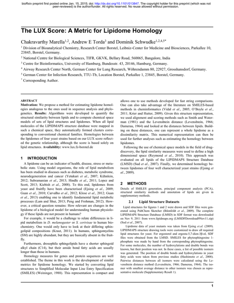
bioRxiv preprint first posted online Jan. 15, 2015; doi: http://dx.doi.org/10.1101/013847. The copyright holder for this preprint (which was not
peer-reviewed) is the author/funder. All rights reserved. No reuse allowed without permission.
The LUX Score: A Metric for Lipidome Homology
Chakravarthy Marella1,2, Andrew E Torda3 and Dominik Schwudke1,2,4,5*
1
Division of Bioanalytical Chemistry, Research Center Borstel, Leibniz-Center for Medicine and Biosciences, Parkallee 10,
23845, Borstel, Germany.
2
National Centre for Biological Sciences, TIFR, GKVK, Bellary Road, 560065, Bangalore, India
3
Centre for Bioinformatics, University of Hamburg, Bundesstr. 43, 20146, Hamburg, Germany.
4
Airway Research Center North, German Center for Lung Research, Wöhrendamm 80, 22927, Grosshansdorf, Germany.
5
German Center for Infection Research, TTU-Tb, Location Borstel, Parkallee 1, 23845, Borstel, Germany.
*
Corresponding Author.
ABSTRACT
Motivation: We propose a method for estimating lipidome homologies analogous to the ones used in sequence analysis and phylogenetics. Results: Algorithms were developed to quantify the
structural similarity between lipids and to compute chemical space
models of sets of lipid structures and lipidomes. When all lipid
molecules of the LIPIDMAPS structure database were mapped in
such a chemical space, they automatically formed clusters corresponding to conventional chemical families. Homologies between
the lipidomes of four yeast strains based on our LUX score reflected the genetic relationship, although the score is based solely on
lipid structures. Availability: www.lux.fz-borstel.de
1
INTRODUCTION
A lipidome can be an indicator of health, disease, stress or metabolic state. Using model organisms, the role of lipid metabolism
has been studied in diseases such as diabetes, metabolic syndrome,
neurodegeneration and cancer (Yetukuri et al., 2007; Kühnlein,
2012; Subramanian et al., 2013; Hindle et al., 2011; Lopez and
Scott, 2013; Kiebish et al., 2008). To this end, lipidomes from
yeast and fruitfly have been characterised (Ejsing et al., 2009;
Guan et al., 2010; Carvalho et al., 2012; Klose et al., 2012; Guan
et al., 2013) enabling one to identify fundamental lipid metabolic
processes (Lam and Shui, 2013; Peng and Frohman, 2012). However, a critical question remains: How relevant are changes in the
lipidome of a biological model for understanding human physiology if these lipids are not present in humans?
For example, it would be a challenge to relate differences in lipid metabolism in D. melanogaster or S. cerivisae to human biochemistry. One would only have to look at their differing sphingolipid compositions (Kraut, 2011). In humans, sphingomyelins
(SM) are highly abundant, but they are basically absent in the fruitfly.
Furthermore, drosophila sphingolipids have a shorter sphingoid
alkyl chain (C14), but their amide bond fatty acids are usually
longer than those in humans.
Homology measures for genes and protein sequences are well
established. The theme in this work is the development of similar
metrics for lipidome homology. We started by converting lipid
structures to Simplified Molecular Input Line Entry Specification
(SMILES) (Weininger, 1988). This representation is compact and
allows one to use methods developed for fast string comparisons.
One can also take advantage of the literature on SMILES-based
methods in cheminformatics (Vidal et al., 2005; O’Boyle et al.,
2011; Krier and Hutter, 2009). Given this structure representation,
we used alignment and scoring methods such as Smith and Waterman (1981) and the Levenshtein distance (Levenshtein, 1966;
Damerau, 1964) and looked at the distances between lipids. Building on these distances, one can represent a whole lipidome as a
dissimilarity matrix. This numerical representation can then be
used for further analyses such as estimating the homology between
lipidomes.
Following the use of chemical space models in the field of drugdiscovery, the lipid similarity measures were used to define a high
dimensional space (Reymond et al., 2010). This approach was
evaluated on all lipids of the LIPIDMAPS Structure Database
(LMSD) (Sud et al., 2007). Finally, we determined homology between lipidomes of four well characterized yeast strains (Ejsing et
al., 2009).
2
METHODS
Details of SMILES generation, principal component analysis (PCA),
structural similarity methods and annotation of lipids are given in
supplementary methods.
2.1
Lipid Structure Datasets
Lipid structures for figures 1 and 2 were drawn and SDF files were generated using PubChem Sketcher (Ihlenfeldt et al., 2009). The complete
LIPIDMAPS Structure Database (LMSD) in SDF format was downloaded
on Nov 9, 2011 from www.lipidmaps.org (LMSDFDownload9Nov11.zip)
(Sud et al., 2007).
Lipidome data of yeast mutants was taken from Ejsing et.al. (2009).
LIPIDMAPS structure drawing tools were customized to draw all required
lipid structures for yeast. For ergosterol and ergosta-5,7-dien-3β-ol, SDF
files were obtained from the LMSD. SMILES for phytosphingosine 1phosphate was made by hand from the corresponding phytosphingosine.
For some molecules, the number of hydroxylations and double bonds was
known, but their position was not. In these cases, a list of possible isomers
was generated. The position of double bonds and hydroxylations in yeast
fatty acids were taken from previous studies (Hashimoto et al., 2008).
Pairwise distances between all isomers were calculated using the Levenshtein distance method (Levenshtein, 1966; Damerau, 1964). The isomer with smallest average distance to other isomers was chosen as representative molecule (Supplementary Result 1).
1
bioRxiv preprint first posted online Jan. 15, 2015; doi: http://dx.doi.org/10.1101/013847. The copyright holder for this preprint (which was not
peer-reviewed) is the author/funder. All rights reserved. No reuse allowed without permission.
C.Marella et al.
2.2
Template-based SMILES
LIPIDMAPS perl scripts were modified to generate a wider spectrum of
lipid structures (Fahy et al., 2007; Sud et al., 2012). These scripts are
available at www.lux.fz-borstel.de. Molecule structures were obtained in
SDF format and subsequently converted to SMILES using the OpenBabel
molecular conversion tool with the default algorithm (O’Boyle et al.,
2011). Characters indicating chirality, cis–trans isomerism and charges
were removed automatically for the yeast lipidome analysis.
2.3
Structural similarity scoring methods
Six similarity scoring methods were tested 1) OpenBabel FP2 Fingerprint
2) LINGO 3) Bioisosteric similarity 4) SMILIGN 5) Smith Waterman
Local Alignment 6) Levenshtein distance. Details are given in supplementary methods. The Levenshtein method was applied for analyzing LMSD
and yeast lipidome (Figures 4, 5 and 6). This algorithm was originally
designed for correcting spelling errors, but the principle can be applied to
compare any pair of strings including SMILES (Levenshtein, 1966;
Damerau, 1964). The source code used in this study is provided in our
website www.lux.fz-borstel.de.
2.4
Lipidome Juxtaposition Score (LUX) Calculation
The LUX score is based on the Hausdorff distance (Jain et al., 1999) and
summarizes the similarity between lipidomes. In pseudocode, the distance
from lipidome A to B is calculated from:
for each lipid in A
find distance d to most similar lipid in B
dsum ∶= dsum + d
n = n +1
return dsum/n
This yields the average shortest distance dAB from A to B. The larger of dAB
and dBA was used as the lipidome homology score (AB). The LUX score is
a simple measure of the homology between a pair of lipidomes. Lower
LUX scores signify higher homology.
2.5
Hierarchical Cluster Analysis
Complete linkage clustering was performed with R, version 2.14.1,
library – ‘stats’ and function ‘hclust’ using the LUX score as the similarity
metric.
Fig. 1. Alignment-based distance calculation algorithms can distinguish isomeric lipid molecules. A. Structure of 17 ceramide molecules consisting of a C16
sphingoid base (light green) and an amide-linked hydroxy fatty acid. The carbon atom number of the hydroxyl group position at the fatty acid chain (red) is
used for naming individual molecules. B. SMILES representation of first and last molecules. Color coding of atoms is identical in SMILES- and structurerepresentations. C-H. Heat map of pairwise distances calculated using Open Babel’s FP2 Fingerprint (C), LINGO (D), Bioisosteric (E), SMILIGN (F),
Smith-Waterman (G) and Levenshtein distance (H) algorithms. Bioisosteric method uses CACTVS canonical SMILES whereas for all other methods
template-based SMILES were used. Color bars in each panel indicate range of distances values of the particular method. Black denotes a distance value of
ZERO, indicating molecular identity. Numbers in rows and columns simultaneously represent the molecule name and the position of hydroxyl group in fatty
acid moiety.
2
bioRxiv preprint first posted online Jan. 15, 2015; doi: http://dx.doi.org/10.1101/013847. The copyright holder for this preprint (which was not
peer-reviewed) is the author/funder. All rights reserved. No reuse allowed without permission.
Lipidome Homology
3
RESULTS
3.1
Alignment-based similarity scoring methods
distinguishes between positional isomers
As a basis for our similarity scoring, we established a templatebased SMILES generation algorithm for lipids. We were able to
write SMILES in a consistent and predictable manner using template-based structure drawing tools (Fahy et al., 2007; Sud et al.,
2012) and OpenBabel default SMILES algorithm (Supplementary
Result 2). We then tested alignment methods and distance metrics,
analogous to those used for protein or nucleotide sequences. Our
quality criterion was based on the methods' sensitivity to small
structural differences commonly found in lipids. The first test dataset consisted of a set of 17 ceramide molecules with the chemical
composition C34H68O4N1. The position of the hydroxyl group was
successively changed from position 2 to 18 at the fatty acid moiety
resulting in 17 isomeric molecules (Figure 1A). All isomers were
converted into SMILES in which the shift of the hydroxyl group
can easily be recognized and we tested six similarity scoring methods (Supplementary Result 2, Figure 1C-H). Three from the literature were used as described under Methods: FP2 Fingerprint
(O’Boyle et al., 2011), LINGO (Vidal et al., 2005) and Bioisosteric similarity (Krier and Hutter, 2009). Three methods were introduced here: the SMILIGN, Smith and Waterman (1981) and Levenshtein (1966) distance (Damerau, 1964).
The first clear result is that a large subset of isomeric structures
cannot be distinguished by either OpenBabel FP2 Fingerprint or
LINGO (Figure 1 C-D). The FP2 Fingerprint algorithm computed
a distance of zero for 78 pairs of ceramide isomers (Figure 1C –
black pixels). LINGO gave a zero distance for 91 pairs of isomers
(Figure 1D). This would only be correct if the molecules were
identical. Both methods segment SMILES into shorter sub-strings
(1-7 character length in Path-length Fingerprint and 4 characters by
LINGO) and apply the Tanimoto coefficient for determining distances. This segmentation into short sub-strings loses the information on the position of the hydroxyl group. In contrast, the Bioisosteric algorithm differentiated all 17 isomeric structures, even
though it uses CACTVS Canonical SMILES. There are no zero
distances off the diagonal (Figure 1E). The Bioisosteric method
also segments SMILES into sub-strings, but in a hierarchical manner, preserving information on the position of the hydroxyl group
(Krier and Hutter, 2009). There is a distinct pattern in the heat map
of the Bioisosteric method characterized by a gradual increase in
distance values for isomers having the hydroxyl group closer to the
terminal methyl carbon. The Bioisosteric method returns a distance
of 0.13 units for the shift of the hydroxyl group from position 5 to
7 (Figure 1E - light green pixel), but returns 0.26 units for position
10 to 12 (Figure 1E - yellow pixel) and for positions 16 to 18, a
distance of 0.41 was calculated (Figure 2E - white pixel). This
dependence of the distance values on the position of the hydroxyl
group leads to an unwanted weighting which is a clear problem
with the approach.
In the SMILIGN algorithm, SMILES strings are treated as if they
were amino acid sequences and a multiple sequence alignment was
calculated with MUSCLE (Edgar, 2004). The pairs of lipids were
rescored using an identity matrix. The SMILIGN method distinguished all 17 ceramide isomers (Figure 1F), but we noticed an
irregular distribution of distance values. For example, comparing
molecule pairs where the hydroxyl group was shifted by one posi-
Fig. 2. The relationship between phosphatidylinositol (PI) molecules is
retained in a two dimensional structural space (The structures and
SMILES are provided in Supplementary Result 1). Pairwise distances
between 16 PIs were calculated with Bioisosteric, SMILIGN, SmithWaterman and Levenshtein methods (A-D). PCA was carried out on the
distance matrices to generate two- and three- dimensional maps.
Approximate contribution of each principal component to the total
variance is shown in brackets. Molecules with double bonds are in grey
and without double bonds are in black. Euclidian distance between the
first molecule PI (10:0/10:0) and all others in the PC1-PC2 plane are
shown as bar graphs on the right panels. Molecules were numbered
according to the length of the sn2 acyl chain length, wherein an underlined
number XX indicate the presence of the double bond.
tion 11-12, 12-13, 13-14 and 14-15 resulted in four different distance values of 0.03, 0.13, 0.25 and 0.06 units respectively. In this
3
bioRxiv preprint first posted online Jan. 15, 2015; doi: http://dx.doi.org/10.1101/013847. The copyright holder for this preprint (which was not
peer-reviewed) is the author/funder. All rights reserved. No reuse allowed without permission.
C.Marella et al.
regard, we identified two problems with the algorithm. First, there
were several misalignments that lead to incorrect distances. Second, one needs 35 characters to represent all the structural details
of all lipid molecules of the LMSD (LipidMaps Structure Database). The software is limited to only 20 characters and too much
information is lost. To overcome these two limitations of
SMILIGN, we tested two pair-wise alignment methods that do not
require conversion to amino acid sequences.
With the Smith-Waterman method, pair-wise alignments are carried out directly with the SMILES strings. All ceramide isomers
were distinguished, but we noticed an anomaly in distance values
for molecules 17 and 18 (Figure 1G). A closer examination of the
pair-wise alignments revealed an inherent issue when applying
local alignment procedure to lipids. In the aligned SMILES pairs
2-17 and 2-18, the hydroxyl groups in the fatty acid were ignored,
while for the pairs 2-15 and 2-16 the characters were retained. The
Smith-Waterman algorithm is designed to find high scoring regions in strings, so differing ends are ignored by design and not by
accident. This means that functional groups at the omega position
are ignored, despite their role in biology (Kniazeva et al., 2004).
Although one could try to adjust parameters, the Smith-Waterman
method is fundamentally not appropriate for this kind of comparison.
Finally, we tested the Levenshtein distance for measuring similarities between lipid molecules (Figure 1H). Unlike SmithWaterman, the Levenshtein approach always aligns all characters
for a given pair of SMILES. This method was the most successful.
It distinguished all ceramide molecules and for each molecule, it
correctly scored and ranked distances up to the molecule’s third
closest isomers. From the fourth closest isomer onwards, a fixed
distance of 0.12 was determined. Unlike other methods, it guarantees a symmetric distance matrix with no unwanted weighting of
groups due to their positions.
These tests of structural similarity measures led to two conclusions. First, the alignment step is necessary. Second, the Levenshtein distance was most likely to be generally applicable for
all molecules in a lipidome.
3.2
From structural similarity to chemical space
A set of distances between n molecules defines an (n − 1) dimensional space. The coordinates of molecule i are simply the distances to all members of the set (including the zero distance to molecule i itself). This is formally a vector space so similar molecules
will have similar coordinates. It is, however, not very compact and
because of structural similarities, coordinates in some dimensions
would be highly correlated with others. Principal component analysis (PCA) was then used to reduce the dimensionality and see
how much information would be lost. The first test was performed
on a set of 16 phosphatidylinositol molecules (Supplementary Result 1).
Considering just the first two principal components was sufficient to highlight problems with some of the distance measures.
For example, the map in Figure 2A shows a clear weakness with
the Bioisosteric method. The extension of the fatty acid chain at
the sn2 position and degree of unsaturation are not accurately rep-
4
Fig. 3. The structural space model clusters thousands of sphingolipids (SP)
according to their chemical relationship. A. Three dimensional map of 3510
SP obtained by PCA from a pair wise distance matrix calculated with
Levenshtein distance. B. Plot of all neutral SP within the same coordinate
system as panel A indicating several associated glycosphingolipid series. C.
Complex glycosphingolipids are highlighted showing the influence of
structural changes in the ceramide backbone and sugar moiety.
resented (Figure 2A, scatter plot). We also computed the Euclidian
distance between molecules in the plane of the first two components. This showed an inconsistent trend in the distance increase
with each structural alteration (Figure 2A, bar graph). Principal
components can often be interpreted in terms of the original descriptors and in the case of SMILIGN, the first two components are
dominated by the extension of the acyl chain at the sn2 position
(Figure 2B). For SMILIGN, the first two principal components are
not sufficient to distinguish molecules that differ only in the presence of a double bond, but the third principal component does
capture it (Supplementary Result 3).
bioRxiv preprint first posted online Jan. 15, 2015; doi: http://dx.doi.org/10.1101/013847. The copyright holder for this preprint (which was not
peer-reviewed) is the author/funder. All rights reserved. No reuse allowed without permission.
Lipidome Homology
In contrast, distances based on Smith-Waterman and Levenshtein
algorithms reflected all gradual structural changes in the molecules
(Figure 2 C,D). In both cases, the projection leads to a set of points
in a ‘U’ shape and, if we take molecule 10 as a reference, stepwise
changes to the chemistry are reflected in distinct shifts in the principal coordinates. We further recognized that the changes in coordinates, when the acyl chain is extended by two methylene groups
(molecules 15-17, 17-19) are about twice as large as the difference
between pairs differing by a single methylene group (Figure 2CD). The first two principal components combined, accounted for
95% of the variability in the underlying data set for SmithWaterman and Levenshtein. Summarizing the results, we see the
Levenshtein method coupled with template-based SMILES as the
best approach for calculating structural differences in small molecule sets. PCA is an appropriate way to reduce dimensionality and
the relation between molecules can be depicted in a PCA map,
which we treat as chemical space.
The set of 16 phosphatidylinositol molecules is useful for highlighting details, but one is interested in using the method on much
larger molecule sets. To this end, we used the 3510 sphingolipids
(SP) from LMSD as a test dataset (Sud et al., 2007). All lipid
structures were converted into template-based SMILES and pairwise distances were computed using the Levenshtein method. Figure 3A shows the position of all molecules in terms of the first
three principal components. There are two clear observations.
First, three principal components account for 99% of the total variance and no two SP have the same coordinates. Second, there was
no biochemical knowledge put into the procedure, but the molecules cluster naturally into chemically similar groups (Figure 3A).
Sphingosines, ceramides and phosphosphingolipids were clustered
separately from the complex glycosphingolipids (GSL). Furthermore, the acidic and neutral GSL where placed in different clusters. Looking at the globo, lacto, neolacto and isoglobo series of
neutral GSL, one can see changes in the sugar moiety and a clear
separation from the simple ‘Glc’ series (Figure 3B). This observation fits the intuitive expectation that the ‘Glc’ series with simple
sugar moieties (glucose, galactose or lactose) should be farther
from lipids with complex sugar composition. We noted that changes in the sugar moiety composition of neutral GSL, which have a
strong impact on biochemical behavior, were separated by a larger
distance compared to changes in the ceramide backbone (Figure
3B). In addition, we were intrigued by the recurring appearance of
geometrical patterns in the form of ‘I’, ‘C’ and ‘L’ shapes and
investigated the structure within these clusters. Within each cluster,
lipids were organized based on changes in the ceramide moiety
(Figure 3C) so that, for example, eight molecules of the isoglobo
series formed a twisted ‘L’ shape with each successive lipid carrying a gradual change in the ceramide backbone (Figure 3C – light
brown colored points). Analogous geometric arrangements were
observed for the globo, lacto and neolacto series (Figure 3C – red,
violet and light-blue points). Next we tested, how all the 30 150
lipids of the LMSD would be organized in a chemical space based
on only the structural similarity. All lipid molecules had unique
coordinates in the computed chemical space, indicating that our
approach can distinguish between all lipid structures within
known, natural lipidomes. With no additional input, the method
grouped lipids into clusters that correspond to the popular lipid
classification of LIPIDMAPS (Figure 4A) (Fahy et al., 2008).
Fig. 4. Spatial distribution of related phosphatidylcholines (PC) moelcules
remains stable in the background of large structure data sets. A. Lipid map
of 30 150 lipid molecules obtained from LMSD. Pair wise distances were
calculated using the Levenshtein method of template-base SMILES. B.
Structures of the 14 PC molecules. Molecules are named based on the
number of carbon atoms of the sn2 acyl chain. C. Two dimensional map of
the selected PC molecules displaying their chemical relation to each other.
Euclidian distances in PC1-PC2 plane between the smallest PC (12) and
all others are shown in the bar graph. D. Spatial distribution of 14 PCs in
the background of 30136 lipids determined from three principal
components and projected on the PC1-PC2 plane. Euclidian distances
between first molecule (12) and all other was determined form the first
two principal components and its trend is shown as bar graph.
Fatty acyls, glycerophospholipids (GPL), sphingolipids (SP) and
polyketides occupied opposite ends of the chemical space. In contrast, glycerolipids and GPL shared a common region because of
their head group similarity. Sterol lipids formed a distinct cluster
due to their unique four-ringed core structure. Prenol lipids were
widely distributed in the chemical space reflecting their varying
chemical composition. For GPL, we observed several distinct clusters, which on closer examination, could be recognized as spatially
separated lipid classes like phosphatidylcholine (PC), phosphatidylserine (PS) and phosphatidylinositols (PI).
5
bioRxiv preprint first posted online Jan. 15, 2015; doi: http://dx.doi.org/10.1101/013847. The copyright holder for this preprint (which was not
peer-reviewed) is the author/funder. All rights reserved. No reuse allowed without permission.
C.Marella et al.
Fig. 5. Lipidome maps highlight relationships between yeast strains. A. All lipids from yeast strains, BY4741, Elo1, Elo2 and Elo3 cultured at 24ºC and
37ºC are combined to create a reference map of the yeast lipidome. Each colored circle corresponds to a unique lipid. B. Comparison of lipidomes from
strains BY4741 and Elo1 (cultured at 24ºC). Arrows in first plot indicate lipids that are present in Elo1, but not in BY4741 and vice versa in the second plot.
C. Comparison of BY4741 and Elo2 lipidomes. A two dimensional Lipidome jUXtaposition (LUX) score is calculated for a pair of lipidomes using
reference-map coordinates (Supplementary Result 1).
3.3
The spatial organization of lipids is robust to
changes in background molecule ensemble
As with the set of PI molecules described above, (Figure 2D) the
PC molecules in the two-dimensional representation form an inverted 'U' pattern (Figure 4C). However, the PC molecules formed
a flipped ‘L’ pattern if all other 30 136 lipids of the LMSD were
present (Figure 4D). In both cases, the sequential arrangement of
the PC-molecules in the two-dimensional chemical space accurately represents the gradual increase in acyl chain length. We also
observed a gradual increase of the Euclidian distance from the
molecule PC (12:0 / 12:0) to all species with longer sn2 acyl chains
(Figure 4 C, D). When we gradually increased the complexity of
the set of lipid molecules, we noticed that the PCA approach could
disturb relationships between structurally similar molecules. In the
case of a set consisting of only GPLs and only GPLs and SPs (data
not shown), we noticed that the distances between molecule 12 and
molecules 21-26 did not reflect the sn2 chain length increase anymore. Interestingly, one can observe that the gradual addition of
more diverse lipid structures spanning a broader chemical variety
compensates for this bias. It seems that the Levenshtein distances
and the projection to a chemical space automatically reconstructs
conventional lipid class definitions. The next natural step is to use
these lipid coordinates to analyze and compare complete lipidomes.
3.4
The Lipidome jUXtaposition (LUX) score, a
single metric for calculating the homology
between lipidomes
The approach to lipidome comparison was then tested on real
data. All lipids from four yeast strains BY474, Elo1, Elo2 and Elo3
6
(Ejsing et al., 2009) were combined, yielding a reference lipidome
with 248 members, each with unique coordinates (Supplementary
Result 1). For clarity, this is shown in a 2D map (Figure 5A),
which is the basis of comparisons of the four strains and two
culturing temperatures (24°C and 37°C). Triacylglycerols (TAG)
occupy the largest area on the map in terms of the variety of
structures. Mannose-bis(inositolphospho)ceramides (M(IP)2C)
form a distinct cluster located in the top-left quadrant of the
reference map. In the top right quadrant of the reference map, there
is a cluster of GPLs consisting of phosphatidic acid (PA),
phosphatidylethanolamines (PE), phosphatidylcholines (PC) and
TAG. The reference lipidome map clearly shows temperature- and
strain-specific changes. The lipidomes of the wild type yeast
strains BY4741 and Elo1 grown at 24°C showed only minor
differences (Figure 5B). In contrast, the lipidome of the Elo2
mutant is very different to the wild type strain BY4741 (Figure
5C). The mutation has led to dramatic changes amongst the
inositol phosphorylceramides seen in the top-left quadrant and the
appearance of new species not present in the wild type. Using this
well-defined lipidome map, one can determine the closest related
lipid in the wild type strain If one calculates the distances that
lipids would have to move to make the members of each pair
overlap, one can use the Hausdorff distance to compare lipidomes
(arrow marked lipids, Fig 5C, D). For that, we chose the
coordinates of all lipids in the two dimensional coordinate system
of the first lipidome and determined the Euclidean distance to its
closest structural neighbor in the second. Subsequently, the
average of all distances was determined, including all distance
values of zero for identical molecular species. Because the
Hausdorff distance depends on the direction of the comparison, we
used the maximum of the two values (max(dAB,dBA)). We named
this measure as the ‘Lipidome jUXtaposition (LUX) score’. This
score is a distance, so larger values indicate more dissimilarity and
bioRxiv preprint first posted online Jan. 15, 2015; doi: http://dx.doi.org/10.1101/013847. The copyright holder for this preprint (which was not
peer-reviewed) is the author/funder. All rights reserved. No reuse allowed without permission.
Lipidome Homology
identity results in a LUX score of zero. From that perspective, one
can see that the LUX score between BY4741 and the Elo2 strain is
three-fold larger than the distance between BY4741 and Elo1
(Figure 5B, C).
Next we evaluated the LUX score by computing a hierarchical
clustered tree of all eight reported lipidomes of yeast (Figure 6A)
and compared it to dendrograms based on the lipid concentrations
(Figure 6B), and simply by counting common lipids (Figure 6C).
That allowed us to test if our approach can correctly depict the
genetic and phenotypic relationship between the yeast strains
reported earlier (Ejsing et al., 2009). The tree computed from the
LUX score indicates that mutation of the Elo1 gene had less
influence on the composition of the lipidome than the temperature
shift. Both strains, BY4741 and Elo1 were closest neighbors to
each other at the culturing temperature of 24°C and 37°C. The
lipidomes from mutant strains Elo2 and Elo3 were located in
separate branches for both temperatures (Figure 6A). This agrees
with the observation that no aberrant phenotype for Elo1 was
observed and that Elo2 and Elo3 had distinct alterations in their
intracellular organization (Ejsing et al., 2009; Oh et al., 1997). A
dendrogram based on the correlation of lipid abundances would
neglect the structural changes in the lipidome completely. This
approach implies that largest difference can be found between
BY4741 and Elo1 mutants grown at 24°C and 37°C (Figure 6B). A
temperature shift induces changes with respect to saturation and
length of the aliphatic chains, but does not necessarily lead to the
synthesis of a new lipids. This response, however dominates the
comparison based on quantities. A dendrogram computed just from
the number of lipids in common failed to capture the relationship
between the mutant strains and the wild type (Figure 6C).
Genotypes and temperature conditions are in disarray in all
branches (Figure 6C). The complexity of the yeast lipidome
comparison is relatively small compared to higher organisms.
Nevertheless, the two-dimensional structural space reflecting 63%
of the overall variability of the dataset (Figure 6A) is sufficient to
determine lipidome homologies based upon the LUX score. We
also note that the tree topology does not change whether one uses
just three principal components (covering 83% of the variability)
or the original pairwise distance matrix (data not shown). This
indicates that our approach enables a simple way to reduce the
complexity of large lipid structure datasets, which can further help
to depict results of a lipidome homology analysis in a well-defined
manner. In this way researchers can report their findings for
interspecies, cell type and cell compartment lipidome analyses in a
defined graphical representation. At the same time, the LUX score
determination workflow can be customized with regards to the
complexity of the lipidome study.
4
DISCUSSION
Our study offers a general approach to characterizing and comparing lipidomes based on the structures of their constituents. It is
certainly useful for making function / phenotype associations and
allows one to correlate changes with habitat, genetic relationships
and environmental stresses. The approach is dependent on the initial SMILES strings which is both an advantage and possible
weakness. One can consider a comparison with small molecule
classification. There, the problem is sometimes easier, especially
when one is dealing with derivatives which are closely related, but
even in small molecule chemoinformatics, there is no universally
accepted scheme (Bender et al, 2009). Optimization of such structures often depends on the interaction sites of a protein and pharmaceutical requirements for administration of drugs (Mohanapriya
and Achuthan, 2012; Ahmed and Ramakrishnan, 2012). In this
study, the analysis does not stop after comparing the details of
individual structures. The larger aim is whole lipidome comparison
and these are sets of structures whose members are functionally
related. In this work, we leverage a SMILES generation scheme
which works well on large sets, but there will probably be pathological examples where it does not perform well. It definitely
seems useful when working with lipids where it reflects 1) chain
length 2) double bond position and 3) bond frequency. However,
lipids are very special with regards to their structural diversity, and
some better similarity metrics might be available in future.
The definition of structural similarity and chemical space model
also concisely depicts the complexity of a lipidome. The projection
down to two- and three-dimensional maps lead to clusters which fit
standard lipid nomenclature. This means that one can intuitively
see qualitative differences between lipidomes. The reference map
for multiple comparisons also shows changes in the overall organization of a lipidome which can support functional association related to membrane organization and metabolic adaptations. The
lipidome comparisons in this study are solely based upon an identity matrix for exchange values which does not account for quantitative changes. This is parsimonious, but obviously not optimal for
comparing biological systems in terms of their lipidomes. In future
work, we will test how quantitative changes should be weighted
with respect to structural changes. We will estimate such weight
measures from well understood model systems based on larger
Fig. 6. The LUX score accounts for genetic alteration of yeast strains. For the yeast strains BY4741 (wild type), Elo1, Elo2 and Elo3 mutant strains cultured
at 24°C and 37°C dendrograms were computed from two dimensional LUX scores (A), comparing concentrations of common lipids (B) and counting the
percentage of common lipids in a pair of lipidomes (C). All dendograms are based on complete linkage using Euclidean distance as the similarity metric.
7
bioRxiv preprint first posted online Jan. 15, 2015; doi: http://dx.doi.org/10.1101/013847. The copyright holder for this preprint (which was not
peer-reviewed) is the author/funder. All rights reserved. No reuse allowed without permission.
C.Marella et al.
data sets that are now becoming available (Voynova et al., 2014;
Tarasov et al., 2014). However, this study shows that the structural
composition of a lipidome is sufficient to recognize the degree of
genetic alteration and growth temperature dependence in yeast
strains in an unsupervised manner. In contrast, all correlation based
methods using lipid quantities as input failed.
The growth in experimental data combined with methods like the
LUX score may provide a basis for disease and trait association
studies as used in genome research.
ACKNOWLEDGEMENTS
The authors are thankful to Eli Lebow, Geoff Hyde, Mukund
Thattai, Sandeep Krishna and Kurt Fellenberg for critical discussions.
Funding: This work was supported by a Wellcome Trust India
Alliance Senior Fellowship to DS and by funds of the German
Research Foundation of the SFB-TR22 consortia. CM was supported by UGC Junior Research Fellowship and NCBS-TIFR
Graduate Student Fellowship.
REFERENCES
Ahmed,S.S.S.J. and Ramakrishnan,V. (2012) Systems biological approach of
molecular descriptors connectivity: optimal descriptors for oral
bioavailability prediction. PloS One, 7, e40654.
Bender,A. et al. (2009) How Similar Are Similarity Searching Methods? A Principal
Component Analysis of Molecular Descriptor Space. J. Chem. Inf.
Model., 49, 108–119.
Carvalho,M. et al. (2012) Effects of diet and development on the Drosophila lipidome.
Mol. Syst. Biol., 8, 600.
Damerau,F.J. (1964) A technique for computer detection and correction of spelling
errors. Commun. ACM, 7, 171–176.
Edgar,R.C. (2004) MUSCLE: multiple sequence alignment with high accuracy and
high throughput. Nucleic Acids Res., 32, 1792–1797.
Ejsing,C.S. et al. (2009) Global analysis of the yeast lipidome by quantitative shotgun
mass spectrometry. Proc. Natl. Acad. Sci. U. S. A., 106, 2136–2141.
Fahy,E. et al. (2007) LIPID MAPS online tools for lipid research. Nucleic Acids Res.,
35, W606–612.
Fahy,E. et al. (2008) Update of the LIPID MAPS comprehensive classification system
for lipids. J. Lipid Res., 50, S9–S14.
Guan,X.L. et al. (2013) Biochemical membrane lipidomics during Drosophila
development. Dev. Cell, 24, 98–111.
Hashimoto,K. et al. (2008) The repertoire of desaturases and elongases reveals fatty
acid variations in 56 eukaryotic genomes. J. Lipid Res., 49, 183–191.
Hindle,S. et al. (2011) Invertebrate models of lysosomal storage disease: what have
we learned so far? Invertebr. Neurosci. IN, 11, 59–71.
Ihlenfeldt,W.D. et al. (2009) The PubChem chemical structure sketcher. J.
Cheminformatics, 1, 20.
Jain,A.K. et al. (1999) Data Clustering: A Review. ACM Comput Surv, 31, 264–323.
Kiebish,M.A. et al. (2008) Cardiolipin and electron transport chain abnormalities in
mouse brain tumor mitochondria: lipidomic evidence supporting the
Warburg theory of cancer. J. Lipid Res., 49, 2545–2556.
Klose,C. et al. (2012) Flexibility of a Eukaryotic Lipidome – Insights from Yeast
Lipidomics. PLoS ONE, 7, e35063.
Kniazeva,M. et al. (2004) Monomethyl branched-chain fatty acids play an essential
role in Caenorhabditis elegans development. PLoS Biol., 2, E257.
Kraut,R. (2011) Roles of sphingolipids in Drosophila development and disease. J.
Neurochem., 116, 764–778.
Krier,M. and Hutter,M.C. (2009) Bioisosteric similarity of molecules based on
structural alignment and observed chemical replacements in drugs. J.
Chem. Inf. Model., 49, 1280–1297.
Kühnlein,R.P. (2012) Thematic review series: Lipid droplet synthesis and metabolism:
from yeast to man. Lipid droplet-based storage fat metabolism in
Drosophila. J. Lipid Res., 53, 1430–1436.
Lam,S.M. and Shui,G. (2013) Lipidomics as a principal tool for advancing biomedical
research. J. Genet. Genomics Yi Chuan Xue Bao, 40, 375–390.
Levenshtein,V.I. (1966) Binary Codes Capable of Correcting Deletions, Insertions and
Reversals. Sov. Phys. Dokl., 10, 707.
Lopez,M.E. and Scott,M.P. (2013) Genetic dissection of a cell-autonomous
neurodegenerative disorder: lessons learned from mouse models of
Niemann-Pick disease type C. Dis. Model. Mech., 6, 1089–1100.
Mohanapriya,A. and Achuthan,D. (2012) Comparative QSAR analysis of cyclo-
8
oxygenase2 inhibiting drugs. Bioinformation, 8, 353–358.
O’Boyle,N.M. et al. (2011) Open Babel: An open chemical toolbox. J.
Cheminformatics, 3, 33.
Oh,C.S. et al. (1997) ELO2 and ELO3, homologues of the Saccharomyces cerevisiae
ELO1 gene, function in fatty acid elongation and are required for
sphingolipid formation. J. Biol. Chem., 272, 17376–17384.
Peng,X. and Frohman,M.A. (2012) Mammalian phospholipase D physiological and
pathological roles. Acta Physiol. Oxf. Engl., 204, 219–226.
Reymond,J.-L. et al. (2010) Chemical space as a source for new drugs.
MedChemComm, 1, 30.
Smith,T.F. and Waterman,M.S. (1981) Identification of common molecular
subsequences. J. Mol. Biol., 147, 195–197.
Subramanian,M. et al. (2013) Altered lipid homeostasis in Drosophila InsP3 receptor
mutants leads to obesity and hyperphagia. Dis. Model. Mech., 6, 734–
744.
Sud,M. et al. (2007) LMSD: LIPID MAPS structure database. Nucleic Acids Res., 35,
D527–D532.
Sud,M. et al. (2012) Template-based combinatorial enumeration of virtual compound
libraries for lipids. J. Cheminformatics, 4, 23.
Tarasov,K. et al. (2014) High-content screening of yeast mutant libraries by shotgun
lipidomics. Mol. Biosyst.
Vidal,D. et al. (2005) LINGO, an efficient holographic text based method to calculate
biophysical properties and intermolecular similarities. J. Chem. Inf.
Model., 45, 386–393.
Voynova,N.S. et al. (2014) Characterization of yeast mutants lacking alkaline
ceramidases YPC1 and YDC1. FEMS Yeast Res.
Weininger,D. (1988) SMILES, a chemical language and information system. 1.
Introduction to methodology and encoding rules. J. Chem. Inf. Comput.
Sci., 28, 31–36.
Yetukuri,L. et al. (2007) Bioinformatics strategies for lipidomics analysis:
characterization of obesity related hepatic steatosis. BMC Syst. Biol., 1,
12.

