Biophysics of blood circulation
advertisement
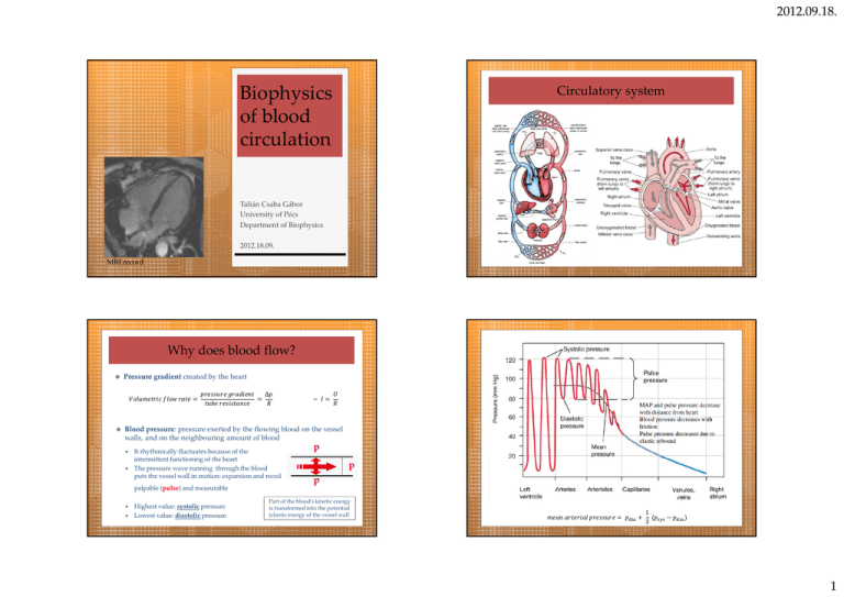
2012.09.18. Biophysics of blood circulation Circulatory system Talián Csaba Gábor University of Pécs Department of Biophysics 2012.18.09. MRI record Why does blood flow? Pressure gradient created by the heart = ∆ = ~ = Blood pressure: pressure exerted by the flowing blood on the vessel walls, and on the neighbouring amount of blood It rhythmically fluctuates because of the intermittent functioning of the heart The pressure wave running through the blood puts the vessel wall in motion: expansion and recoil palpable (pulse) and measurable Highest value: systolic pressure Lowest value: diastolic pressure p p p Part of the blood’s kinetic energy is transformed into the potential (elastic energy of the vessel wall 1 = + ("#" − ) 3 1 2012.09.18. Blood pressure In every 10 cm ∆ = ∆ℎ ∙ ()*++ ∙ = 0,1 ∙ 1.065 = 1.0457 = 8, 9:;<< 1 ∙ 9,81 5 2 Systolic (mmHg) Diastolic (mmHg) Left ventricle 120 0 Aorta 120 75 Venae cavae ~0 ~0 Right atrium 4,5 -4,5 Right ventricle 35 0 Pulmonary artery 35 7-9 Pulmonary vein 7 7 Left atrium 4,5 -4,5 Head arteries (~ +50cm) ~ 80 Foot arteries (~ -100cm) ~ 200 Peripheral circulation ∆ = ( ∙ ∙ ∆ℎ Peripheral resistance Function of peripheral circulation: maintain a steady, unidirectional and laminary (relatively slow) flow = (Q =) 2. Law of Hagen-Poiseuille Laws of hydrodynamics can be applied pressure gradient ∆U = V ∙ W R ∙ S ∆ ∙ 8∙T resistance stroke volume 1. Continuity equation = = 5 >= ∙ ?= = >5 ∙ ?5 A = @ B= A > = @ > B= > ∙ ? = Σ> ∙ ?̅ = constant It can be influenced by: Elasticity of the vessels Intermittent work of the heart Changes in blood volume (lymph formation) Σ>KL**M# O 750 >+MN Resistance is different in the various kinds of vessels and depends on pressure fall viscosity of blood cross-section of the vessels Distributive vessels Resistance vessels Diffusive vessels Capacitance vessels high pressure high resistance low velocity, filtration low pressure aorta, arteries arterioles capillaries veins, lymph vessels 2 2012.09.18. Pressure fall/gradient Laws of Kirchoff: serial coupling MX" = = + 5 + ⋯ + A parallel coupling 1 1 1 1 = + + ⋯+ MX" = 5 A Many small parallel sub-circuits Lower resultant resistance Local, independent regulation of the flow Better oxygen supply Capillaries in serial coupling only at specific places (e.g. 2 blood circuits!) Aneurism Viscosity Viscosity transforms kinetic energy into heat and lowers blood pressure positive feedback Average viscosity of blood ~4,5 mPa·s A1 v1 incr. A2 p1 decr. v2 continuity equation It depends on 1. haematocrit (e.g. leukemia) 2. concentration of plasma proteins (e.g. globulinemia) 3. deformability of the cellular components (rbc) incr. p2 law of Bernoulli suspension of similar sized solid particles is hard as brick, viscosity of 95% rbc-suspension is only ~20 mPa·s 3 2012.09.18. Cross-section of the vessels 4. Aggregation property of rbc (raises viscosity) 5. flow velocity = (Q =) R ∙ S ∆ ∙ 8∙T = = Z5 = = [\5 rouleaux Most important way to regulate intensity of current and blood pressure: Neural and hormonal (chatecolamines) Segré-Silberberg effect 6. Vessels’ cross-section (Fåhræus-Lindqvist effect) In vessels with substantial smooth muscle content: cell-free zone muscular arteries and arterioles „plasma skimming” Heart cycle Generation of impulse Isovolumetric contraction Ejection (rapid and reduced) Ventricular diastole 0,5s Flow is normally under critical velocity, laminary At some spaces (valves, stenosis) can speed up causing turbulence It can be heard as various sounds (heart sounds and murmurs, Korotkov sound) Ventricular systole 0,3s Isovolumetric relaxation Rapid filling Diastasis Atrial systole Atrial systole 0,1s Atrial diastole 0,7s 4 2012.09.18. Heart cycle Pump function Cardiac output = stroke volume x heart rate Heart rate = end-diastolic volume – end-systolic volume 120 − 50 = 8_<` 70 ℎ x75 = 5.250 ℎ Daily ~ 108.000 heartbeats (75x60x24) ~ 7.560 L blood ejected (5,25x60x24) Yearly ~ 40 million heartbeats ~ 2,76 million L (2.760m3) blood ejected http://library.med.utah.edu/kw/pharm/hyper_heart1.html Components of stroke volume Ejection fraction (EF) Ratio of stroke volume and end-diastolic volume 70 O 60% 120 Preload ab = End-diastolic pressure or volume in the ventricle Load/stretch of the heart muscle (cell) before contraction Effect of the blood returning to heart („filling pressure”) Increases sarcomer length Determined by the venous pressure and venous return Afterload Tension in the ventricle wall needed to eject blood Pressure in the ventricle needed to open the semilunar valve Determined by the systemic resistance/aortic pressure and pulmonary pressure 5 2012.09.18. Heart muscle Components of stroke volume Contractility Force exerted by the myocytes during contraction Frank-Starling law of the heart: stroke volume is directly proportional to end-systolic ventricular volume higher preload causes a greater stretch of the myocytes (increased sarcomer length) enhancing contractility 1,6µm 1,0µm Work of the heart Volume-pressure loop A. B. C. D. Closure of mitral valves Opening of aortic valve Closure of aortic valve Opening of mitral valves pe O 100Hgmm O 13,5kPa pe O 3Hgmm O 0,5kPa staticcomponent = m ∙ ∆n [ kineticcomponent = o ∙ pe Z Z q = 13 ∙ 102 7 ∙ 70 ∙ 10rs q + 0,5 ∙ 0,071 ∙ (0,5 )5 = 910mJ + 9mJ O uZ_ov Additional effect: stretch of the muscle fibre enhances troponin-C sensitivity for Ca that increases the number of actin-myosin cross-bridges rightatrium O 160mJtotal O [, [v power: P = 1,1J O [, }~ 0,8s 6 2012.09.18. Summary Gross anatomy of the cardiovascular system Blood pressure Peripheral resistance THANK YOU FOR YOUR ATTENTION! Pressure fall Viscosity Cross-section of the vessels Phases of the heart cycle Cardiac output Work of the heart 7 2012.09.18. 8

