GE Healthcare
BrainWave
Fast, easy fMRI analysis.
Functional Magnetic Resonance Imaging (fMRI) is fast becoming the
diagnostic tool of choice for mapping neuro function in the brain.
Non-invasive, no ingestion or infusion of tagged material required,
and radiation free, fMRI maps neurological function through blood
oxygen level dependent (BOLD) MRI scans of the brain. This technique
measures heightened oxygen levels within a region of interest in
response to external stimuli.
Analyzing maps generated by this technique yields valuable
information that can help guide surgical intervention procedures.
Consequently, technology that helps develop accurate and specific
exam reports is essential.
Provides retrospective motion
correction for patient motion.
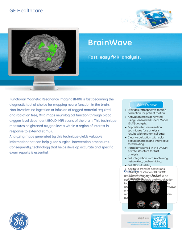
Activation maps generated
using Generalized Lineal Model
(GLM) analysis.
Sophisticated visualization
techniques fuse analysis
results with anatomical data.
Clear visualization with color
activation maps and interactive
thresholding.
Paradigms saved in the DICOM
private structure for fast
analysis.
Full integration with AW filming,
networking, and archiving.
Full DICOM fidelity.
Ability to transfer activations
Overview
into high resolution 3D DICOM
data setsimaging
for neurological
BrainWave
software is an
applications.
easy-to-use analysis and visualization
tool for functional brain image data
acquired with BrainWave RT. BrainWave
enables processing, analysis, 3D
rendering and display of results from
BOLD MRI scans.
Visit us:
www.gehealthcare.com/aw/
applications/brainwave/
Features
• BrainWave PA lets you interactively
view and edit fMRI data. It can
reside on the scanner or on your
AW station
• Software reads volumetric T1weighted acquisitions in addition to
fMRI acquisitions.
• Processing steps for fMRI data
include:
- Six parameter rigid body motion
correction via registration to the
first volume
- Spatial filtering
- Segmentation of the anatomical
data so that outer layers of nonbrain signal are removed.
- Registration of fMRI data sets to
the structural data set
- Calculation of statistical maps
corresponding to task-based
activation
- Formation of Burnt In Pixel (BIP)
maps outlining the area of
activation in white on the
structural scan
- Calculation of DTI fiber tracks
(option with BrainWave Fusion)
View functional data overlaid on
the structural image in 2D or 3D
renderings. Add annotations or
change color and opacities.
• BrainWave Fusion (option) lets you
display white matter tracks with
functional data on a highresolution anatomical image.
System Requirements
• Volume Share 5 or above
• One or two display monitors
• BrainWave PA requires BrainWave
RT
• BrainWave Fusion requires
BrainWave PA and DTI FiberTrak.
Minimum hardware required:
• HP Z800: 12 GB RAM or above
Device Description
The GE BrainWave Option(s) for MRI
systems is a modification to the GE
Functional Brain Mapping Imaging
Option for MRI systems. The GE
BrainWave Option(s) produces
images highlighting changes in blood
oxygen level dependent images over
time. These differences correspond
to changing stimuli presented to
patients which are synchronized with
scanning. The resulting parametric or
activation images are superimposed
on structural images from the same
patient. This device can be used to
acquire, process, and display the
results of BOLD MR studies with or
without external stimulation
hardware.
Indications for Use
The GE Functional Brain Mapping
Option is a software and hardware
package that can be used to acquire,
process, and display the results of
BOLD MRI scan studies taken in the
presence of synchronized stimuli
presented to a person being
scanned. When interpreted by a
trained physician, these results may
be useful in the determination of a
course of treatment.
Regulatory Compliance
This product complies with the
European CE Marking regulation for
Medical Devices Directive: Directive
93/42/EEC, dated 14 June 1993.
© 2012 General Electric Company.
All rights reserved. Data subject to change.
GE and GE Monogram are trademarks of General Electric Company.
* Trademark of General Electric Company.


