From www.bloodjournal.org by guest on September 30, 2016. For personal use only.
National Cancer Institute-Sponsored Working Group Guidelines for
Chronic Lymphocytic Leukemia: Revised Guidelines for
Diagnosis and Treatment
By Bruce D. Cheson, John M . Bennett, Michael Grever, Neil Kay, Michael J. Keating, Susan O'Brien, and Kanti R. Rai
I
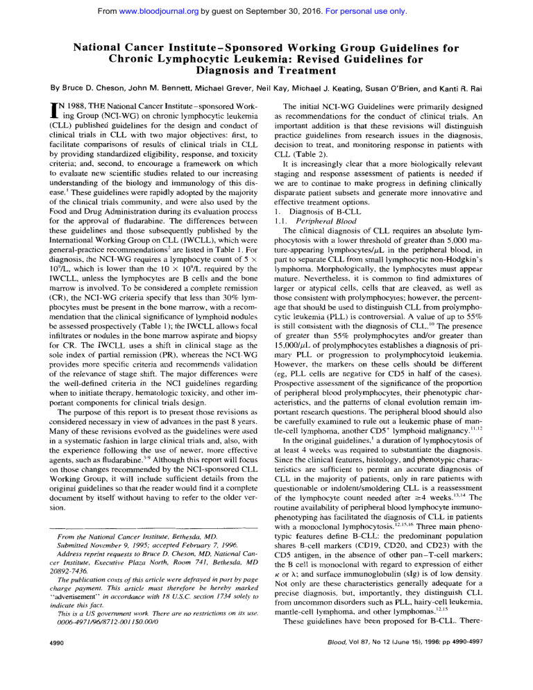
N 1988, THENational Cancer Institute-sponsored WorkThe initial NCI-WC Guidelines were primarily designed
ing Group (NCI-WC) on chronic lymphocytic leukemia as recommendations for the conduct
ofclinicaltrials. An
(CLL) published guidelines for the design and conduct of
importantadditionisthattheserevisionswill
distinguish
clinical trialsin CLL with twomajor objectives: first, to
practiceguidelines from researchissuesin
the diagnosis,
facilitate comparisons of results of clinicaltrials in CLL
decision to treat, and monitoring response in patients with
by providing standardized eligibility, response, and toxicity
CLL (Table 2).
criteria; and, second, to encourage a framework on which
It is increasingly clear that a more biologically relevant
to evaluate new scientific studies related to our increasing
staging andresponse assessment of patients is needed if
understanding of the biology and immunology of this diswe are to continue to make
progress in defining clinically
ease.' These guidelines wererapidly adopted by the majority
disparate patient subsets and generate more innovative and
of the clinical trials community, and were also used by the
effective treatment options.
Food and Drug Administration during its evaluation process
1. Diagnosis of B-CLL
for theapproval of fludarabine. The differencesbetween
1.1. Peripheral
Blood
these guidelinesand thosesubsequentlypublished
by the
The clinical diagnosis of CLL requires an absolute lymInternational Working Group on CLL (IWCLL),
which were
phocytosis with a lower threshold of greater than 5,000 mageneral-practice recommendations' are listed in Table 1. For
ture-appearinglymphocytes/wL in the peripheralblood, in
diagnosis, the NCI-WC requires a lymphocyte count of 5 X
part to separate CLL fromsmall lymphocytic non-Hodgkin's
loy& which is lower than the 10 X 109/Lrequired by the
lymphoma. Morphologically, the lymphocytes must appear
IWCLL, unless the lymphocytes are B cells and the bone
mature.Nevertheless,itis
common to find admixtures of
marrow is involved. To be considereda complete remission
largeror atypicalcells, cells that are cleaved,aswell
as
(CR), the NCI-WC criteria specify that less than 30% lymthose consistent with prolymphocytes; however, the percentphocytes must be present in the bone marrow, with a recomage that should be used to distinguish CLL from prolymphocytic leukemia (PLL) is controversial. A value of up to 55%
mendation that the clinical significance of lymphoid nodules
is still consistent with the diagnosis of CLL.'" The presence
be assessed prospectively (Table 1); the IWCLL allows focal
infiltrates or nodulesin the bone marrowaspirate and biopsy
of greater than 55% prolymphocytesand/or greater than
for CR. The IWCLL
usesashift
in clinical stageas the
15,OOO/pL of prolymphocytes establishes a diagnosis of primary PLL or progression to prolymphocytoidleukemia.
sole index of partial remission (PR), whereas the NCI-WC
However,themarkers
on thesecells should be different
provides more specific criteria and recommends validation
(eg, PLL cells are negative for CD5 in half of the cases).
of the relevance of stage shift. The major differences were
Prospective assessment of the significance of the proportion
the well-definedcriteria in theNCIguidelinesregarding
of peripheral blood prolymphocytes, their phenotypic charwhen to initiate therapy, hematologic toxicity, and other imacteristics, and the patterns of clonal evolution remain important components for clinical trials design.
portant research questions. The peripheral blood should also
The purpose of this report is to present those revisions as
be carefully examined to rule out a leukemic phase of manconsidered necessary in view of advances in the past 8 years.
tle-cell lymphoma, another CD5' lymphoid malignancy.","
Many of these revisions evolved as theguidelines were used
In the original guidelines,' a duration of lymphocytosis of
in a systematic fashion in large clinical trials and, also, with
at least 4 weeks was required to substantiate the diagnosis.
the experience following the use of newer, more effective
Since the clinical features, histology, and phenotypic characagents, such asfludarabine."' Although this report will focus
teristics are sufficient to permit an accurate diagnosis of
on those changes recommended by the NCI-sponsored CLL
CLL in the majority of patients, only in rare patients with
Working Group, it will includesufficient details from the
questionable or indolent/smoldering CLL is a reassessment
original guidelines so that the reader wouldfind it a complete
of thelymphocyte count neededafter 2 4 weeks."." The
document by itself without having to refer to the older verroutine availability of peripheral blood lymphocyteimmunosion.
phenotyping has facilitated the diagnosis of CLL in patients
with a monoclonal lymphocytosis.'2.'5.'6 Threemain phenotypic featuresdefineB-CLL:the
predominantpopulation
From the National Cancer Institute, Bethesda, MD.
shares B-cellmarkers (CD19, CD20, and CD23) with the
Submitted November 9, 1995; accepted February 7, 1996.
Address reprint requests to Bruce D.Cheson, MD, National CanCD5 antigen, in the absence of other pan-T-cell markers;
cer Institute, Executive Plaza North, Room 741, Bethesda, M D
the B cell is monoclonal with regard to expression of either
20892-7436.
K or h; and surface immunoglobulin (slg) is of low density.
The publication costsof this article were defrayed in part by page
Not only are these characteristics generally adequate for a
charge payment. This article must therefore be hereby marked
precisediagnosis,but,importantly,
they distinguish CLL
''advertisement" in accordance with 18 U.S.C. section 1734 solely to
from
uncommon
disorders
such
as
PLL,
hairy-cell leukemia.
indicate this fact.
mantle-cell lymphoma, and other lymphomas.'z~''
This is a US government work. Tltere clre no restrictions on its use.
These guidelines have been proposed for B-CLL. There0006-4971/96/8712-001I$0.00/0
4990
Blood, Vol 87,No 12 (June 15), 1996: pp 4990-4997
From www.bloodjournal.org by guest on September 30, 2016. For personal use only.
4991
NCI-WGCLLGUIDELINES
Table 1. Comparison of NCI-Working Group and IWCLL Guidelines for CLL
NCI
Variable
Diagnosis
Lymphocytes ( x 109/L)
Atypical cells (%) (eg. prolymphocytes)
Duration of lymphocytosis
Bone marrow lymphocytes W O )
Staging
Eligibility for trials
Response criteria
CR
Physical exam
Symptoms
Lymphocytes ( x 109/L)
Neutrophils ( x 109/L)
Platelets ( x 109/L)
Hb (g/dL)
Bone marrow lymphs (%)
PR
Physical exam (nodes, and/or liver, spleen)
Plus 2 1 of:
Neutrophils ( x 109/L)
Platelets ( X i09/L)
Hemoglobin (g/dL)
Duration of CR or PR
Progressive disease
Physical exam (nodes, liver, spleen)
Circulating lymphocytes
Other
Stable disease
IWCLL
-
<55
None required
210 + B-phenotype or bone marrow
involved
< I O + both of above
Not stated
Not stated
230
>30
Modified Rai, correlate with Binet
Active disease (details in document)
IWCLL
A: lymphs >50 x 109/Ldoubling time < l 2
mo diffuse marrow
B, C: all patients
Normal
None
54
21.5
>l00
> 11 (untransfused)
<30; no nodules
Normal
None
14
>1.5
> 100
Not stated
Normal, allowing nodules or focal infiltrates
250% decrease
Downshift in stage
>5; rl B-cell marker (CD19, CD20,
CD23) + CD5
21.5
>l00
> l 1 or 50% improvement
2 2 mo
250% increase or new
250% increase
Richter's syndrome
All others
fore, the following lymphoid malignancies are specifically
excluded from protocol studies directed at patients with BCLL: T-CLL, prolymphocytic leukemia (B and T cell),
hairy-cell leukemia and variant forms, splenic lymphoma
with villous lymphocytes, large granular lymphocytosis, SCzary-cell leukemia, adult T-cell leukemidymphoma, and
leukemic manifestations of non-Hodgkin's lymphomas, including follicular center-cell and mantle-cell lymphoma
types,12,15.17
1.2. Bone Marrow Examination
A bone marrow aspirate and biopsy are generally not required to make the diagnosis of CLL. Nevertheless, CLL is
a disease of the bone marrow, and it is appropriate to evaluate
a major site of involvement. The aspirate smear must show
230% of all nucleated cells to be lymphoid. A bone marrow
examination also provides useful prognostic information by
determining whether there is diffuse or nondiffuse involvement,lXand permits an assessment of the erythroid precursors
and megakaryocytes.
1.3. Immunophenotype
As noted earlier, a thorough immunophenotypic profile
of the malignant lymphocytes from the peripheral blood is
necessary for the initial diagnosis of the patient with CLL.
1.4. MolecularBiology/Cytogenetics
Not only do cytogenetic analyses provide useful prognos-
Not stated
Upshift in stage
No change in stage
tic information, but they help identify potentially important
nonrandom genetic alterations and o n c ~ g e n e s . " ~Sequenl~-~~
tial analysis of established genetic alterations may also be
helpful in evaluating the evolution of the disease process.
However, given the expense and limited availability of these
studies, they should be restricted to a research setting in
which to evaluate their potential prognostic and biologic
importance.
2. Clinical Staging
We recognize that there are two somewhat different major
staging methods that are currently in use throughout the
world: the Rai systemz6and the Binet system.27In 1981, the
IWCLL recommended that the two systems be integrated so
that each of the Binet stages be subclassified with the Rai
stage. However, the IWCLL-integrated system has not received widespread usage, and physicians continue to use
either the Rai or Binet method in both patient care and in
clinical trials. For clinicians using the Rai classification, we
recommend the use of the modified version, which reduces
the number of prognostic groups from five to three." These
two systems are outlined following.
2.1. Rai System
In the three-stage Rai system low risk encompasses Rai
stage 0, with the clinical features of lymphocytosis in blood
and bone marrow only. Intermediate risk encompasses stage
From www.bloodjournal.org by guest on September 30, 2016. For personal use only.
4992
CHESONET
Table 2. Recommendations Regarding Evaluation and Monitoring
of CLL Patients
General
Recommendation
Pretreatment evaluation
History and physical
Examination of PBS
lmmunophenotyping of PBLs
Bone marrow at diagnosis
BM prior to therapy
Cytogeneticimolecular studies
CT scans, MRI, lymphangiogram gallium
scan
Indications for treatment
Treat with stage 0-1
Treat for activeiprogressive disease
(newly dx)
Treat without activeiprogressive disease
(newly dx)
Treat without activeiprogressive disease
(relapsedirefractory)
Treat beyond maximum response
Response assessment
CBC, differential
Bone marrow
Phenotype
CytogeneticsiFISH
Practice*
Clinical
Trial
rc
rc
V
+
V V
X
X
X
V
*
v
X
X
X
*
rc
V
+
V
X
X
*
+
For purposes of this discussion, general practice is defined as the
use of accepted treatment options for a CLL patient not enrolled on
a clinical trial.
Abbreviations: V , always; X, not generally indicated; f, desirable;
*, if a research question; V, if a study not performed recently,
eg,
at diagnosis; PBS, peripheral blood smear; PBLs, peripheral blood
lymphocytes; dx, diagnosis; MRI, magnetic resonance imaging; FISH,
fluorescence in situ hvbridization.
I, with lymphocytosis and enlarged nodes, and stage11, with
lymphocytosis
plus
splenomegaly
andor hepatomegaly
(nodes positive or negative). High risk encompasses stage
111, with lymphocytosis plus anemia, and stage
IV, with lymphocytosis and thrombocytopenia.
2.2. Binet Staging System
Staging is based on the number of involved areas, and
the level of hemoglobin (Hb) and platelet count. Whether
significant adenopathy (>1 cm in diameter) is bilateral or
unilateral is recorded.
Area of involvement considered for staging
(1) Head and neck, includingtheW2ldeyer
ring (this
counts as one area even
if morethan one group of
nodes are enlarged).
(2) Axillae (involvement of both axillae countsasone
area).
(3)Groins,including
superficial femorals (involvement
of both groins counts as one area).
(4) Palpable spleen.
( 5 ) Palpable liver (clinically enlarged).
Stage A. Hb 210 g/dL and platelets 2 1 0 0 X 10’/L and
up to two of the above involved.
Stage B. Hb 210 g/dL and platelets ~ 1 0 X0 109/Land
organomegaly greater than that defined for stage A, ie,
three or more areas of nodal or organ enlargement.
AL
Stage C. All patients,irrespective of organomegaly in
whom Hb less than 10 g/dL and/or platelets less than 100
x 10”L.
3. EligibilityCriteria for Clinical Trials
3. I . ClinicalStage
The stage of CLL eligible for a clinical trial should reflect
the therapeutic objectives, anticipated toxicities, and desired
end results foreach study. For example, a phase I study
should involve only patients in advanced stages (Rai high
risk, poor prognosis), while phase I1 and, particularly, phase
111 studies may also include patients in the intermediate-risk
group. Decisions will be based on pilot data from phase I
and early phase I1 trials with the particular agent or regimen.
0 disease shouldgenerallynot
be
PatientswithRaistage
entered into clinical trials. Other requirements for eligibility
for clinical trials with respect to age, clinical stage, performance status, organ function, and status of
disease activity
should be defined for each study.
3.2. Perjbrmance
Status
For phase I clinical trials, only patients with an Eastern
Cooperative Oncology Group (ECOG)
performancestatus
(PS) 0 to 2 should be eligible. For phase I1 and 111 clinical
trials,patientswith
PS 0 to 3may be eligible; however,
these limits may be individualized on the basis of the drugs
or therapies being tested. For trials in which it appears reasonable to include patients with PS 3, yet where there is a
concern over the potential toxicities of an agent or therapy,
it may be advisable to initially start those patients at a lower
dose of the treatment (eg, SO% reduction) and to gradually
increase the dose over subsequent courses to
the standard
dose. if the treatment is well tolerated and toxicity is within
an acceptable range. This approach should be individualized
for each relevant protocol and may not be appropriate for
some therapies(eg,high-dosetherapywithstem-cellsupport).
3.3. Organ Funciion Eligibility for Clinical Trials
Most chemotherapy agentspossess the potential for toxicity to the liver, kidneys, heart,
lungs, central or peripheral
nervoussystem, or other organsystems. Therefore,organ
function requirements must be guided by the known toxicities of each drug based on observations from animal studies
and previous therapeutic trials. Normal function for organs
for which there is a well-recognized, specific toxicity must
be required. Otherwise, as a general principle:
3.3.1. Baseline liver enzymes (ie,transaminase levels)
should be no worse than 1 .S times the upper range of normal
values. Serum bilirubin concentration should be 5 2 . 0 mg/
dL, unless resulting from documented hemolysis.
3.3.2. Baseline renalfunction(ie,bloodureanitrogen
[BUN], creatinine) should be no worse than 1.5 times the
upper range of normal values.
3.3.3.Baselinerequirements
for otherstudies (eg. systolic ejection fraction, pulmonary
function tests) should be
decided individually for each study.
3.4. InfectionStatus
3.4.1. Patientswithactiveinfectionsrequiringsystemic
antibioticsshould be excluded from B-CLL
clinicaltrials
until resolution of infection.
3.4.2.Patients
whoarehuman immunodeficiencyvirus
From www.bloodjournal.org by guest on September 30, 2016. For personal use only.
NCI-WG CLLGUIDELINES
(H1V)-positive should be excluded because of their poor
tolerance to chemotherapy and the potential risks from the
immunosuppressive effects of new agents such as fludarabine.29
3.5. Second Malignancies
Patients with a second malignancy, other than non-basalcell carcinoma of the skin or in situ carcinoma of the cervix,
should not be entered onto a CLL clinical trial unless the
tumor was treated with curative intent at least 2 years previously.
3.6. Required Pretreatment Evaluation
As already noted, the parameters that are considered necessary for a complete pretreatment evaluation differ whether
the patient is being treated in a general practice setting or
on a clinical research protocol (Table 2). In general, and
where feasible, these studies should be quantified within 48
hours of placing a patient on a treatment protocol (except for
bone marrow aspirate and biopsy) (see later), and computed
tomography (CT) scans (see later). They should also be repeated at appropriate intervals to assess the maximum response to therapy.
3.61. Complete blood cell count (CBC; white blood cell
count, hemoglobin and hematocrit, platelet count) and differential, including both percent and absolute number of lymphocytes and prolymphocytes, and reticulocyte count.
3.62. Unilateral bone marrow aspirate and biopsy should
be performed within 2 weeks prior to entering the study,
unless a previous diagnostic specimen was diffusely involved and there has been no intervening systemic therapy.
It is preferable to evaluate the bone marrow at diagnosis for
prognostic purposes; however, it ismandatoryin
clinical
trials, and highly desirable in clinical practice, to perform a
unilateral bone marrowaspirate and biopsy prior to treatment
to provide a baseline for further response assessment. If a
repeat bone marrow is obtained, it should be reviewed along
with the original diagnostic sample.
3.63. Lymph node evaluation
3.53 1. Physical examination should record the diameter,
in two planes, of the largest palpable nodes in each of the
following sites: cervical, axillary, supraclavicular, inguinal,
and femoral.
3.632. Chest radiograph.
3.633. CT scans are generally not necessary in the initial
evaluation of patients with CLL, but should only be performed if clinically indicated. A chest CT may be useful if
the chest radiograph shows hilar adenopathy. These should
be obtained within 2 weeks prior to entering the protocol.
3.634. Lymph nodebiopsy is generally not indicated,
unless such tissue is necessary for companion scientific studies.
3.64. Liver and spleen size should be assessed by physical examination. CT scans should only be performed if clinically indicated or if part of a research question (see section
3.433).
3.65. Serum chemistries (eg, creatinine, bilirubin).
3.66. Assessment of PS (ECOG).
3.67. Baseline immunobiologic, cytogenetic, and molecular assessment for CLL trials (see Table 2,and earlier).
Those that should be performed on all patients include serum
4993
immunoglobulin determination, including quantitative immunoglobulins and immunoelectrophoresis, direct and indirect antiglobulin (Coombs’ test), and immunophenotypic
evaluation of the B-cell clone (see earlier).
4. Indications for Treatment
4.1. Primary Treatment Decisions
Once the diagnosis of CLL has been made, the treating
physician is faced with the decision of not only how to treat
the patient, but when to initiate therapy. Criteria for initiating
treatment may be quite different between clinical practice
and clinical trial conduct. A subset of patients are considered
as having smoldering CLL; they include those with Rai stage
0 (Binet A),with a nondiffuse pattern of bonemarrow
involvement, a serum Hb concentration 213.0 g/dL, peripheral blood lymphocytes less than 30 X 109/L,and a lymphocyte doubling time longer than 12 month^.".'^ Therapy
should notbe offered to these patients untilthey exhibit
clear evidence of disease progression. Other newly diagnosed patients with early stage disease (Rai 0 to I, Binet A),
should be monitored without therapy until evidence of disease progression. Studies from both the French Cooperative
Group on CLL and the Cancer and Leukemia Group B
(CALGB) in patients with early-stage disease confirm that
early therapy of patients with early-stage disease does not
prolong ~ u r v i v a l , ~but
~ ~may
”
beassociated with an increased
frequency of fatal epithelial cancers.30However, these studies were conducted with alkylator-based regimens, and the
potential benefit of earlier therapy using nucleoside analog
therapy is an important research question.
Whereas most patients with Rai stages I11 and IV require
treatment at presentation, many can still be monitored without therapyuntilthey
exhibit evidence of progressive or
symptomatic disease.
Active disease should be clearly documented for protocol
therapy. The following criteria must be met:
(1) A minimum of any one of the following disease-re-
lated symptoms must be present:
(a) Weight loss 210% within the previous 6 months.
(b) Extreme fatigue (ie, ECOG PS 2 or worse; cannot
work or unable to perform usual activities).
(c) Fevers of greater than 100.5”Ffor 2 2 weeks without evidence of infection.
(d) Night sweats without evidence of infection.
(2) Evidence of progressive marrow failure as manifested
by the development of, or worsening of, anemia and/
or thrombocytopenia
(3) Autoimmune anemia andor thrombocytopenia poorly
responsive to corticosteroid therapy
(4) Massive (ie, >6 cm below the left costal margin) or
progressive splenomegaly
( 5 ) Massive nodes or clusters (ie, > 10 cm in longest diameter) or progressive lymphadenopathy
(6) Progressive lymphocytosis with an increase of >50%
over a 2-month period, or an anticipated doubling time
of less than 6 months
(7) Marked hypogammaglobulinemia or the development
of a monoclonal protein in the absence of any of the
above criteria for active disease is not sufficient for
protocol therapy
From www.bloodjournal.org by guest on September 30, 2016. For personal use only.
4994
Patients with CLL may present with a markedly elevated
leukocyte count; however, the symptoms referable to leukocyte aggregates that develop in patients with acute leukemia
rarely occur in patients with CLL. Therefore, the absolute
lymphocyte count should not be used as the sole indicator
for treatment, but should be included as a part of the total
clinical picture, which includes the lymphocyte doubling
time (see earlier).
4.2. Second-LineTreatment Decisions
Treatment of CLL is generally palliative in intent; therefore, patients who have relapsed may be followed without
therapy until they experience disease-related symptoms or
progressive disease, with deterioration of blood counts, discomfort from lymphadenopathy or hepatosplenomegaly, recurrent infections, or associated autoimmune disorders. A
possible exception is allogeneic bone marrow transplantation. Recent data suggest that, in selected patients, allogeneic
bonemarrow transplantation or high-dose chemotherapy
with autologous stem-cell support may be reasonable treatment options, particularly in the context of a clinical research
proto~ol.~*-~~
The acceptable extent of prior therapy for protocol entry
must be decided separately for each study.
(A) For all phase I11 therapeutic trials, it is recommended
that onlythose patients who have not received previous cytotoxic or biological therapy be eligible. Itis appropriate to
include patients who have received previous corticosteroids
if this is compatible with the therapeutic objectives of the
trial. However, it maybe necessary to analyze these previously treated patients as a separate group.
(B) For phase I and I1 studies, we recommendthatno
more than two types of prior therapy (eg, fludarabine, chlorambucil with or without prednisone) be allowed for entering
patients. Certain trials may require previously untreated patients; this will be determined separately depending on the
objectives of the study.
5. Definition of Response
Assessment of response should include a careful physical
examination and evaluation of the peripheral blood and bone
marrow. The response criteria in the original NCI-WG guidelines have been retained (Table 3).
5. 1. Complete remission requires all of the following for
a period of at least 2 months:
5. 1 I . Absence of lymphadenopathy by physical examination and appropriate radiographic techniques.
5. 12. No hepatomegaly or splenomegaly by physical examination, or appropriate radiographic techniques if in a
clinical trial.
5.13. Absence of constitutional symptoms.
5. 14. Normal CBC as exhibited by:
5. 141. Polymorphonuclear leukocytes 2 1,50O/pL.
5.142. Platelets > lOO,OOO/pL.
5.143. Hemoglobin > 11.0 g/dL (untransfused).
5. 15. Bone marrow aspirate and biopsy should beperformed 2 months after clinical and laboratory results demonstrate that all of the requirements listed in 5.1 1 to 5.14 have
been met to demonstrate that a CR has been achieved. The
marrow sample must be at least normocellular for age, with
less
than
30%
of
nucleated cells being lymphocytes.
CHESON ET AL
Table 3. Grading Scale for Hematological Toxicity in CLL Studies
Decrease in Platelets* or Hbt (nadir)
From Pretreatment
value
(Yo)
No change10%
11%-24%
25%-49%
50%-74%
275%
Grade*
ANCIpLI (nadir)
0
22,000
~ ~ 1 , 5 and
0 0 <2,000
2 1,000 and < 1,500
2500 and <1,000
<500
1
2
3
4
* If. at any level of decrease the platelet count is <2O,OOO/pL, this
will be considered grade 4 toxicity, unless a severe or life-threatening
decrease in the initial platelet count (eg, ~20,00O/pL) was present
pretreatment, in which case the patient is inevaluable for toxicity referable to platelet counts.
t Baseline and subsequent Hb determinations must be performed
before any given transfusions.
Grades: 1, mild; 2, moderate; 3, severe; 4, life-threatening; 5, fatal.
Death occurring as a result of toxicity at any level of decrease from
pretreatment will be recorded as grade V.
§ If the absolute neutrophil count (ANC) reaches less than l,OOO/pL,
it should be judged to be grade 3 toxicity. Other decreases in the
white blood cell count, or in circulating granulocytes, are not to be
considered, since a decrease in the white blood cell count is a desired
therapeutic end point. A gradual decrease in granulocytes is not a
reliable index in CLL for stepwise grading of toxicity. If the ANC was
less than l.OOO/pL prior to therapy, the patient is inevaluable for toxicity referable to the ANC.
*
Lymphoid nodules should be absent. If the bone marrow is
hypocellular, a repeat determination should bemadein 4
weeks. Samples should be re-reviewed in conjunction with
the prior pathology.
5.16. For patients who fulfill all of the previous criteria
for a CR, an abdominal CT scan may be performed to confirm this clinical and hematologic impression if clinically
indicated or if required testing for a clinical research study.
5.2. PRis considered in a broad sense to enable the
detection of agents with biological effect. To be considered
a PR, the patient must exhibit 5.21 and 5.22 and/or 5.23 (if
abnormal prior to therapy), aswell as one or more of the
remaining features for atleast 2 months. In addition, the
presence or absence of constitutional symptoms will also be
recorded.
5.21. 250% decrease in peripheral blood lymphocyte
count from the pretreatment baseline value.
5.22. 2 5 0 % reduction in lymphadenopathy.
5.23. 250% reductionin the size of the liver and/or
spleen.
5.24. Polymorphonuclear leukocytes 2 1,5OO/pL or 50%
improvement over baseline.
5.25. Platelets >lOO,OOO/pL or 50% improvement over
baseline.
5.26. Hemoglobin > 11.0 g/dL or50%improvement
over baseline without transfusions.
5.27. In a subset of patients who are otherwise in a
complete remission, bone marrow nodules can be identified
histologically. It is, unfortunately, difficult with currently
available techniques to determine the clonality of these nodules. The original NCI-WG guidelines suggested thatpatients with a CR and persistent nodules should be analyzed
From www.bloodjournal.org by guest on September 30, 2016. For personal use only.
NCI-WG CLL GUIDELINES
carefully to compare their outcome relative to others who
are more conventionally classified as a CR or PR.’ Robertson
et a13’ have since demonstrated that patients with a nodular
CR had a shorter time to disease progression compared with
patients with a CR. Therefore, nodular CRs should be reported separately from CRs, and should not be used to inflate
the percentage of CRs. We recommend that they be referred
to as nodular PRs (nPR) and included with the PRs.
5.28. A controversial issue is how best to categorize the
response of patients who fulfill all the criteria for a CR, but
who have a persistent anemia or thrombocytopenia apparently unrelated to disease activity and more likely the consequence of persistent drug toxicity. The long-term outcome
of these patients may differ from the more routine complete
responders. Therefore, these patients should not be considered CRs or a separate response category, but should be
considered PRs. However, they should be monitored prospectively to better characterize their outcome, and may be
described within the context of results of clinical trials.
5.3. Progressive disease will be characterized by at least
one of the following:
5.31. 250% increase in the sum of the products of at
least two lymph nodes on two consecutive determinations 2
weeks apart (at least one node must be 2 2 cm); appearance
of new palpable lymph nodes.
5.32. 250% increase in the size of the liver and/or
spleen as determined by measurement below the respective
costal margin; appearance of palpable hepatomegaly or splenomegaly, which was not previously present.
5.33. 250% increase in the absolute number of circulating lymphocytes to at least 5,0oO/pL.
5.34. Transformation to a more aggressive histology (eg,
Richter’s syndrome or PLL with >55% prolymphocytes).
5.35. In the absence of progression, as defined earlier,
the presence of a 2 2 g/dL decrease in Hb, or 250% decrease
in platelet count, and/or absolute granulocyte count will not
exclude a patient from continuing the study. Each protocol
will define the amount of drug(s) to be administered with
such hematological parameters. Bone marrow aspirate and
biopsy are strongly encouraged to better define the cause of
the suppressed counts.
5.4. Patients who have not achieved a CR or a PR, or
who have notexhibited PD, will be considered to have stable
disease.
5.5. Responses that should be considered clinically beneficial include CR, nPR and PR; all others, eg, stable disease,
nonresponse, progressive disease, and death from any cause,
should be rated as a treatment failure.
5.6. Because current criteria for response are arbitrary
and often not validated by prospective studies, alternative
criteria may also be evaluated; however, to ensure comparability with other studies, these should be studied within the
framework of the current schema and be well defined, with
adequate rationale. Should such a schema be determined
to have important clinical relevance following prospective
evaluation, it will be considered for incorporation into criteria for future studies.
5.7. Duration of response should be measured from the
time the patient has exhibited the features of maximum re-
4995
sponse until evidence of progressive disease. Survival duration should be measured from the time of entry onto the
clinical trial.
6. Prognostic Factors Requiring Stratification
6.1. Previous treatment versus no previous treatment in
studies for which prior therapy is allowed.
6.2. If more than one clinical stage is allowed, patients
should be stratified for stage (eg, if intermediate and poor
risk are eligible, intermediate v poor), depending on the nature of the study and the available patient resources.
6.3. Application of New Prognostic Factors
In the interval since the initial publication of the guidelines, several modifications have been recommended.
6.31. Decrease in lymphocyte count: In several recent
studies, a decrease in the peripheral blood lymphocyte count
has been used as the primary index of response? Although
this parameter may identify a therapy that has lymphocytotoxic activity, there is no evidence that it has long-term
clinical implications. It has, therefore, not been incorporated
into the current response criteria.
6.32. Immunobiological assessment
6.321. Quantification of the serum immunoglobulin concentration in responders is recommended at the time of maximal clinical response, but it is not an established indicator
of response.
6.322. Repeat immunophenotyping atthetimeof a response is not part of standard practice. Moreover, progression of disease after a CR should not be based purely on the
basis of a small number of clonal cells identified using flow
cytometric determinations.
6.323. In the clinical trials setting, not only should the
peripheral blood smear and bone marrow aspirate and biopsy
be carefully examined, but immunophenotype, cytogenetics,
(including fluorescent in situ hybridization [FISH]), and molecular biologic studies provide important data and should
be performed at diagnosis, at the time of maximal response,
and at recurrence if part of a research question.
6.324. Serum &microglobulin is recommended as an
inexpensive prognostic marker.37
6.325. Other optional studies that maybeof
interest
include markers of B-cell proliferation such as Ki-67, which
might identify alterations in the malignant cell population,
soluble CD23, adhesion molecules, or molecular analysis
of specific genes (eg, oncogenes, tumor-suppressor genes).
These scientific parameters that assess thebiology of the
malignant clone may help us to identify new therapeutic
strategies.
6.33. Minimal residual disease: The optimal approach
to the patient with minimal residual disease remains another
important research issue. Careful assessment for minimal
residual disease as determined by flow cytometry, cytogenetics, or similar studies is not indicated outside of a research
study at the time of CR and at recurrence. Additional treatment decisions on the basis of minimal residual disease remains an issue for clinical investigation.
7. Assessment of Toxicity
An evaluation of potential treatment-induced toxicity in
patients with advanced malignancies may be quite difficult,
requiring careful consideration of both the manifestations of
From www.bloodjournal.org by guest on September 30, 2016. For personal use only.
4996
the underlying disease, as well as adverse reactions to the
therapies under study. Moreover, some of the conventional
criteria for toxicity are not applicable to studies involving
patients with hematological malignancies in general, or CLL
in particular. An example is hematological toxicity; patients
with advanced CLL may exhibit a deterioration in blood
counts, which may represent either treatment-related toxicity
or progressive bone marrow failure from the disease itself.
This discrimination may become increasingly difficult as
new agents are tested earlier in their development at a point
where the complete spectrum of their toxicities has not yet
been elaborated.
A few guidelines are presented recognizing that evaluation
methodswillbe
determined to a large extent within the
therapy involved.
7.1. HematologicalToxicity
As is the case with virtually all of the hematological malignancies, an evaluation of hematological toxicity in patients
with CLL must consider the high frequency of hematological
compromise at the initiation of therapy. Therefore, the standard criteria used for solid tumors cannot be applied directly;
many patients would be considered to have grade I1 to IV
hematological toxicity at presentation.
Also, in the past, the peripheral blood neutrophil level has
rarely been used as a criterion for dose modification since
these values were felt to be unreliable in CLL. However, the
increasing use of more effective therapeutic agents, particularly those with neutropenia as a dose-limiting toxicity (eg,
nucleoside analogs), has resulted in clinically significant myelosuppression. Therefore, we have proposed a new dosemodification scheme for quantifying hematological deteriorationin patients with CLL, which includes alterations in
the dose of myelosuppressive agents based on the absolute
neutrophil count (Table 3).
7.2. Infectious Complications
In CLL, as with many other hematological malignancies,
it may be difficult to distinguish between the occurrence of
infections related to the disease itself or to the consequences
of therapy. However, such an analysis is of value when
comparing the results of various treatments, particularly with
immunosuppressive agents such as the nucleoside analogs.*’
The etiology of the infection should be reported and categorized as bacterial, viral, or fungal, and proven or probable.
The severity of infections should be quantified asminor
(requiring either oral antimicrobial therapy or symptomatic
care alone), major (requiring hospitalization and systemic
antimicrobial therapy), or fatal (death as a result of the infection).
1.3. Nonhematological Toxicities
Other nonhematological toxicities should be graded according to the NCI Common Toxicity Criteria.38
8. Reporting of Clinical Response Data
Clear and careful reporting of data is an essential part of
any clinical trial. In clinical studies involving previously
treated patients, patients who are relapsed or refractory
should be clearly distinguished. Relapse is defined as a patient who has previously achieved the clinicopathologic criteria to be classified as a CR or PR, but, after a period of
2 6 months, demonstrated evidence of disease progression
CHESON ET AL
(Table 1). For those patients who have relapsed, it is also
usefulto describe the quality and duration of their prior
response. Refractory disease refers to the clinical situation
in which a patient fails to achieve at least a PR or progresses
while on therapy.
ACKNOWLEDGMENT
The following colleagues are credited for their active participation
in the original versionof the Guidelines: Charles A. Schiffer, Martin
M. Oken, David H. Boldt, Sanford J. Kempin, and Kenneth A. Foon.
REFERENCES
1. Cheson BD, Bennett JM, Rai KR, Grever MR, Kay NE, Schiffer CA, Oken MM, Keating MJ, Boldt DH, Kempin SJ, Foon KA:
Guidelines for clinical protocols for chronic lymphocytic leukemia:
Report of theNCI-sponsoredWorkingGroup.AmerJHematol
29: 152, 1988
2. InternationalWorkshoponChronicLymphocyticLeukemia:
Chroniclymphocyticleukemia:Recommendationsfordiagnosis,
staging, and response criteria. Ann Intern Med
110:236, 1989
3. KeatingMJ,Kantarjian H, TalpazM,RedmanJ,KollerC,
Barlogie B, Valasquez W, Plunkett W, Freireich EJ, McCredie KB:
Fludarabine:A
new agent with majoractivityagainstchronic
lymphocytic leukemia. Blood 74: 19, 1989
4. KeatingMJ,Kantarjian H, O’Brien S, KollerC,Talpaz M,
Schachner J, Childs CC, Freireich EJ, McCredie KB: Fludarabine:
A new agent with marked cytoreductive activity in untreated chronic
lymphocytic leukemia. J Clin Oncol 9:44,
1991
5. Hiddemann W, Rottmann R, Wormann B, Thiel A, Essink M,
Ottensmeier C, Freund M, Buchner T, van de Loo J: Treatment of
advanced chronic lymphocytic leukemia by fludarabine. Results of
a clinical phase-I1 study. Ann Hematol 63:1, 1991
S, Ranson M, Smith OP, Mehta
6. Whelan JS, Davis CL, Rule
AB, Catovsky D, Rohatiner AZS, Lister TA: Fludarahine phosphate
for the treatment of low grade lymphoid malignancy. Br J Cancer
64: 120, 1991
7. Sorensen JM, Vena D, Fallavollita A, Cheson BD: Treatment
of refractory chronic lymphocytic leukemia (CLL) with fludarabine
monophosphate(FAMP).ProcAmSocClinOncol11:264,1992
(abstr 867)
8. Keating MJ, O’Brien S, Kantarjian H, Plunkett W, Estey E,
Koller C, Beran M, Freireich EJ: Long-term follow-up of patients
with chroniclymphocyticleukemia
treated withfludarabineasa
single agent. Blood 81:2878, 1993
9. O’Brien S, Kantarjian H, Beran M, Smith T, Koller C, Estey
E, Robertson LE, Lemer S, Keating M: Results of fludarahine and
prednisone therapy in 264 patients with chronic lymphocytic leukemia with multivariate analysis-derived prognosticmodel for response
to treatment. Blood 82:1695, 1993
IO. Melo JV, CatovskyD, Galton DAG: The relationship between
I.
chroniclymphocyticleukaemiaandprolymphocyticleukaemia.
Clinical and laboratory features of 300 patients and characterization
of an intermediate group. Br J Haematol 63:377, 1986
1 1. Catovsky D: Diagnosisandtreatment
of CLL variants,in
Cheson BD (ed):ChronicLymphocyticLeukemia:ScientificAdvances and Clinical Developments. New York, NY, Dekker, 1993,
p 369
12. Harris NL, Jaffe ES, Stein H, Banks PM, Chan JKC, Cleary
ML,DelsolG,DeWolf-PeetersC,
Falini B,Gatter KC, Grogan
TM,IsaacsonPG,KnowlesDM,MasonDY,Muller-Harmelink
H-K. Pileri SA, PirisMA,RalfkiaerE,WarnkeRA:Arevised
European-American classification of lymphoid neoplasms: A proposal from
the
International Lymphoma
Study
Group.
Blood
84:1361,1994
From www.bloodjournal.org by guest on September 30, 2016. For personal use only.
NCI-WG CLL GUIDELINES
13. French Cooperative Group on Chronic Lymphocytic Leukaemia: Natural history of stage A chronic lymphocytic leukaemia untreated patients. Br J Haematol 76:45, 1990
14. Molica S: Progression and survival studies in early chronic
lymphocytic leukemia. Blood 78:895, 1991
15. Bennett JM, Catovsky D, Daniel M-T, Flandrin G, Galton
DAG, Gralnick HR, Sultan C: Proposals for the classification of
chronic (mature) B and T lymphoid leukaemias. J Clin Pathol
42567, 1989
16. Reinisch W, Willheim M, Hilgarth M, Gasche C, Mader R,
Szepfalusi S, Steger G, Berger R, Lechner K, Boltz-Nitulescu G,
Schwarzmeier JD: Soluble CD23 reliably reflects disease activity in
B-cell chronic lymphocytic leukemia. J Clin Oncol 12:2146, 1994
17. Hoyer JD, Ross CW, Li C-Y, Witzig TE, Gascoyne R D ,
Dewald GW, Hanson CA: True T-cell chronic lymphocytic leukemia: A morphologic and immunophenotypic study of 25 cases. Blood
86: 1163, 1995
18. Rozman C, Montserrat E, Rodriguez-Fernhndez JM, Ayats
R, Vallespi T, Parody R, Rios A, Prados D, Morey M, Gomis F,
Alcali A, Gutitrrez M, Maldonado J, Gonzalez C, Giralt M, Hernandez-Nieto L, Cabrera A, Fernandez-Raiiada JM: Bone marrow histologic pattern-Thebest
single prognostic parameter in chronic
lymphocytic leukemia: A multivariate survival analysis of 329 cases.
Blood64:642, 1984
19. Escudier SM, Pereira-Leahy JM, DrachW, Weier HU, Goodacre AM, Cork MA, Trujillo JM, Keating MJ, Andreef M: Fluorescent in situ hybridization and cytogenetic studies of trisomy 12 in
chronic lymphocytic leukemia. Blood 81:2702, 1993
20. Bentz M, Huck K, du Manoir S, Joos S, Werner CA, Fischer
K, Dohner H, Lichter P Comparative genomic hybridization in
chronic B-cell leukemias shows a high incidence of chromosomal
gains and losses. Blood 85:3610, 1995
21. Hanada M, Delia D, Aiello A, Stadtmauer E, Reed JC: bcl2 gene hypomethylation and high-level expression in B-cell chronic
lymphocytic leukemia. Blood 82: 1820, 1993
22. El Rouby S, Thomas A, Costin D, Rosenberg CR, Potmesil
M, Silber R, Newcomb EW: p53 Gene mutation in B-cell chronic
lymphocytic leukemia is associated with drug resistance and is independent of MDRlNDR3 gene expression. Blood 82:3452, 1993
23. Dohner H, Fischer K, Mentz M, Hansen K, Benner A, Cabot
G, Diehl D, Schlenk R, Coy J, Stilgenbauer S, Volkmann M, Galle
PR, Poustka A, Hunstein W, Lichter P: p53 gene deletion predicts
for poor survival and non-response to therapy with purine analogs
in chronic B-cell leukemias. Blood 85: 1580, 1995
24. Stilgenbauer S , Dohner H, Bulgay-Morschel M, Weitz S,
Bentz M, Lichter P: High frequency of monoallelic retinoblastoma
gene deletion in B-cell chronic lymphoid leukemia shown by interphase cytogenetics. Blood 81:2118, 1993
25. Brown AG, Ross F M , Dunne EM, Steel M, Weir-Thompson
EM: Evidence for a new tumor suppressor locus (DBM) in human
4997
B-cell neoplasia telomeric to the retinoblastoma gene. Nature Gen
3:67, 1993
26. Rai KR, Sawitsky A, Cronkite EP, Chanana AD, Levy RN,
Pasternack BS: Clinical staging of chronic lymphocytic leukemia.
Blood 46:219, 1975
27. Binet JL, Lepomer M, Dighiero G, Charron D, DAthis P,
Vaughier G, Beral HM, Natal1 JC, Raphael M, Nizet B, Follezou
JY: A clinical staging system for chronic lymphocytic leukemia.
Prognostic significance. Cancer 40:855, 1977
28. Rai KR: A critical analysis of staging in CLL, in Gale RP,
Rai KR (eds): Chronic Lymphocytic Leukemia. Recent Progress and
Future Direction. New York, NY, Liss, 1987, p 253
29. Cheson B: Immunologic and immunosuppressive complications of purine analogue therapy. J Clin Oncol 13:2431, 1995
30. French Cooperative Group on Chronic Lymphocytic Leukemia: A randomized clinical trial of chlorambucil versus COP in stage
B chronic lymphocytic leukemia. Blood 75: 1422, I990
3 I . Shustik C, Mick R, Silver R, Sawitsky A, RaiK, Shapiro
L: Treatment of early chronic lymphocytic leukemia: Intermittent
chlorambucil versus observation. Hematol Oncol 6:7, 1988
32. Rabinowe SN, Soiffer RJ, Gribben JG, Daley H, Freedman
AS, Daley J, Pesek K, Neuberg D, Pinkus G, Leavitt PR, Spector
NA, Grossbard ML, Anderson K, Robertson MJ, Mauch P, ChaytMarcus K, Ritz J, Nadler LM: Autologous and allogeneic bone
marrow transplantation for poor prognosis patients withB-cell
chronic lymphocytic leukemia. Blood 82: 1366, 1993
33. Khouri IF, Keating MJ, Vriesendorp HM, Reading CL, Przepiorka D,Huh YO, Anderson BS, vanBesien KW, Mehra RC,
Giralt SA, Ippoliti C , Marshall M, Thomas MW, O’Brien S, Robertson LE, Deisseroth AB, Champlin RE: Autologous and allogeneic
bone marrow transplantation for chronic lymphocytic leukemia: Preliminary results. J Clin Oncol 12:748, 1994
34. Michallet M, Archimbaud E, Bandini G, Rowlings P, Deeg
HJ, Gahrton G. Montsenat E, Rozman C, Gratwohl A, Gale R P
HLA-identical sibling bone marrow transplantation in younger patients with chronic lymphocytic leukemia. Ann Intern Med 124:31 l ,
I996
35. Robertson LE, Huh YO, Butler JJ, Pugh WC, Hirsch-Ginsberg C, Stass S, Kantarjian H, Keating MJ: Response assessment
in chronic lymphocytic leukemia after fludarabine plus prednisone:
Clinical, pathologic, immunophenotypic, and molecular analysis.
Blood 80:29, 1992
36. Juliusson G, Liliemark J: High complete remission rate from
2-chloro-2’-deoxyadenosinein previously treated patients with Bcell chronic lymphocytic leukemia: Response predicted by rapid
decrease in blood lymphocyte count. J Clin Oncol 11 :679, 1993
37. Keating MJ, Lerner S , Kantarjian H, Freireich W,O’Brien
S: The serum beta2-microglobulin (beta2M) level is more powerful
than stage in predicting response and survival in chronic lymphocytic
leukemia (CLL). Blood 86:606a, 1995 (abstr, suppl 1)
38. Wittes RE (ed): Manualof Oncologic Therapeutics: 1991/
1992. Philadelphia, PA, Lippincott, 1991, p 446
From www.bloodjournal.org by guest on September 30, 2016. For personal use only.
1996 87: 4990-4997
National Cancer Institute-sponsored Working Group guidelines for
chronic lymphocytic leukemia: revised guidelines for diagnosis and
treatment
BD Cheson, JM Bennett, M Grever, N Kay, MJ Keating, S O'Brien and KR Rai
Updated information and services can be found at:
http://www.bloodjournal.org/content/87/12/4990.citation.full.html
Articles on similar topics can be found in the following Blood collections
Information about reproducing this article in parts or in its entirety may be found online at:
http://www.bloodjournal.org/site/misc/rights.xhtml#repub_requests
Information about ordering reprints may be found online at:
http://www.bloodjournal.org/site/misc/rights.xhtml#reprints
Information about subscriptions and ASH membership may be found online at:
http://www.bloodjournal.org/site/subscriptions/index.xhtml
Blood (print ISSN 0006-4971, online ISSN 1528-0020), is published weekly by the American
Society of Hematology, 2021 L St, NW, Suite 900, Washington DC 20036.
Copyright 2011 by The American Society of Hematology; all rights reserved.

