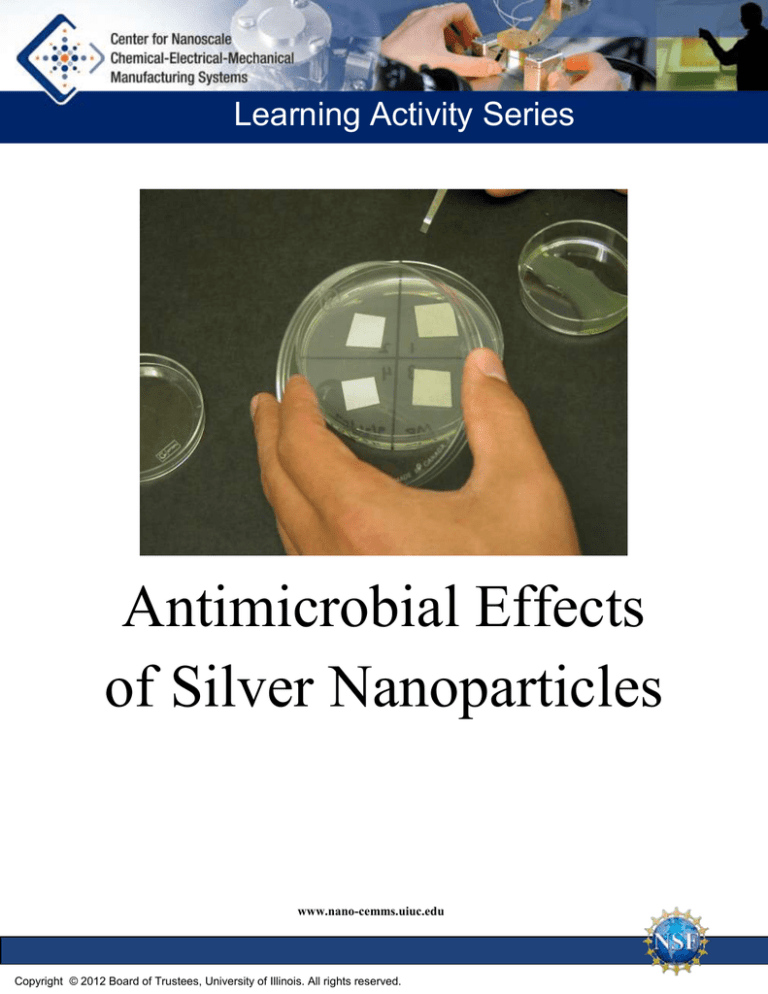
Learning Activity Series
Antimicrobial Effects
of Silver Nanoparticles
www.nano-cemms.uiuc.edu
Copyright © 2012 Board of Trustees, University of Illinois. All rights reserved.
Antimicrobial Effects of Silver Nanoparticles
Description:
Students make silver nanoparticles using a quick,
simple and safe procedure. They then design
experiments to test the effectiveness of the
nanoparticles as an antimicrobial agent.
Prerequisites:
Students should have an introductory knowledge of
nanotechnology that can be provided by the
Introduction to Nanotechnology kit.
The Gold and Silver Nanoparticles lab is a good
companion activity.
Instruction Time:
Approximately three 50-minute class periods. The
first period is for the PowerPoint presentation, the
second is for the lab, the third is for collecting data.
Audience:
Middle or high school General Science, Biology,
or Chemistry students.
Lesson Objectives:
Students will make silver nanoparticles and design
an experiment to test the effectiveness of silver
nanoparticles as an antimicrobial agent.
National Science Education Standards:
Content Standard A: Abilities Necessary to Do
Scientific Inquiry.
Content Standard B: Structure and Properties of
Matter.
Content Standard C: The Cell
Content Standard E: Understandings about
Science and Technology.
Contents
2
Instructional Method
2
Background Information
2
Overview
3
Materials
3
Safety
3
Preparation
4
Procedure
7
ESEM Images
11
Presentation Details
A-1
Student Lab
A-3
Student Lab Key
19
Notes
Illinois State Learning Standards:
11.A.4b Conduct controlled experiments or
simulations to test hypotheses.
12.B.4b Simulate and analyze factors that
influence the size and stability of populations.
12.C.5b Analyze the properties of materials in
relation to their physical and/or chemical
structures.
13.B.5b Analyze and describe the processes and
effects of scientific and technological
breakthroughs.
1
Univ
Instructional Method:
The instructor gives a PowerPoint presentation about
the anti-bacterial properties of silver nanoparticles.
Students then do a lab where they make and test the
effectiveness of nanoparticles.
Background Information:
Nanotechnology is an emerging industry which is
bringing us exciting new products and promises to
change the way we live and work in the future. Several
new products are using silver nanoparticles to generate
antimicrobial surfaces. Silver nanoparticles are
integrated into fabrics to prevent clothes from
developing foul odors, doorknobs have silver
nanoparticles embedded in their surfaces—even silver
nanoparticle-treated pacifiers are on the market. For a
list of hundreds of nanotechnology products using
silver nanoparticles see http://nanotechproject.org/44.
Scientists are very interested in nanoparticles as it
pertains to nanotechnology. Nanoparticles are often
defined as having dispersed particles in the size range 1-100 nm. Gold nanoparticles are finding
applications in cancer treatment, and silver nanoparticles are found to have antimicrobial properties.
Though there is a serious lack of information to describe the mechanism in which the silver nanoparticles
actually prevent bacterial growth, most research points to interactions with the bacterial cell wall, and
regulation of materials across the membrane. This could also be why there are different results in gram
positive and gram negative bacterial strains. E. coli is a common gram negative bacteria used in
microbiology, and is safely cultured and maintained. For quick results E. coli should grow overnight
when incubated at 37°C.
Overview:
In this lab students will make a silver colloidal mixture. Silver solution will be reduced and stabilized
with sodium citrate in a boiling water bath. The colloidal dispersions formed will be poured into petri
dishes, soaked up into filters, and placed on top of a bacterial agar plate. The silver colloid should be
yellow in color. Silver colloidal particles are 20-50 nm in size. If the silver nanoparticles prevent bacterial
growth, there should be a ring of inhibition around the location where the filters soaked with
nanoparticles are placed on the bacterial agar plate.
2
Univ
Materials:
1 mM silver nitrate
E. coli bacterial culture
1% sodium citrate
Coffee filter/filter paper
Small test tube
Q-tip swab
250-mL beaker
Disposable transfer pipettes
Incubator
Agar plate
Hot plate
Scissors
Tongs
Tweezers
Small containers for soaking filter paper
Test tube rack
Safety:
Goggles, gloves and aprons should be worn as in all chemistry laboratory activities. The hot water baths
should be handled with care to avoid burns. Any liquids spilled on skin can be washed off with water.
This lab uses Escherichia coli, E. coli, a gram-negative rod-shaped bacterium that is part of the normal
intestinal fauna in mammals. Some strains, particularly O157:H7, are pathogenic to humans; most strains,
however, are benign. E. coli it has become the “workhorse” of microbiology because it can be easily
cultured. E. coli strains available for use in laboratories and classrooms grow very well on petri dishes but
very poorly in intestines. The E.coli used in this lab is nonpathogenic; it would not likely live in a human
intestine even if ingested in large amounts.
Preparation:
Procure sterile bacterial media. Culture bacteria 1-2 days before activity. Get the PowerPoint presentation
or overheads ready. Make a hot water bath using 100 mL beakers half filled with boiling distilled water.
Set out the solutions and equipment.
You will need to prepare the following:
• Sterile media (2-3 days before lab)
• Liquid bacteria culture (1-2 days before lab)
• Hot water baths consisting of 100 mL beakers half-filled with boiling distilled water on a hot
plate
You will need to prepare stock solutions of:
• 1 mMAgNO3(.34 g of AgNO3 in 2000 mL of distilled water)
• 1% Na3C6H5O7 (0.5 g of the solid in 50 mL of distilled water)
3
Univ
Procedure:
Make Silver Nanoparticles
1. Add 2 mL of 1mM silver nitrate to a small test
tube.
2. Place this test tube in a 250-mL beaker of
boiling water. While waiting start on Plate
Bacteria and Add Variables section below.
3. Leave the test tube in the boiling water bath for
10 minutes.
4. Add 7 drops of 1% sodium citrate to the test
tube containing the hot silver nitrate.
5. Continue to heat until the silver nitrate solution
changes color (yellowish). ~15 minutes for silver
6. Remove the test tube and set it in a test tube
rack to cool.
4
Univ
Soak Filter in Nanoparticles
7. While waiting for the silver nanoparticles to form,
cut the filter paper (or coffee filters) into small
squares about 2 cm across.
8. Place the filter paper squares in a small
container and pour the test tube of silver
nanoparticles over them. Let the filter paper
squares soak for about 10 minutes. While
waiting, start on the next step.
Plate Bacteria and Add Variables
9. Mark the bottom of an agar plate with your
initials, divide the bottom into sections and label
each of the sections. Remember to set up your
plate with a control for comparison.
10. Put 1 to 2 drops of bacterial culture on the agar
plate using a 1-mL disposable transfer pipette.
5
Univ
Plate Bacteria and Add Variables
11. Spread the drops of bacteria culture on the agar
plate using a Q-tip swab.
12. Place your nanoparticle-soaked filter paper
squares and your control(s) in the designated
areas.
Incubate and Check Results
13. Incubate your agar plate for 24 hours at 37ºC.
14. Examine the petri plate. Record results.
6
Univ
ESEM Images:
These images were taken of bacterial cells using an ESEM microscope. It is impossible to see our
nanoparticles using a light microscope since they are smaller than the wavelength of light. Instead of
focusing light, a SEM uses a beam of electrons which allows us to see objects smaller than the
wavelength of light.
E.coli Images:
Healthy E. coli cells 1000x (no nanoparticles)
Healthy E. coli cells 10000x (no nanoparticles)
E. coli is known as the “workhorse” of microbiology
because it is easy, safe, and easy to manipulate.
Healthy E. coli cells 20000x (no nanoparticles)
Healthy E. coli cells 50000x (no nanoparticles)
Notice the biofilm that E. coli makes; the weblike
structure shown is a mucus barrier that covers the
surface of E. coli. It looks very similar to a web if dry.
7
Univ
E.coli Images:
Unhealthy E. coli cells 8000x (with nanoparticles)
Unhealthy E. coli cells 20000x (with nanoparticles)
There are not as many cells present since you can
see the honeycomb lattice of the filter.
There are nanoparticles interacting with the surface
of bacteria. Bacteria look deflated and elongated.
Unhealthy E. coli cells 50000x (with nanoparticles)
Unhealthy E. coli cells 20000x (with nanoparticles)
The bacteria surfaces do not look the same as the
healthy cells.
More cells are deflated and elongated. The
elongation is a sign of cell distress; the cell cannot
divide or doesn’t recognize when it should divide.
8
Univ
B. subtilis Images:
Healthy B. Subtilis 65000x (no nanoparticles)
Unhealthy B. Subtilis 65000x (with nanoparticles)
Another rod-shaped bacteria. This one is grampositive. B. subtilis is an endospore-forming soil
bacteria.
These cells do not look shriveled or deflated, but
you can notice nanoparticles on the surface of the
cells.
S. lutea Images:
Healthy S. Lutea 35000x (no nanoparticles)
Unhealthy S. Lutea 35000x (with nanoparticles)
A gram-positive micrococcus bacteria. They grow in
clusters of 4 and are often found on the skin of
mammals.
Noticeable surface differences between these cells
and healthy cells.
9
Univ
Sock Images
Fibers of “silver nanoparticle treated” socks 350x
Fibers of “silver nanoparticle treated” socks 88x
Denser elements (silver) are highlighted.
Denser elements (silver ) are highlighted.
Fibers of a regular white sock 160x
Fibers of a regular white sock 80x
10
Univ
Presentation Details:
Slide 1 (Antibacterial Properties of Silver Nanoparticles): Our focus today and tomorrow will be on
the antimicrobial properties of silver nanoparticles.
Slide 2 (Objective): Nanotechnology is a field that involves controlling matter on an atomic or molecular
scale to make functional devices. During this presentation you will learn about some nanotechnology
products. Even though the products come in standard sizes, they have all been altered using nanometersized particles of silver which have special properties.
Slide 3-4 (Motivation): It is often difficult to define size, especially without a point of comparison, but
the nanoscale is usually defined as being in the range of 1 to 100 nanometers. The size scale and
accompanying graphics on this slide allow you to compare the size of items you can see, such as an ant
and a strand of human hair (macroscale), with things you can only see with the help of an optical
microscope (microscale), such as blood cells. You can also compare these items with things that you can
only detect with a scanning electron microscope (nanoscale), such as strands of DNA, molecules, and
individual atoms. This last category of items that can only be detected with a very powerful microscope
can be classified as nanoscale.
Slide 5-6 (Motivation): A lot of nanotechnology products are being developed both internationally and
domestically. In the slides that follow, you will learn about only a few of these products. There are
thousands of companies and research laboratories who are working in the area of nanotechnology
development. However, as you learn about products, keep in mind that in many cases products have not
yet been regulated or approved by the government. Nanotechnology is a very exciting field, but with
changes happening so quickly there may be some societal, health and financial implications.
Slide 7 (Food Containers): Generally both new and leftover foods will stay fresh for only a couple of
days, even when they are kept in the refrigerator. This is unfortunate since certain foods such as fresh
produce can be quite expensive. With “Fresh Box” food containers, you can keep foods fresh, healthy and
tasty for much longer. This prevents food from having to be thrown away and saves money in the process.
These special airtight containers utilize a silver nanoparticle technology that can decrease bacteria levels
by as much as 99.9%.
Slide 8 (Baby Bottles): Most parents are especially concerned with hygiene when it comes to their
child’s health. A Korean company has developed a special milk bottle and mug for babies. The company
describes the hygienic, medical, scientific and ergonomic properties of the product on their website. This
product claims to be enhanced using a silver nano poly technology, a system that prevents 99.9% of
germs through an anti-bacterial deodorizing function.
Slide 9 (Toothpaste): This toothpaste contains a silver powder with properties that sterilize and disinfect
bacteria and prevent various oral problems. Gum disease, including gingivitis and periodontitis, is a
serious bacterial infection that can lead to tooth loss. This product can be used by people suffering from
11
Univ
these conditions to promote healing and decrease inflammation. It is also possible that some people may
use this product as a preventative measure.
Slide 10 (Toothbrush): This toothbrush, developed by Songsing Nanotechnology Company, suppresses
the growth and spread of bacteria. It can be used for a comparable amount of time as regular toothbrushes
and maintains its antiseptic properties.
Slide 11 (Cutting Board): A Korean company by the name of Nano Silver Clean has created a product
somewhat similar to the food storage containers. This cutting board is embedded with silver nanoparticles
such that this product has a 99.9% antibacterial effect. The company website explains that all surfaces of
their products are made using pure silver. Unlike some of the other companies, Nano Silver Clean’s
website contains the result of research that compared the bacteria growth rate of standard products to nano
products.
Slide 12 (Computer Mouse): IOGEAR’s Personal Security Mouse is coated with a titanium Dioxide
(TiO2) and a silver (Ag) nano-particle compound. The coating uses two mechanisms to deactivate
enzymes and proteins to prevent a wide spectrum of bacteria, fungi and algae from surviving on the
surface of the mouse.
Slide 13 (Antibacterial Athletic Socks): These socks made by Sharper Image are fairly typical sports
socks (cushioned, fitted, quarter-length). However, the standard fabric has been interwoven with a cotton
material containing millions of invisible (to the naked eye) silver nanoparticles. The socks are advertised
as being non toxic and non allergenic.
http://www.avid4men.co.uk/nano_tech_sock.htm
Slide 14 (Cotton Sheets): If you are looking for health benefits or a more restful night of sleep, these
sheets may be the solution. The sheets are 100% cotton and have been treated with SilverSure to help in
the fight against cross infection of superbugs such as MRSA.
Slide 15 (Washing Machine): Globally recognized for its high-tech and futuristic appliances, Samsung’s
Silver Nano C1235A 5.2 kg Drum Type washing machine incorporates a technology called Silver Wash,
an advanced washing technology that kills bacteria faster and helps to sterilize your clothes. Samsung’s
website explains that by using a cleaning solution containing dissolved silver ions, this product is capable
of affecting “your clothes at an almost molecular level.” The sterilization is listed as 99.9% and the antibacterial effect will last up to one month.
http://ww2.samsung.co.za/silvernano/silvernano/washingmachine.html
Slide 16 (Today’s Activities): Students will first make nanoparticles to test the effectiveness of silver
nanoparticles and an antimicrobial agent, They will soak the nanoparticles in filters. The filters will allow
the nanoparticles to slowly diffuse away from the filters and stop the bacteria from growing next to the
filter –that is, if the nanoparticles work as an antimicrobial agent. Students will inoculate the plates, place
12
Univ
the soaked filters on them, and check the results after growing the bacteria overnight at 37C (human body
temperature).
Slide 17 (How to Make Silver Nanoparticles): We will start with individual atoms of silver and stick
them together to make our silver nanoparticles. To get these atoms of silver we will start with a
compound called silver nitrate. It has a silver atom bonded to a nitrate group.
(It is not critical that you measure exactly 5 ml, in fact, you can just eye-ball how full to make your test
tubes. The reaction will work if you are off even by quite a lot.)
Slide 18 (How to Make Silver Nanoparticles): We are going to heat this compound up so it can react
quickly with another substance. (Give appropriate warnings about hot materials.)
Heat the test tubes containing their solutions in a boiling water bath. It is a good idea to use distilled water
in the water bath because salts will form larger pieces of gold that are purple or blue instead of the ruby
red color that should form. Results will not be affected even if some boiling distilled water bubbles into
the test tube.
(Alternately, the students can microwave their solutions for a few seconds in a small flask. Do not boil the
solution—just heat it to right before the boiling point.)
Slide 19 (How to Make Silver Nanoparticles): Sodium citrate will free the silver atoms from the silver
nitrate. Use a disposable pipette to add only ½ a milliliter of the sodium citrate. It is not important to get
exactly ½ a milliliter— it can be slightly less or slightly more.
Slide 20 (How to Make Silver Nanoparticles): After the solution heats for about 20 minutes, the silver
will start to form colloids and change to a yellow color. Be sure to leave it in the boiling water bath for a
couple extra minutes.
(Provide appropriate warnings about removing the hot test tubes from the boiling water bath.)
Slide 21 (How to Make Silver Nanoparticles): Here is the procedure on one slide.
Slide 22 (Growth of Bacteria): A colony is a large number of bacteria growing from a single cell—each
of the dots” on the top plate are a colony. As the bacteria divide, they grow outward and increase in
number. The bacterial cells can be seen as a dot, just like sand on a beach can be seen from an airplane –
individual bacterial cells are not seen, but the large number growing next to each other are seen as a
colony.
A lawn consists of many bacteria cells growing together on the plate. Individual colonies are not seen,
instead a smooth lawn of bacteria can be viewed.
13
Univ
Slide 23 (Bacterial Antibiotic Sensitivity): On this lawn of bacteria you can see regions where no
growth occurs. These regions can be seen around disks soaked with antibiotics. The antibiotic molecules
diffuse out from the disk and inhibit the growth of bacterial cells. This is seen as a cleared area of no
growth around the disk.
As the distance from the disk increases, the diffusion of antibiotic molecules decreases. At some point,
there are so few antibiotic molecules that bacteria can grow. This is seen as the growth of a lawn of
bacteria.
Slide 24 (Procedure): We need to prepare our disks to test the antimicrobial properties of the silver
nanoparticles. Cut a filter paper into small pieces and soak them in the silver nanoparticles for about 10
minutes.
Slide 25 (Procedure): (It is helpful to pass around a fresh agar plate for students to touch. (It will become
contaminated and will later be discarded.)) Students can gain a sense of how hard they can push on the
plate without ripping the agar, which will be useful for them to know when they inoculate their own plate.
When students get their experimental plate, they will label and divide the plate into sections by marking it
with a sharpie pen on the bottom of the plate.)
Plates are inoculated by putting about two drops of a liquid culture of bacteria on the plate and then
spreading the drop over the plate with a sterile Q-tip or a bacteria spreader sterilized by flaming.
Slide 26 (Procedure): After the liquid bacteria media sits on the plate for a few minutes to allow the
media to be absorbed into the agar, the prepared filter papers are placed on the plates. At least one of the
filter squares should have been soaked in silver nanoparticles. The others can be treated several ways as
controls (for example, they could be dry or soaked in sterile water).The plates should be incubated at 37 C
overnight.
Slide 29 (Results): Both of the left filter squares are controls that were soaked in water. The right filters
have both been soaked in silver nanoparticles and show a halo where no bacteria grew around the filter
squares.
Slide 30 (Results): A close-up of a filter square soaked in silver nanoparticles displays the halo where no
bacteria could grow.
Slide 31 (Results): The left filter square is a control – bacteria grew right up to the filter square. The right
filter square shows a halo zone where no bacteria could grow because of the diffusing silver
nanoparticles.
Slide 32 (Results): Sample 1 is a dry unsoaked filter. Sample 2 is a filter soaked in sterile water. Sample
3 was soaked in silver nanoparticles diluted 1:1 with sterile water. Sample 4 was soaked in full strength
silver nanoparticles.
14
Univ
Slide 33 (Results): Close-up of previous slide
Slide 34 (Results): Previous slide opened and viewed from the top.
Slide 35 (Results): Close-up of previous slide
15
Univ
Antimicrobial Effects of Silver Nanoparticles – Student Lab
Purpose:
Nanotechnology is an emerging industry that is bringing us exciting new products and promises to change
the way we live and work in the future. Several new products use silver nanoparticles to generate
antimicrobial surfaces. Silver nanoparticles are integrated into fabrics to prevent clothes from developing
foul odors. Doorknobs have silver nanoparticles embedded in their surfaces. Even silver nanoparticletreated pacifiers are on the market.
Scientists are very interested in nanoparticles as they pertain to nanotechnology. Nanoparticles are often
defined as having dispersed particles in the size range 1-100 nm. Gold nanoparticles are finding
applications in cancer treatment, and silver nanoparticles are found to have antimicrobial properties.
Though there is a serious lack of information to describe the mechanism in which the silver nanoparticles
actually prevent bacterial growth, most research points to interactions with the bacterial cell wall and
regulation of materials across the membrane. This could also be why there are different results in gram
positive and gram negative bacterial strains. E. coli is a common gram negative bacteria used in
microbiology and is safely cultured and maintained.
Safety:
Goggles, gloves and aprons should be worn as in all chemistry laboratory activities. The hot water baths
should be handled with care to avoid burns. Any liquids spilled on skin can be washed off with water.
Materials:
1 mM silver nitrate
38.8 mM (1%) sodium citrate
Hot plate
Small test tubes
250 mL beaker
Disposable transfer pipettes
Agar plates
Scissors
Tweezers
E. coli bacteria culture
Filter paper
Q-tip swabs
Incubator
Small plastic petri dishes
A-1
Procedure:
1. Make colloidal silver using the following method:
a. Add 2 mL of silver nitrate to a small test tube.
b. Place this test tube in a 250 mL beaker of hot water.
c. Leave in the beaker of hot water for about 10 minutes.
d. Add 7 drops of sodium citrate to the test tube containing hot silver nitrate.
e. Continue to heat until the silver nitrate solution turns color (yellowish).
f. Remove test tubes and set in a test tube rack to cool.
2. Cut filter paper into small squares.
3. Place filter paper squares in a small petri dish and pour the test tube of colloidal silver over the
squares.
4. Let the filter paper squares soak for about 10 minutes (can be even longer).
5. While waiting for the filter paper to soak, mark the bottom of a petri plate with initials, divide the
bottom into sections and label each of the sections. Remember to set up control for comparison.
6. Use a 1 mL disposable transfer pipette and spread 1 drop of the bacteria culture on the agar plate
using a Q-tip swab.
7. After 10 minutes of soaking, place filter paper squares in the designated areas.
8. Incubate the petri plates overnight at 37 degrees Celsius.
9. On the second day of this activity, the petri plates should be removed from the incubator and
examined
.
Analyze Data:
1. Draw your petri plate; be sure to include your labels.
2. Describe the bacterial growth in each labeled section.
3. How do you explain any differences that you observe?
1A-2
Univ
Questions:
1. Are products containing silver nanoparticles justified in claiming that these products have
antimicrobial effects?
2. What conclusions can you draw about the effect of silver nanoparticles to bacterial cell growth
based on these images?
SEM images of Healthy E. coli (left) and E. coli cultured in the presence of silver nanoparticles (right)
2
Univ
Antimicrobial Effects of Silver Nanoparticles – Student Lab Key
Purpose:
Nanotechnology is an emerging industry that is bringing us exciting new products and promises to change
the way we live and work in the future. Several new products use silver nanoparticles to generate
antimicrobial surfaces. Silver nanoparticles are integrated into fabrics to prevent clothes from developing
foul odors. Doorknobs have silver nanoparticles embedded in their surfaces. Even silver nanoparticletreated pacifiers are on the market.
Scientists are very interested in nanoparticles as they pertain to nanotechnology. Nanoparticles are often
defined as having dispersed particles in the size range 1-100 nm. Gold nanoparticles are finding
applications in cancer treatment, and silver nanoparticles are found to have antimicrobial properties.
Though there is a serious lack of information to describe the mechanism in which the silver nanoparticles
actually prevent bacterial growth, most research points to interactions with the bacterial cell wall and
regulation of materials across the membrane. This could also be why there are different results in gram
positive and gram negative bacterial strains. E. coli is a common gram negative bacteria used in
microbiology and is safely cultured and maintained.
Safety:
Goggles, gloves and aprons should be worn as in all chemistry laboratory activities. The hot water baths
should be handled with care to avoid burns. Any liquids spilled on skin can be washed off with water.
Materials:
1 mM silver nitrate
38.8 mM (1%) sodium citrate
Hot plate
Small test tubes
250 mL beaker
Disposable transfer pipettes
Agar plates
Scissors
Tweezers
E. coli bacteria culture
Filter paper
Q-tip swabs
Incubator
Small plastic petri dishes
A-4
Procedure:
1. Make colloidal silver using the following method:
a. Add 2 mL of silver nitrate to a small test tube.
b. Place this test tube in a 250 mL beaker of hot water.
c. Leave in the beaker of hot water for about 10 minutes.
d. Add 7 drops of sodium citrate to the test tube containing hot silver nitrate.
e. Continue to heat until the silver nitrate solution turns color (yellowish).
f. Remove test tubes and set in a test tube rack to cool.
2. Cut filter paper into small squares.
3. Place filter paper squares in a small petri dish and pour the test tube of colloidal silver over the
squares.
4. Let the filter paper squares soak for about 10 minutes (can be even longer).
5. While waiting for the filter paper to soak, mark the bottom of a petri plate with initials, divide the
bottom into sections and label each of the sections. Remember to set up control for comparison.
6. Use a 1 mL disposable transfer pipette and spread 1 drop of the bacteria culture on the agar plate
using a Q-tip swab.
7. After 10 minutes of soaking, place filter paper squares in the designated areas.
8. Incubate the petri plates overnight at 37 degrees Celsius.
9. On the second day of this activity, the petri plates should be removed from the incubator and
examined
.
Analyze Data:
1. Draw your petri plate; be sure to include your labels.
Answers may vary.
2. Describe the bacterial growth in each labeled section.
Answers should describe each quadrant labeled in question 1.
3. How do you explain any differences that you observe?
Answers may vary, should include information from teacher presentations and background information.
1A-5
Univ
Questions:
1. Are products containing silver nanoparticles justified in claiming that these products have
antimicrobial effects?
Answers may vary, but students should support their conclusions with data from the lab.
2. What conclusions can you draw about the effect of silver nanoparticles to bacterial cell growth
based on these images?
Research leads us to believe that the nanoparticles are interacting with the cell wall of the bacterium and
preventing a bacterium from regulating transport across that cell membrane. The density of cells and the
shapes of the cells are good indicators of cell health
SEM images of Healthy E. coli (left) and E. coli cultured in the presence of silver nanoparticles (right)
2A-6
Univ
Notes:
Established in 2003, the Center for Nanoscale Chemical-Electrical-Mechanical Manufacturing Systems (NanoCEMMS) is funded by the National Science Foundation. Partnering Institutions include the University of Illinois,
North Carolina Agriculture and Technical State University, Stanford University, University of Notre Dame,
University of California – Irvine, and Northwestern University. Researchers are developing a nanomanufacturing
system that will build ultrahigh-density, complex nanostructures. The Center’s research will ultimately result in a
new way of working and has the potential to create millions of jobs for American workers. Our nation’s school
children must be prepared to assume the new roles that will be the inevitable outcome of these emerging
technologies.
This learning module is one of a series that is designed to interest middle and high school students in pursuing
this new field. The Center also offers ongoing professional development for teachers through a continuous series
of workshops and institutes. To sign up for a workshop or to order more learning modules, visit our website at
http://www.nano-cemms.illinois.edu.
For more information, contact: Center for Nanoscale Chemical-Electrical-Mechanical Manufacturing Systems; University of Illinois
at Urbana-Champaign, 4400 Mechanical Engineering Laboratory, 105 South Mathews Avenue, MC-244, Urbana, IL 61801
Phone: 217.265.0093 Email: nano-cemms@illinois.edu Website: http://www.nano-cemms.illinois.edu
22
Univ


