Document 16063546
advertisement
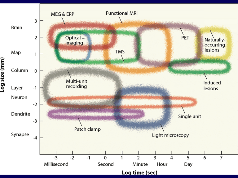
Cognitive Neuroscience MRI vs. fMRI MRI studies brain anatomy. Functional MRI (fMRI) studies brain function. Source: Jody Culham’s fMRI for Dummies web site Brain Imaging: Anatomy CAT Photography PET MRI Source: modified from Posner & Raichle, Images of Mind The major methods • • • • Single-unit recording Lesion studies Transcranial magnetic stimulation (TMS) Neurosurgery-related methods – Direct cortical stimulation – Split-brain – WADA • Functional imaging – Electromagnetic: EEG, MEG – Hemodynamic: PET, fMRI Single unit recording • • Used extensively in animal studies A microelectrode is inserted into brain tissue and recordings of action potentials can be made from nearby neurons, ideally a single neuron. – Recordings are typically extracellular • • • The animal can then be presented with various sensory stimuli, or trained to perform some task, and the effects on neural activity can be monitored Advantages: great spatial and temporal resolution Disadvantages: sampling only a very small fraction of a functional neural system Lesion studies • Correlation of functional deficits with regions of damage • Both human and animal studies – In animals, lesions can be made experimentally – In humans, lesions are causes by “experiments of nature” Lesion studies (con’t) • Common types of lesions in humans – Stroke (A “brain attack”) • Ischemic: blockage of blood flow in an artery • Hemmorrhagic: rupture of an artery – Trauma • Open vs. closed head injury – Tumor – Degenerative disease (e.g., alzheimers disease) • In general, the more focal the lesion, the easier it is to link the site of damage to a behavioral deficit Transcranial Magnetic Stimulation • A method for producing temporary focal brain “lesion” (disruption), via stimulation with a strong magnetic field. • With milder fields, can produce “excitation” or facilitation effects. Neurosurgery Methods • Direct cortical stimulation – Delivery of a small electric current directly on the cortical surface – Causes temporary disruption or facilitation of function in cortex being stimulated – Used clinically to map function, so that critical regions can be avoided during tissue resection – Can be done intra-operatively, or more commonly now, via chronically implanted electrode grids Neurosurgery methods (con’t) • Split-brain – Sectioning of corpus callosum as a treatment for medically intractable epilepsy – Can study the separate contributions of the left and right hemispheres to various abilities/tasks W. W. Norton Neurosurgery methods (con’t) • WADA procedure – Injection of sodium amytal (a barbituate), into one and then the other carotid artery temporarily (5-10min) puts half the brain to sleep allowing neurologists to assess function in the awake hemisphere W. W. Norton Neurosurgery methods (con’t) • General considerations – Advantages: better experimental control in some situations, e.g., temporary lesions can be very focal and reversible – Disadvantages: all subjects in these subjects are undergoing these procedures because they have a neurological disorder, therefore it is not clear how generalizable the results are. Cognitive Neuroscience PET and fMRI Activation Source: Posner & Raichle, Images of Mind Cognitive Neuroscience Cognitive Neuroscience • PET scans = Positron Emission Tomography scans • Rely upon electrical activity calling for local increase in oxygen and energy • Based upon detected photons emitted from electron-positron annihilations. • Slow, some risk, costly Cognitive Neuroscience Cognitive Neuroscience Cognitive Neuroscience Cognitive Neuroscience Cognitive Neuroscience Cognitive Neuroscience Cognitive Neuroscience fMRI Setup fMRI Experiment Stages: Prep 1) Prepare subject • Consent form • • Safety screening Instructions 2) Shimming • putting body in magnetic field makes it non-uniform • adjust 3 orthogonal weak magnets to make magnetic field as homogenous as possible 3) Sagittals Note: That’s one g, two t’s Take images along the midline to use to plan slices Source: Jody Culham’s fMRI for Dummies web site fMRI Experiment Stages: Anatomicals 4) Take anatomical (T1) images • high-resolution images (e.g., 1x1x2.5 mm) • • 3D data: 3 spatial dimensions, sampled at one point in time 64 anatomical slices takes ~5 minutes Source: Jody Culham’s fMRI for Dummies web site Slice Terminology VOXEL (Volumetric Pixel) Slice Thickness e.g., 6 mm In-plane resolution e.g., 192 mm / 64 = 3 mm 3 mm 6 mm SAGITTAL SLICE IN-PLANE SLICE Number of Slices e.g., 10 Matrix Size e.g., 64 x 64 Field of View (FOV) e.g., 19.2 cm Source: Jody Culham’s fMRI for Dummies web site 3 mm fMRI Experiment Stages: Functionals 5) Take functional (T2*) images • images are indirectly related to neural activity • • • • usually low resolution images (3x3x5 mm) all slices at one time = a volume (sometimes also called an image) sample many volumes (time points) (e.g., 1 volume every 2 seconds for 150 volumes = 300 sec = 5 minutes) 4D data: 3 spatial, 1 temporal … first volume (2 sec to acquire) Source: Jody Culham’s fMRI for Dummies web site Activation Statistics Functional images ~2s ROI Time Course fMRI Signal (% change) Time Condition Statistical Map superimposed on anatomical MRI image Time Region of interest (ROI) ~ 5 min Source: Jody Culham’s fMRI for Dummies web site Statistical Maps & Time Courses Use stat maps to pick regions Then extract the time course Source: Jody Culham’s fMRI for Dummies web site 2D 3D Source: Jody Culham’s fMRI for Dummies web site Design Jargon: Runs session: all of the scans collected from one subject in one day run (or scan): one continuous period of fMRI scanning (~5-7 min) experiment: a set of conditions you want to compare to each other condition: one set of stimuli or one task Note: Terminology can vary from one fMRI site to another (e.g., some places use “scan” to refer to what we’ve called a volume). 4 stimulus conditions + 1 baseline condition (fixation) A session consists of one or more experiments. Each experiment consists of several (e.g., 1-8) runs More runs/expt are needed when signal:noise is low or the effect is weak. Thus each session consists of numerous (e.g., 5-20) runs (e.g., 0.5 – 3 hours) Source: Jody Culham’s fMRI for Dummies web site Design Jargon: Paradigm paradigm (or protocol): the set of conditions and their order used in a particular run run epoch: one instance of a condition first “objects right” epoch second “objects right” epoch volume #1 (time = 0) Time epoch 8 vol x 2 sec/vol = 16 sec volume #105 (time = 105 vol x 2 sec/vol = 210 sec = 3:30) fMRI Equipment Magnet (4T) Gradient Coil 4T magnet RF Coil gradient coil (inside) Head Coil Source: Joe Gati, photos Surface Coil Source: Jody Culham’s fMRI for Dummies web site What Does fMRI Measure? • Big magnetic field – protons (hydrogen molecules) in body become aligned to field • RF (radio frequency) coil – radio frequency pulse – knocks protons over – as protons realign with field, they emit energy that coil receives (like an antenna) • Gradient coils – make it possible to encode spatial information • MR signal differs depending on – concentration of hydrogen in an area (anatomical MRI) – amount of oxy- vs. deoxyhemoglobin in an area (functional MRI) BOLD signal Blood Oxygen Level Dependent signal neural activity blood flow oxyhemoglobin T2* MR signal Source: fMRIB Brief Introduction to fMRI Magnet Safety The whopping strength of the magnet makes safety essential. Things fly – Even big things! Source: www.howstuffworks.com Source: http://www.simplyphysics.com/ flying_objects.html Source: Jody Culham’s fMRI for Dummies web site Cognitive Neuroscience Cognitive Neuroscience Cognitive Neuroscience Cognitive Neuroscience Cognitive Neuroscience Cognitive Neuroscience Cognitive Neuroscience Cognitive Neuroscience Cognitive Neuroscience Cognitive Neuroscience Cognitive Neuroscience Cognitive Neuroscience Cognitive Neuroscience Cognitive Neuroscience Cognitive Neuroscience Cognitive Neuroscience Cognitive Neuroscience Cognitive Neuroscience Cognitive Neuroscience Cognitive Neuroscience Cognitive Neuroscience Cognitive Neuroscience Cognitive Neuroscience Cognitive Neuroscience Cognitive Neuroscience Cognitive Neuroscience Cognitive Neuroscience Cognitive Neuroscience Cognitive Neuroscience QuickTime™ and a YUV420 codec decompressor are needed to see this picture. fMRI Activation Flickering Checkerboard OFF (60 s) - ON (60 s) -OFF (60 s) - ON (60 s) - OFF (60 s) Brain Activity Time Source: Kwong et al., 1992 Cognitive Neuroscience Methods QuickTime™ and a YUV420 codec decompressor are needed to see this picture. Cognitive Neuroscience How can we define regions? 1. Talairach coordinates 2. Anatomical localization 3. Functional localization • Region of interest (ROI) analyses • already covered in Design lectures so will not be reconsidered here Talairach Coordinate System Individual brains are different shapes and sizes… How can we compare or average brains? Talairach & Tournoux, 1988 • squish or stretch brain into “shoe box” • extract 3D coordinate (x, y, z) for each activation focus Note: That’s TalAIRach, not TAILarach! Source: Brain Voyager course slides Rotate brain into ACPC plane Corpus Callosum Fornix Find anterior commisure (AC) Find posterior commisure (PC) ACPC line = horizontal axis Note: official Tal sez use top of AC and bottom of PC Pineal Body “bent asparagus” Source: Duvernoy, 1999 Deform brain into Talairach space Mark 8 points in the brain: • anterior commisure • posterior commisure • front • back • top • bottom (of temporal lobe) • left • right Squish or stretch brain to fit in “shoebox” of Tal system y<0 AC=0 y y>0 z y>0 ACPC=0 y<0 x Extract 3 coordinates Left is what?!!! Neurologic (i.e. sensible) convention • left is left, right is right L R - Note: Make sure you know what your magnet and software are doing before publishing left/right info! + x=0 Radiologic (i.e. stupid) convention • left is right, right is left R L + x=0 Note: If you’re really unsure which side is which, tape a vitamin E capsule to the one side of the subject’s head. It will show up on the anatomical image. How to Talairach For each subject: • • • Rotate the brain to the ACPC Plane (anatomical) Deform the brain into the shoebox (anatomical) Perform the same transformations on the functional data For the group: Either a) Average all of the functionals together and perform stats on that b) Perform the stats on all of the data (GLM) and superimpose the statmaps on an averaged anatomical (or for SPM, a reference brain) Averaged anatomical for 6 subjects Averaged functional for 7 subjects Talairach Atlas Brodmann’s Areas Brodmann (1905): Based on cytoarchitectonics: study of differences in cortical layers between areas Most common delineation of cortical areas More recent schemes subdivide Brodmann’s areas into many smaller regions Monkey and human Brodmann’s areas not necessarily homologous Talairach Pros and Cons Advantages • widespread system • allows averaging of fMRI data between subjects • allows researchers to compare activation foci • easy to use Disadvantages • based on the squished brain of an elderly alcoholic woman (how representative is that?!) • not appropriate for all brains (e.g., Japanese brains don’t fit well) • activation foci can vary considerably – other landmarks like sulci may be more reliable Anatomical Localization Sulci and Gyri gray matter (dendrites & synapses) BANK white matter (axons) FISSURE FUNDUS Source: Ludwig & Klingler, 1956 in Tamraz & Comair, 2000 Variability of Sulci Source: Szikla et al., 1977 in Tamraz & Comair, 2000 Variability of Functional Areas Watson et al., 1995 -functional areas (e.g., MT) vary between subjects in their Talairach locations -the location relative to sulci is more consistent Source: Watson et al. 1995 Cortical Surfaces segment gray-white matter boundary render cortical surface inflate cortical surface sulci = concave = dark gray gyri = convex = light gray Advantages • surfaces are topologically more accurate • alignment across sessions and experiments allows task comparisons Source: Jody Culham Cortical Inflation Movie QuickTime™ and a YUV420 codec decompressor are needed to see this picture. Movie: unfoldorig.mpeg http://cogsci.ucsd.edu/~sereno/unfoldorig.mpg Source: Marty Sereno’s web page Cortical Flattening 2) make cuts along the medial surface (Note, one cut typically goes along the fundus of the calcarine sulcus though in this example the cut was placed below) 1) inflate the brain 3) unfold the medial surface so the cortical surface lies flat 4) correct for the distortions so that the true cortical distances are preseved Source: Brain Voyager Getting Started Guide Spherical Averaging Future directions of fMRI: Use cortical surface mapping coordinates Inflate the brain into a sphere Use sulci and/or functional areas to match subject’s data to template Cite “latitude” & “longitude” of spherical coordinates QuickTime™ and a YUV420 codec decompressor are needed to see this picture. Movie: brain2ellipse.mpeg http://cogsci.ucsd.edu/~sereno/coord1.mpg Source: Marty Sereno’s web page Source: Fischl et al., 1999 Spherical Averaging Source: MIT HST583 online course notes Functional imaging • Electroencephalography (EEG) – Scalp electrodes measure the summed electrical activity of large populations of synchronously active neurons – Can look at the changes in this signal as a function of mental activity • Changes in synchrony of different populations of neurons • Changes in morphology of EEG signals that are time-locked to an event (e.g., a perceptual stimulus), this is called event-related potentials (ERPs) W. W. Norton W. W. Norton Cognitive Neuroscience Cognitive Neuroscience Cognitive Neuroscience Cognitive Neuroscience Cognitive Neuroscience Cognitive Neuroscience Cognitive Neuroscience Cognitive Neuroscience Cognitive Neuroscience Cognitive Neuroscience Cognitive Neuroscience Cognitive Neuroscience Magnetoencephalography Magnetoencephalography Front Magnetoencephalography Magnetoencephalography MEG Results Right hippocampal activation during place learning was observed in 2/7 children with FA Right hippocampal activation during place learning was observed in 6/7 controls Magnetic Resonance Spectroscopy Cognitive Neuroscience Cognitive Neuroscience Cognitive Neuroscience Cognitive Neuroscience
