DEEP VENOUS THROMBOSIS AND PULMONARY EMBOLISM
advertisement
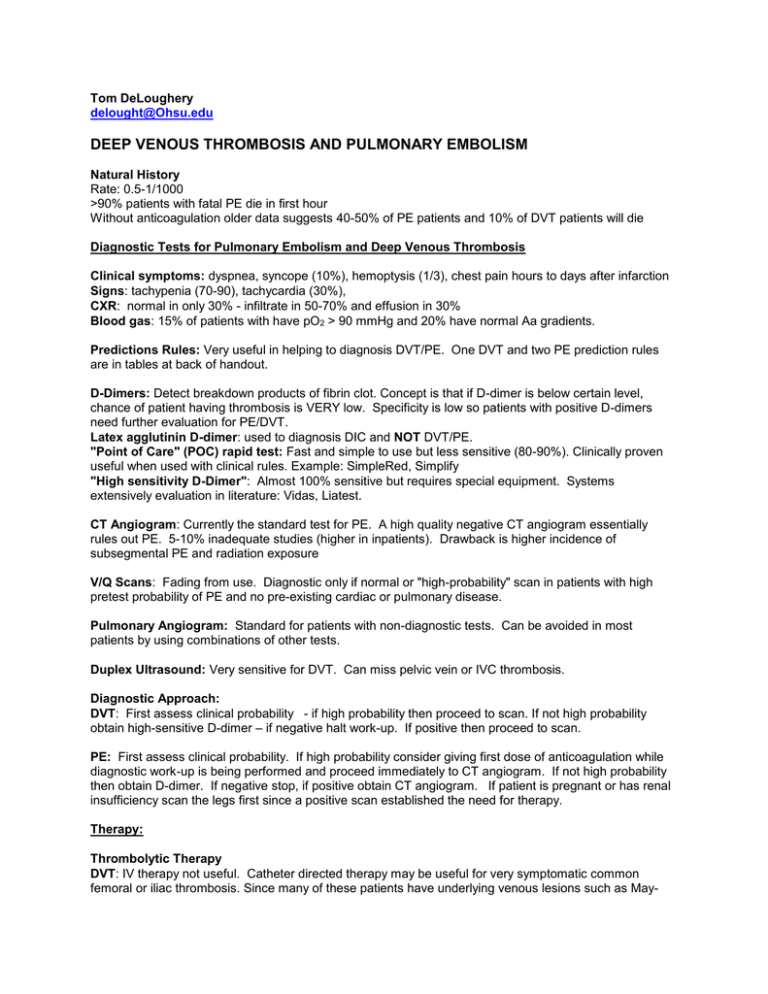
Tom DeLoughery delought@Ohsu.edu DEEP VENOUS THROMBOSIS AND PULMONARY EMBOLISM Natural History Rate: 0.5-1/1000 >90% patients with fatal PE die in first hour Without anticoagulation older data suggests 40-50% of PE patients and 10% of DVT patients will die Diagnostic Tests for Pulmonary Embolism and Deep Venous Thrombosis Clinical symptoms: dyspnea, syncope (10%), hemoptysis (1/3), chest pain hours to days after infarction Signs: tachypenia (70-90), tachycardia (30%), CXR: normal in only 30% - infiltrate in 50-70% and effusion in 30% Blood gas: 15% of patients with have pO2 > 90 mmHg and 20% have normal Aa gradients. Predictions Rules: Very useful in helping to diagnosis DVT/PE. One DVT and two PE prediction rules are in tables at back of handout. D-Dimers: Detect breakdown products of fibrin clot. Concept is that if D-dimer is below certain level, chance of patient having thrombosis is VERY low. Specificity is low so patients with positive D-dimers need further evaluation for PE/DVT. Latex agglutinin D-dimer: used to diagnosis DIC and NOT DVT/PE. "Point of Care" (POC) rapid test: Fast and simple to use but less sensitive (80-90%). Clinically proven useful when used with clinical rules. Example: SimpleRed, Simplify "High sensitivity D-Dimer": Almost 100% sensitive but requires special equipment. Systems extensively evaluation in literature: Vidas, Liatest. CT Angiogram: Currently the standard test for PE. A high quality negative CT angiogram essentially rules out PE. 5-10% inadequate studies (higher in inpatients). Drawback is higher incidence of subsegmental PE and radiation exposure V/Q Scans: Fading from use. Diagnostic only if normal or "high-probability" scan in patients with high pretest probability of PE and no pre-existing cardiac or pulmonary disease. Pulmonary Angiogram: Standard for patients with non-diagnostic tests. Can be avoided in most patients by using combinations of other tests. Duplex Ultrasound: Very sensitive for DVT. Can miss pelvic vein or IVC thrombosis. Diagnostic Approach: DVT: First assess clinical probability - if high probability then proceed to scan. If not high probability obtain high-sensitive D-dimer – if negative halt work-up. If positive then proceed to scan. PE: First assess clinical probability. If high probability consider giving first dose of anticoagulation while diagnostic work-up is being performed and proceed immediately to CT angiogram. If not high probability then obtain D-dimer. If negative stop, if positive obtain CT angiogram. If patient is pregnant or has renal insufficiency scan the legs first since a positive scan established the need for therapy. Therapy: Thrombolytic Therapy DVT: IV therapy not useful. Catheter directed therapy may be useful for very symptomatic common femoral or iliac thrombosis. Since many of these patients have underlying venous lesions such as May- Thurner syndrome, venoplasty or venous stenting can be done with the lytic therapy. PE: Consider only in the patient with refractory hypotension. Current data does NOT support use in patients with right heart dysfunction but no hypotension. Surgical or catheter directed embolectomy: consider in patient with proven PE and refractory hypotension. Inferior Vena Cava Filters: prevents most but not all PE. Since most indications are short-term consider use of retrievable filter. Will increase risk of future DVT. Uses: Prevention of PE in patient who has DVT but cannot be anticoagulated. Prevention of PE in patient who has DVT, can be anticoagulated but very ill. Key is to still anticoagulate the patient as soon as it is feasible to do so. Can remove filter with patient anticoagulated Compression Stockings: Knee-high stockings 30-40 mmHg compression does not prevent postphlebitic syndrome but may help pain in some patients. Best rest: Shown in multiple clinical trials NOT to be useful. Patients with PE/DVT should be encouraged to ambulate as this has been shown to decrease symptoms. In or Outpatient Therapy: Multiple clinical trials have shown that patients with DVT/PE can be treated as an outpatient UNLESS: 1) Patients who need to be hospitalized for other reasons than there DVT/PE such as MI etc... 2) Patients with active bleeding or considered to be at high risk for bleeding. 3) Patients who are hemodynamic instability or a requirement for oxygen therapy to maintain normal oxygen saturation. 4) Patients with contraindications to heparin. Another risk stratification tool is the Pulmonary Embolism Severity Index Predictors Points Assigned Demographics Age in years Age in years Male sex +10 Comorbid Conditions Cancer +30 Heart Failure +10 Chronic Lung Disease +10 Clinical Findings Pulse ≥ 100 +20 Systolic Blood Pressure < 100 mmHg +30 Respiratory rare ≥ 30 +20 Temperature < 36 C +20 Altered mental status +60 O2 saturation < 90% +20 PESI Score ≤65 66-85 86-105 106-125 125 Class I II III IV V Risk Low Low High High High 30 day Mortality range 0-1.1 0-3.1 0-6.5 3.4-10.4 9.2-24.5 (Arch IM 170:1383, 2010, J Thom Haem 8:1509, 2010, 8:517, 2010) Heparin LMWH has been shown to be as effective if not more effective for all venous thrombosis ranging from sub-massive PE to superficial DVT. Dosing: Enoxaparin 1mg/kg/12 hours (1.5mg/kg/24 hours for low risk patients) Fondaparinux: 7.5 mg/day (if < 50kg: 5mg/day, if > 100 kg: 10 mg/day) • Contraindicated in renal disease • Limited Pregnancy data Monitoring: most patients do not need monitoring for routine therapy. Therapeutic range best established for enoxaparin - 0.7-1.1 anti-Xa units performed FOUR hours after injection. Adjustments for special patients: Obesity: no adjustment or capping of dose for weight. Check level after third dose. Renal disease: Clearance 10-30: 0.65mg/kg/12 hours, < 10: 1mg/kg/24 hours. Check level after third dose. Pregnancy: LMWH has been established to be both effective and safer in pregnancy than standard heparin. Dose for body weight and check levels after third dose then every month. Can use LMWH or warfarin with breast feeding. Direct Oral Anticoagulants All shown safe and effective in DVT/PE - Xa inhibitors safer than LMWH/Warfarin Apixaban - 10mg bid x 7 days then 5mg bid Dabigatran - heparin for 5 days then 150mg bid Edoxaban - heparin for 5 days then 60mg/day Rivaroxaban - 15 mg bid x 21 days then 20mg/day Warfarin: start evening of PE/DVT diagnosis. Most patients start with two doses of 5 mg/day. In younger patients (<40) consider 10 mg and in the very old (>75) use 2.5mg. All patients should receive at least FIVE days of heparin. Consider long term LMWH in cancer patients. Reversal of Anticoagulation LMWH – Protamine effective. Time after dose 0-4 hours: 1 mg protamine/1mg of enoxaparin or 100units other LMWH and repeat with 50% dose in 4 hours. Time 4-8 hours 0.5 mg of protamine/ 1mg of enoxaparin or 100 units other LMWH Fondaparinux: Protamine NOT effective. If life threatening bleeding is present use rVIIa. Direct oral anticoagulants - no difference in bleeding outcomes when compared to warfarin. PCC 50 units/kg Warfarin: PO vitamin K effective within 6-24 hours. IV rapid (4-6 hours) but most be given over 1 hour. Sub-q or IM NOT recommended due to erratic onset of action. Not Bleeding: Goal to get INR back in therapeutic range INR 4.5-10: 1 mg po vitamin K INR > 10 2.5 mg po vitamin K Bleeding: Goal to reverse INR (short term risk of bleeding >>risk of thrombosis) INR 2-4.5: 1-2.5 mg vitamin K ± 15 ml/kg of FFP INR 4.5 - 10: 2.5 - 5 mg vitamin K ± 15 ml/kg of FFP INR > 10: 5 - 10 mg of vitamin K ± 15 ml/kg of FFP Intracranial hemorrhage: 4-factor PCC If INR 2-4: 25 units/kg (not to exceed 2500 units) If INR 4-6: 35 units/kg (not to exceed 3500 units) If INR > 6: 50 units/kg (not to exceed 5000 units) Duration of Therapy Superficial Venous Thrombosis: most respond to NSAID/heat but data show that at least a 10-12 day course (up to 42 days) of prophylactic LMWH or fondaparinux is more effective that NSAIDs and should be considered in patients with extensive (> 5cm), greater saphenous, or painful SVT. Upper Extremity DVT: Catheter related: In PICC deep venous thrombosis hold anticoagulation unless very symptomatic (pain limited use of arm, thrombus extension into SVC) and then just for 4-6 weeks. Removal of PICC is only treatment associated with resolution of thrombosis. Remember the basilic and cephalic are superficial veins. For tunneled catheters consider removal and 1-3 months therapy depending on bleeding risk and extent of thrombosis. If catheter is not removed then need 3 months of therapy. Spontaneous: 3 month of therapy. Consider catheter directed thrombolytic therapy for extensive thrombosis especially if in a dominant arm or a young patient - both for symptom relief and to find any anatomical lesions Muscular Calf Vein (soleus or gastrocnemius) Thrombosis: 10 days of therapeutic LMWH or direct oral anticoagulants. Calf Vein Thrombosis: 6 weeks of therapy. Proximal Vein Thrombosis (popliteal vein and above): Duration of therapy influenced by number of thrombosis and presence of provoking factors. Catheter directed therapy is useful for symptomatic common femoral or more proximal thrombosis. Since many of these patients have underlying venous lesions such as May-Thurner syndrome, venoplasty or venous stenting can be done with the lytic therapy. Provoked first DVT or PE: 3 months. • Provoking factors: trauma, surgery, bedrest > 72 hours, pregnancy, estrogen, very long (> 10 hours) plane flights. Idiopathic first DVT or PE: Strongly consider indefinite therapy with warfarin INR 2-3 or direct oral anticoagulants. High risk (10-25%) of recurrence in next 2 years without anticoagulation. Two or more Lower extremity proximal DVT or PE: Indefinite anticoagulation . Pregnancy: LMWH has been established to be both effective and safer in pregnancy than standard heparin. Dose for body weight and check levels after third dose then every month. Can use LMWH or warfarin with breast feeding. Duration – entire course of pregnancy and at least 6 weeks after delivery – total should be at least three months. Thrombophilia • Inherited hypercoagulable states – raises risk of first DVT but not a predictor of recurrence. Multiple guidelines recommend not checking in provoked thrombosis • Severe acquired states – antiphospholipid antibody syndrome, myeloproliferative disease, PNH, cancer – consider long term anticoagulation Cancer Use of LMWH at least 3-6 month should be considered especially if lung cancer or pancreatic cancer. Long term LMWH is mandatory for warfarin failures. Note that studies have shown that incidentally discovered PE in cancer patients has the same adverse outcome as symptomatic PE and requires aggressive therapy. Visceral Vein Thrombosis: Portal vein thrombosis - unless discovered incidentally while screening cirrhotics for hepatomas all require anticoagulation. If provoked by surgery or infections 3 months otherwise indefinite. Consider JAK2/PNH screening in idiopathic cases. Budd-Chiari - indefinite anticoagulation and JAK2/PNH screening. DVT Prophylaxis: Risk Assessment: Low-Risk Patients Fully mobile medicine patients Procedures lasting less than 30 minutes Medium Risk Patients Most medical or general, urological, or surgical patients High Risk Patients Previous history of venous thrombosis (or strong family history) Pelvic or abdominal surgery for malignancy. Lower limb orthopedic surgery Trauma patients Surgery in patients with other risk factors-previous pulmonary embolism, CHF, cancer Recommendations: Low-Risk Patients Early Ambulation Medium Risk Patients Standard Heparin 5000 TID or LMWH or Intermittent Pneumatic Compression Stocks (IPC) if high risk of bleeding High Risk Patients Apixaban 2.5 mg bid Enoxaparin 40mg/day Fondaparinux 2.5 mg/day Rivaroxaban 10mg/day Warfarin INR 2-3 Special Situations: Neurosurgery: non-malignant: IPC Malignant: IPC plus LMWH High risk Medical or ICU: LMWH DVT predication Rule: Variable Points Active Cancer +1 Paralysis or recent plaster immobilization of lower extremity +1 Recently bedridden for > 3 days or major surgery within 4 weeks +1 Local tenderness +1 Calf Swelling greater than 3cm than asymptomatic side (measured 10 cm below tibial tuberosity) +1 Pitting edema in symptomatic leg +1 Dilated superficial veins (non-varicose) in symptomatic leg only +1 Alternative diagnoses as or more likely than DVT -2 Low probability <0, Moderate probability 1-2 and high probability >3 Table 2: Clinical Probability Score for Pulmonary Embolism (1) Variable Points Clinical signs and symptoms of DVT +3 Pe as likely or more likely than alternative diagnosis +3 Immobilization or surgery in past four weeks 1.5 Previous PE or DVT 1.5 Heart rate more than 100/min 1.5 Hemoptysis 1 Active Cancer 1 Low probability <2, moderate probability 2-6 and high probability > 6 Wells Ann Int Med 135: 108, 2001 Clinical Probability Score for Pulmonary Embolism (2) Wicki Arch Int Med 161:92, 2001 Variable Points Previous DVT or PE +2 Heart rate > 100 +1 Recent Surgery +3 Age: 60-79 +1 >80 +2 PaCO2 < 36 mmHg +2 36-40 mmHg +1 P02 <50 mmHg +4 50-59 mmHg +3 60-69 mmHg +2 70-79 mmHg +1 Atelectasis +1 Elevated Hemi-diaphragm +1 Low Probability 0-4, Intermediate probability 5-8, High >9 Nomograms for Warfarin Loading Ma Crowther, L Harrison and J Hirsh Annals of Internal Medicine 127:333, 1997 5 Mg Warfarin Nonomgram Day INR Dosage (Mg) Day 5.0 1 < 1.5 5.0 2 1.5-1.9 1 2 3 4 5 6 10 mg warfarin nonomgram INR Dosage (Mg) 10.0 < 1.5 7.5-10.0 2.5 1.5-1.9 2.5 2.0-2.5 1.0-2.5 2.0-2.5 1.0-2.5 >2.5 0.0 >2.5 0.0 <1.5 5.0-10.0 <1.5 5.0-10.0 1.5-1.9 2.5-5.0 1.5-1.9 2.5-5.0 2.0-2.5 0.0-2.5 2.0-2.5 0.0-2.5 2.5-3.0 0.0-2.5 2.5-3.0 0.0-2.5 >3.0 0.0 >3.0 0.0 <1.5 10.0 <1.5 10.0 1.5-1.9 5.0-7.5 1.5-1.9 5.0-7.5 2.0-3.0 0.0-0.5 2.0-3.0 0.0-0.5 >3.0 0.0 >3.0 0.0 <1.5 10.0 <1.5 10.0 1.5-1.9 7.5-10.0 1.5-1.9 7.5-10.0 2.0-3.0 0.0-5.0 2.0-3.0 0.0-5.0 >3.0 0.0 >3.0 0.0 <1.5 7.5-12.5 <1.5 7.5-12.5 1.5-1.9 5.0-10.0 1.5-1.9 5.0-10.0 2.0-3.0 0.0-7.5 2.0-3.0 0.0-7.5 >3.0 0.0 >3.0 0.0 3 4 5 6 Maintenance Warfarin Adjustment Nonomgram (Hatheway and Goodnight) INR Dose Change 1.1-1.4 Day 1: Add 10-20% Twd* Weekly: Increase TWD by 10-20% Return: 1 Week 1.5-1.9 Day 1: Add 5-10% of TWD Weekly: Increase Twd by 5-10% Return: 2 Weeks 2.0-3.0 No Change Return: 4 Weeks 3.1-3.9 Day 1: Subtract 5-10% TWD Weekly: Reduce TWD by 10-20% Return: 2 Weeks 4.0-5.0 Day 1: No Warfarin Weekly: Reduce TWD by 10-20% Return: 1 Week > 5.0 Stop Warfarin until INR <3.0 Decreased TWD by 20-50% Return Daily *TWD = Total Weekly Dose REFERENCES: GENERAL REFERENCE Antithrombotic therapy for VTE disease: Antithrombotic Therapy and Prevention of Thrombosis, 9th ed: American College of Chest Physicians Evidence-Based Clinical Practice Guidelines Chest. 2012 Feb;141(2 Suppl):e419S-94S. Antithrombotic and Thrombolytic Therapy, 8th Ed: ACCP Guidelines Chest 2008 Jun; 133 (Suppl) : 67S-968S. References Ageno W, Dentali F, Squizzato A. How I treat splanchnic vein thrombosis. Arch Intern Med. 2002 Dec 9-23;162(22):2521-3. Arnason T, Wells PS, Forster AJ. Appropriateness of diagnostic strategies for evaluating suspected venous thromboembolism. Thromb Haemost. 2007 Feb;97(2):195-201. Blood. 2014 Dec 11;124(25):3685-91 Dalen JE.The uncertain role of thrombolytic therapy in the treatment of pulmonary embolism. Jones MA, Lee DY, Segall JA, Landry GJ, Liem TK, Mitchell EL, Moneta GL. Characterizing resolution of catheter-associated upper extremity deep venous thrombosis. J Vasc Surg. 2010 Jan;51(1):108-13 Kearon C, Ginsberg JS, Kovacs MJ, Anderson DR, Wells P, Julian JA, MacKinnon B, Weitz JI, Crowther MA, Dolan S, Turpie AG, Geerts W, Solymoss S, van Nguyen P, Demers C, Kahn SR, Kassis J, Rodger M, Hambleton J, Gent M; Extended Low-Intensity Anticoagulation for ThromboEmbolism Investigators. Comparison of low-intensity warfarin therapy with conventional-intensity warfarin therapy for long-term prevention of recurrent venous thromboembolism. N Engl J Med. 2003 Aug 14;349(7):631-9 Konstantinides SV, Torbicki A.Management of pulmonary embolism: recent evidence and the new European guidelines.Eur Respir J. 2014 Dec;44(6):1385-90 Lee AY, Levine MN, Baker RI, Bowden C, Kakkar AK, Prins M, Rickles FR, Julian JA, Haley S, Kovacs MJ, Gent M; Randomized Comparison of Low-Molecular-Weight Heparin versus Oral Anticoagulant Therapy for the Prevention of Recurrent Venous Thromboembolism in Patients with Cancer (CLOT) Investigators.Low-molecular-weight heparin versus a coumarin for the prevention of recurrent venous thromboembolism in patients with cancer.N Engl J Med. 2003 Jul 10;349(2):146-53 Masuda EM, Lee RW, Okazaki IJ, Benyamini R, Kistner RL, Thrombophilia testing has limited usefulness in clinical decision-making and should be used selectively. J Vas Surg 3:228-235, 2015 Pinede L, Ninet J, Duhaut P, Chabaud S, Demolombe-Rague S, Durieu I, Nony P, Sanson C, Boissel JP; Investigators of the "Durée Optimale du Traitement AntiVitamines K" (DOTAVK) Study.Comparison of 3 and 6 months of oral anticoagulant therapy after a first episode of proximal deep vein thrombosis or pulmonary embolism and comparison of 6 and 12 weeks of therapy afte Schwarz T, Schmidt B, Beyer J, Schellong SM.Therapy of isolated calf muscle vein thrombosis with low-molecular-weight heparin. Blood Coagul Fibrinolysis. 2001 Oct;12(7):597-9. Stein PD, Sostman HD, Bounameaux H, Buller HR, Chenevert TL, Dalen JE, Goodman LR, Gottschalk A, Hull RD, Leeper KV Jr, Pistolesi M, Raskob GE, Wells PS, Woodard PK.vChallenges in the diagnosis of acute pulmonary embolism.vAm J Med. 2008 Jul;121(7):565-71 Superficial Thrombophlebitis Treated By Enoxaparin Study Group.A pilot randomized doubleblind comparison of a low-molecular-weight heparin, a nonsteroidal anti-inflammatory agent, and placebo in the treatment of superficial vein thrombosis. Arch Intern Med. 2003 Jul 28;163(14):1657-63. Tapson VF. Acute pulmonary embolism. N Engl J Med. 2008 Mar 6;358(10):1037-52 Torbicki A. Acute and long term management of pulmonary embolism. Heart. 2010 Sep;96(17):1418-24. van Es N, Coppens M, Schulman S, Middeldorp S, Büller HR.Direct oral anticoagulants compared with vitamin K antagonists for acute venous thromboembolism: evidence from phase 3 trials. Blood. 2014 Sep 18;124(12):1968-75. Vedovati MC, Germini F, Agnelli G, Becattini C.Direct Oral Anticoagulants in Patients With VTE and Cancer: A Systematic Review and Meta-analysis. Chest. 2015 Feb 1;147(2):475-83 Wang TF, Squizzato A, Dentali F, Ageno W. The role of thrombolytic therapy in pulmonary embolism: evidence-based focused review. Blood. 2015 Jan 28. Wells PS, Owen C, Doucette S, Fergusson D, Tran H.Does this patient have deep vein thrombosis? JAMA. 2006 Jan 11;295(2):199-207
