Scanning Probe Microscopy History
advertisement
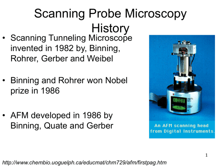
Scanning Probe Microscopy History • Scanning Tunneling Microscope invented in 1982 by, Binning, Rohrer, Gerber and Weibel • Binning and Rohrer won Nobel prize in 1986 • AFM developed in 1986 by Binning, Quate and Gerber 1 http://www.chembio.uoguelph.ca/educmat/chm729/afm/firstpag.htm Hierarchy of Techniques Scanning Probe Microscopy • Scanning Tunneling Microscopy • Atomic Force Microscopy • Contact Mode • Tapping Mode • Non-contact Mode 2 http://www.chembio.uoguelph.ca/educmat/chm729/afm/firstpag.htm 3 STM It ~ Ve-cd • Feedback loop keeps current constant • therefore d is constant • Sample must conduct electricity • Capable of detecting atomic scale defects 4 AFM Contact Mode • Scan tip along surface Hook’s law F=-kx • Maintains constant cantilever deflection (force) using a split photo diode k limits sensitivity (want low k) • Samples can be in liquid state • Works in ambient conditions 5 AFM Tapping Mode • Cantilever oscillates at or below resonance frequency • Maintains constant tip movement • amplitude • Position of scanner stored to create image • Tip must breakthrough water layer without getting stuck • Also works in ambient or liquid 6 Silicon Nitride Probe Spring Constant (k) 0.58, 0.32, 0.12, 0.06 N/m (1) Nominal Tip Radius of Curvature 20 - 60nm Cantilever Lengths 100 & 200μm Cantilever Configuration V-shaped Reflective Coating Gold Sidewall angles 35° on all 4 sides (1)Calculated spring constant values are based on the 0.6μm silicon nitride thickness; however, this value can actually vary from 0.4μm to 0.7μm. Thickness is cubed in the spring constant calculation, thus, actual values can vary substantially. 7 Silicon Probe 8 AFM Imaging and Tip Shape The radius of curvature of the tip limits the resolution of the image that can be taken 9 AFM Imaging and Tip Shape The probe cannot image a sidewall that is steeper than the angle of the tip Silicon nitride probe 10 AFM Imaging and Tip Shape Silicon probe 11 Surface Roughness Measurements Rz : average difference in height between the five highest peaks and five lowest valleys relative to the mean plane Image Ra : average of the absolute values of the surface height deviations measured from the mean plane 1 N R a Z j Z avg N j 1 Z N Image Rq : Root mean square average of height deviations taken from the mean plane. Rq i 1 Z avg 2 i N 12 NanoScope Software 6.13 User Guide, Section 6.4.3 AFM height image of a semiconducting polymer thin film on SiO2 AFM images of SiO2 substrates for FET’s Uncleaned SiO2 5 nm Mean Roughness: 0.44 nm 0 nm -5 nm 5 nm 0 nm -5 nm No detergent, No Plasma SiO2 Mean Roughness: 0.19 nm 6nm tall particle on surface 8/4/08 AFM images of SiO2 substrates for FET’s 5 nm No detergent, Airplasma 5min SiO2 0 nm Mean Roughness: 0.27 nm -5 nm 5 nm 0 nm -5 nm detergent, air plasma 5min SiO2 Mean Roughness: 0.17 nm 6nm tall particles on surface 8/4/08
