Bacterial Transformation with pGLO, Part 1 O
advertisement
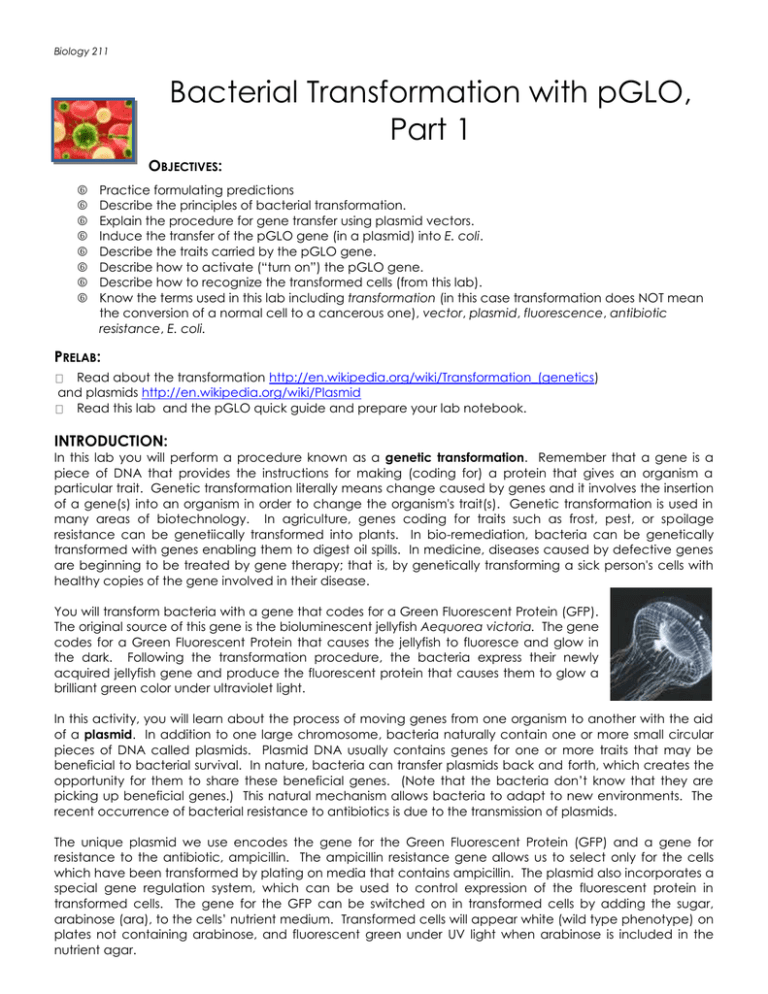
Biology 211 Bacterial Transformation with pGLO, Part 1 OBJECTIVES: Practice formulating predictions Describe the principles of bacterial transformation. Explain the procedure for gene transfer using plasmid vectors. Induce the transfer of the pGLO gene (in a plasmid) into E. coli. Describe the traits carried by the pGLO gene. Describe how to activate (“turn on”) the pGLO gene. Describe how to recognize the transformed cells (from this lab). Know the terms used in this lab including transformation (in this case transformation does NOT mean the conversion of a normal cell to a cancerous one), vector, plasmid, fluorescence, antibiotic resistance, E. coli. PRELAB: Read about the transformation http://en.wikipedia.org/wiki/Transformation_(genetics) and plasmids http://en.wikipedia.org/wiki/Plasmid Read this lab and the pGLO quick guide and prepare your lab notebook. INTRODUCTION: In this lab you will perform a procedure known as a genetic transformation. Remember that a gene is a piece of DNA that provides the instructions for making (coding for) a protein that gives an organism a particular trait. Genetic transformation literally means change caused by genes and it involves the insertion of a gene(s) into an organism in order to change the organism's trait(s). Genetic transformation is used in many areas of biotechnology. In agriculture, genes coding for traits such as frost, pest, or spoilage resistance can be genetiically transformed into plants. In bio-remediation, bacteria can be genetically transformed with genes enabling them to digest oil spills. In medicine, diseases caused by defective genes are beginning to be treated by gene therapy; that is, by genetically transforming a sick person's cells with healthy copies of the gene involved in their disease. You will transform bacteria with a gene that codes for a Green Fluorescent Protein (GFP). The original source of this gene is the bioluminescent jellyfish Aequorea victoria. The gene codes for a Green Fluorescent Protein that causes the jellyfish to fluoresce and glow in the dark. Following the transformation procedure, the bacteria express their newly acquired jellyfish gene and produce the fluorescent protein that causes them to glow a brilliant green color under ultraviolet light. In this activity, you will learn about the process of moving genes from one organism to another with the aid of a plasmid. In addition to one large chromosome, bacteria naturally contain one or more small circular pieces of DNA called plasmids. Plasmid DNA usually contains genes for one or more traits that may be beneficial to bacterial survival. In nature, bacteria can transfer plasmids back and forth, which creates the opportunity for them to share these beneficial genes. (Note that the bacteria don’t know that they are picking up beneficial genes.) This natural mechanism allows bacteria to adapt to new environments. The recent occurrence of bacterial resistance to antibiotics is due to the transmission of plasmids. The unique plasmid we use encodes the gene for the Green Fluorescent Protein (GFP) and a gene for resistance to the antibiotic, ampicillin. The ampicillin resistance gene allows us to select only for the cells which have been transformed by plating on media that contains ampicillin. The plasmid also incorporates a special gene regulation system, which can be used to control expression of the fluorescent protein in transformed cells. The gene for the GFP can be switched on in transformed cells by adding the sugar, arabinose (ara), to the cells’ nutrient medium. Transformed cells will appear white (wild type phenotype) on plates not containing arabinose, and fluorescent green under UV light when arabinose is included in the nutrient agar. Biology 211 Transformation You will be provided with the tools and a protocol for performing genetic transformation in Escherichia coli. These steps are intended to introduce the plasmid DNA into the E. coli cells and provide an environment for the cells to express their newly acquired genes. Many species of bacteria have special membrane proteins for the uptake of DNA from the external environment. E. coli does not appear to have these types of membrane proteins; however, placing E. coli in a relatively high concentration of calcium ions and performing a procedure called “heat shock” will stimulate these cells to take up pieces of foreign DNA. The transfomation consists of three main steps 1. Use a transformation solution of CaCl2 (calcium chloride) 2. Carry out a procedure referred to as heat shock 3. In order for transformed cells to grow in the presence of ampicillin you must provide them with nutrients and a short incubation period to recover from heat shock and to begin expressing their newly acquired genes! Biology 211 pGLO From Wikipedia, the free encyclopedia http://en.wikipedia.org/wiki/PGLO The pGLO plasmid is an engineered plasmid used in biotechnology as a vector for creating genetically modified organisms. GFP was isolated from the jelly fish Aequorea victoria. The plasmid contains three genes of interest to us bla which codes for the enzyme beta-lactamase giving the transformed bacteria resistance to the betalactam family of antibiotics (such as of the penicillin family). The presence of this gene is what allows us to select for the bacteria that have incorporated our plasmid. Beta lactamase will break down ampicillin in the plates and allow the bacteria that have this gene to survive. Any bacteria that have the plasmid should be able to grow on LB/Amp plates Any bacteria that do not have the plasmid will not grow on LB/Amp plates GFP This is the gene for the Green Fluorescent protein itself. The expression of this gene is under control of a bidirectional promoter that also controls the gene for ara C. ara C The ara C gene product is a repressor that STOPS the expression of the Green Fluoresent Protein. GFP can be induced in bacteria containing the pGLO plasmid by growing them on +arabinose plates Arabinose binds to the ara C gene product and removes the repression – allows GFP to be produced. Biology 211 Any bacteria that have the plasmid will produce GFP only on LB/ara plates – green colonies Exercise B: Bacterial Transformation (in lab session) MATERIALS & PROCEDURES 1. Follow the procedures in the “Transformation Kit-Quick Guide” provided in lab. Be sure to record your protocol in your lab notebook!! 2. The plates will be incubated for 24-48 hours, and then placed in a refrigerator to slow the growth of the bacteria. You will than observe the plates in the next lab period to collect your data. 3. Before leaving lab today, make sure that all contaminated items are placed in the biohazard bucket and take care to properly disinfect your lab bench. Data Table Expected Result Growth Green under fluoresecent light Actual Result Growth Green under # of colonies fluorescent light + pGLO on LB/amp + pGLO on LB/amp/ara - pGLO on LB/amp - pGLO on LB POSTLAB: You do not have a formal “post lab” for this week’s work! (But do complete the pre-lab for part 2 before your next lab session!) Bacterial Transformation with pGLO, Part 2 INTRODUCTION: Recall that in your last laboratory session, you performed a transformation, hopefully introducing the pGLO plasmid into E. coli. In this session, we’ll examine our bacterial plates to determine whether or not this transformation was successful. Fortunately, this is an easy task. The Green Florescent Protein (GFP) emits green light when excited by UV light. This allows researchers like ourselves to quickly determine whether or not the protein is being expressed. For this reason, GFP is commonly used in research labs around the world. PROCEDURE: 1. Retrieve your plates from the incubator or fridge. Count the colonies formed on each plate and record this result in an organized table in your lab notebook. Note that if you have a very large number of colonies, you may count either a half or a quarter of the plate and estimate the total plate count from that number. In some cases, you may have so many growing bacterial colonies on your plate that they don’t have the space or resources to form distinct colonies; this is referred to as a bacterial “lawn”. 2. Use one of the UV light boxes in the lab to carefully examine your bacterial plates under UV light. To best see the colonies, you should place the petri dish right side up in the light box, and carefully remove the Biology 211 lid during viewing. Record your observations in the data table you created for your bacterial counts in step 1. 3. When you have finished with your plates, please be sure they are placed in a biohazard bucket and that your work space is properly disinfected. NOTE: LB is the nutrient mixture that is added to the plate agar to feed the bacteria. POSTLAB: Prepare a “Results” and “Discussion” section for this experiment. Your “Results” should contain an organized data table, complete with a descriptive title. Like all “Results” sections in scientific writing, it should also contain a brief (2-3 sentence) description of the results. Your “Discussion” section should comment on your interpretation of the results and any other implications. In particular, be sure your Discussion answers the following questions (in paragraph form): Was your genetic transformation successful? How do you know? Are your results consistent with the predictions you made in your pre-lab? If not, why? Consider the following two pairs of plates. What do the results obtained from these plates tell you about your experiment? 1. -DNA LB and -DNA LB/amp 2. +DNA LB/amp and -DNA LB/amp
